Polysaccharide Microparticles Containing Biological Agents: Their Preparation and Applications

a technology of polysaccharide microparticles and biological agents, applied in the field of polysaccharide microparticles containing biological agents : their preparation and application, can solve the problems of low loading capacity of proteins, compromised system, and inability to load proteins into fine polysaccharide particles, and achieve the effect of facilitating uptak

Inactive Publication Date: 2008-10-09
BIODVERY PHARMATECH LTD
View PDF11 Cites 23 Cited by
  • Summary
  • Abstract
  • Description
  • Claims
  • Application Information

AI Technical Summary

Benefits of technology

[0012]For treatment of tumors using antibodies, the antibody can be loaded using this invention in sustained-release polymer microspheres that can be injected in the region of a solid tumor for localized and prolonged antibody action. For targeting to certain organs which are not feasible for site-specific injection (such as liver), the antibody-carrying microspheres can be made in a size which facilitate up-take by reticular endothelial system of the organ.

Problems solved by technology

Loading proteins into fine polysaccharide particles can be a formidable task due to the fact that reported particle-forming methods generally involve the use of organic solvents, interfacial tension (such as water-oil interfaces or water-air interfaces and other hydrophilic / hydrophobic interfaces), shear stress, elevated temperatures, and other conditions that are hazardous to the loaded proteins.
This system, however, is compromised with some drawbacks.
If solubility of the protein is low, the ratio of polysaccharide / protein will be too high so that loading capacity for proteins will be too low (since the concentration of polysaccharide must be high enough for phase separation).

Method used


Image

  • Polysaccharide Microparticles Containing Biological Agents: Their Preparation and Applications
  • Polysaccharide Microparticles Containing Biological Agents: Their Preparation and Applications
  • Polysaccharide Microparticles Containing Biological Agents: Their Preparation and Applications

Examples


example 1

Preparation of Polysaccharide Glassy Particles (AqueSpheres) Using Low Temperature Aqueous-Aqueous Emulsification

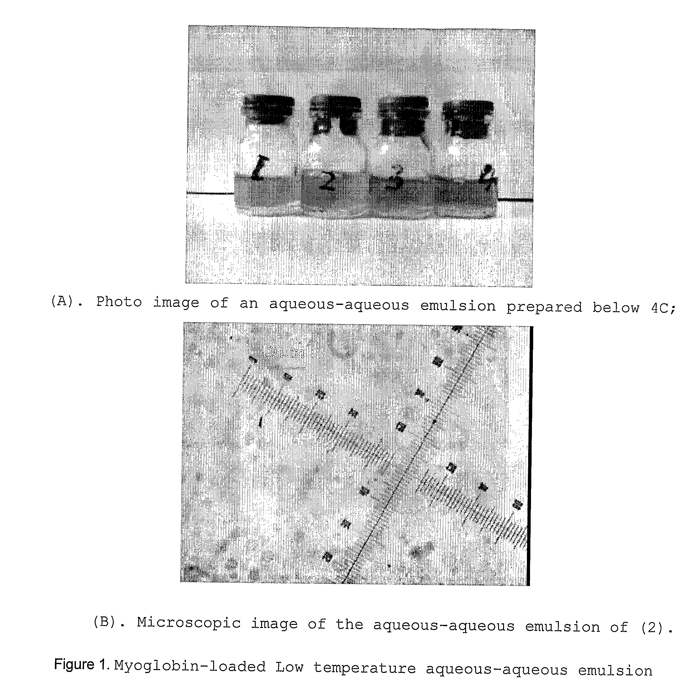
[0080]A fairly stable aqueous-aqueous emulsion was prepared by simply mixing a 10 w / w % dextran solution (containing 2 w / w % myoglobin) with a 10 w / w % PEG solution at 0-4° C. A small molecular sugar, trehalose (1 w / w %) was added in the dextran solution as protein stabilizer for lyophilization. FIG. 1 shows photo and microscopic images of the aqueous-aqueous stored in a refrigerator for 1 hour. The sample was removed from the refrigerator and images were quickly taken before the dispersed phase fuses at elevated temperature.

[0081]The size of the dispersed dextran phase (the droplets) ranged between 3-7 μm in diameter, similar to those prepared using polyelectrolyte stabilizers at room temperature [5,8]. This emulsion sample was then frozen at −20° C, followed by lyophilization and dichloromethane-washing to remove the continuous PEG phase. Photo and microscopic images of...

example 2

Preparation of Polysaccharide Glassy Particles (AqueSpheres) Using Freezing-Induced Phase Separation

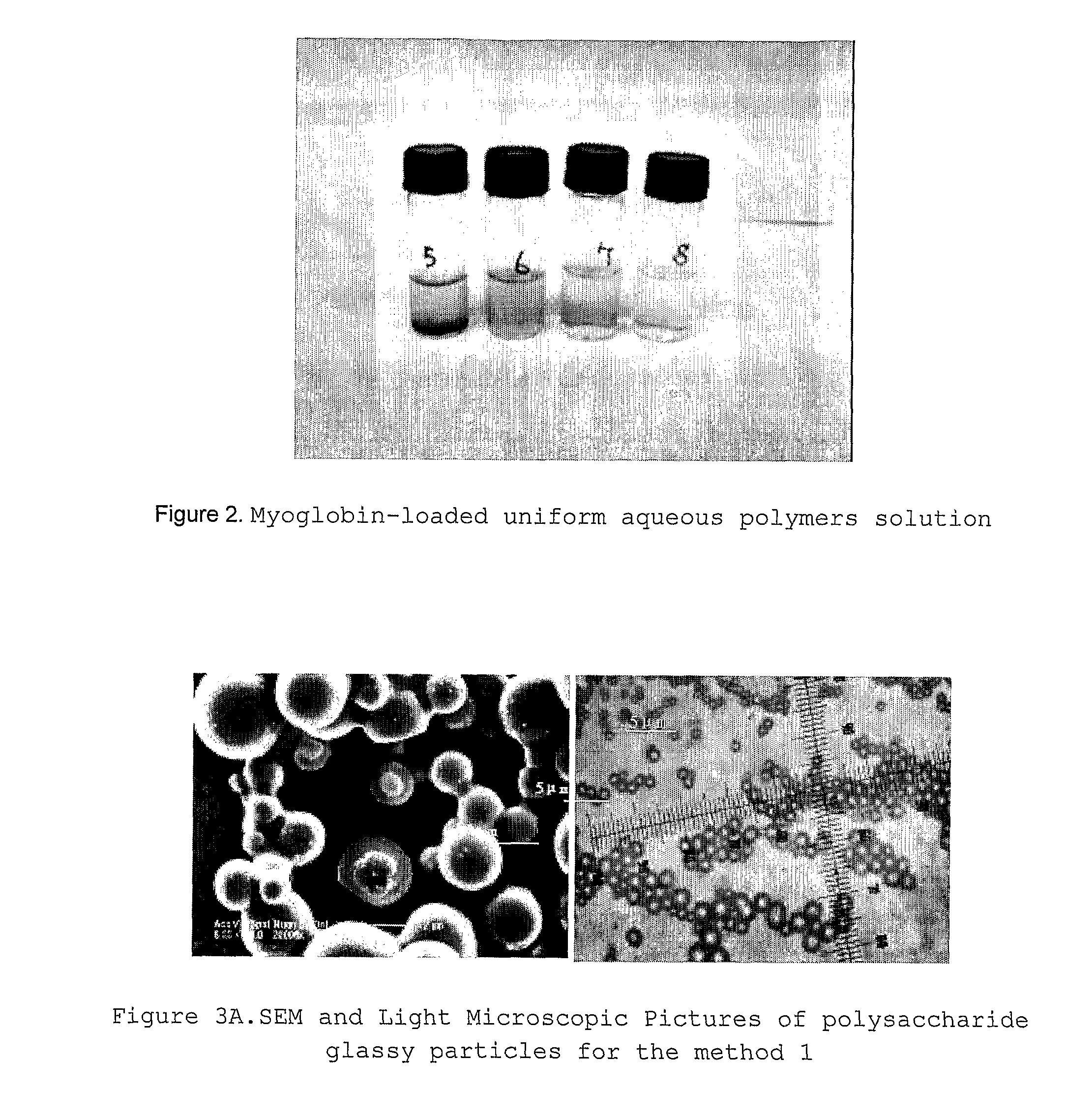
[0082]For a solution system containing dextran and PEG, its phase separation is a function of temperature, and concentration and molecular weight of the hydrophilic polymers [11]. In order to prepare a solution which is single phase at room temperature but becomes two phases (dispersed and continuous phases), a series of solutions containing dextran, PEG and myoglobin but different in concentrations were prepared at room temperature. A small molecular sugar, trehalose (1 w / w %) was added in the dextran solution as protein stabilizer for lyophilization. FIG. 2 shows photographs of the mixed solutions of various concentrations of dextran, PEG and myoglobin (See the figure legend for FIG. 1). Among these samples, that formed by mixing two solutions each containing 10 w / w % dextran and 10 w / w % PEG separated to two phases. All the other samples with lower concentration of dextran and PEG...

example 3

Encapsulation of AqueSpheres into PLGA Microspheres

[0084]Protein containing fine polysaccharide glassy particles (AqueSpheres) may easily encapsulated in PLGA microspheres using a solid-in-oil-in-water (S-O-W) method used in our previous invention [8]. In brief, AqueSphares were suspended in a dichloromethane solution of PLGA, then the suspension was added into a containing polyvinyl alcohol 0.5-1.5 w / w %) and NaCl (5-10 w / w %) at 1 / 6 valume ratio under stirring. Once a composite double emulsion was formed, this S-O-W system was immediately added large cold water (˜0-10 ° C.) under gentle stirring for preliminary hardening, followed by aging and rinsing (to remove polyvinyl alcohol and NaCl). The PLGA microspheres obtained were lyophilized again to remove water and solvent residues prior to storage.

[0085]FIG. 5A shows the optical microscopic images of the PLGA droplets before (left) and after (right) preliminary hardening. FIG. 5B and 5C show electroscopic images of the hardened PLG...


PUM

PropertyMeasurementUnit
diameteraaaaaaaaaa
diameteraaaaaaaaaa
particle sizeaaaaaaaaaa

Abstract

A method of preparing polysaccharide glassy microparticles which are less than 10 μum in diameter and contain structurally delicate agents, such as proteins, peptides, gene materials, vaccines, antibodies, viruses and liposomes using low-temperature aqueous-aqueous emulsification (free of polyelectrolytes) and freezing-induced phase separation. When delicate agents are added to a polysaccharide-PEG two phase system followed by homogenization (or other shear adding process), the agents partition into the polysaccharide dispersed phase preferentially. These processes help to avoid aggregation of proteins caused by interaction with charged polyelectrolytes used for stabilizing the polysaccharide dispersed phase in our previously reported aqueous-aqueous emulsion. When this system is frozen and lyophilized, glassy particles less than 10 μm in diameter containing delicate agents can be formed. These fine polysaccharide particles protect proteins within their hydrophilic glassy matrix, and can therefore be easily suspended in hydrophobic polymer solutions and formulated to various forms of sustained release devices such microsphere, sheets, fibers, coating layers, and scaffolds. The particles can also be dispersed in hydrophilic gels to improve releasing kinetics and to deliver vaccines and antibodies for immune therapy.

Description

CROSS REFERENCES AND RELATED APPLICATIONS[0001]This application claims priority of US provisional application 60 / 712,548 and U.S. Pat. Nos. 9,805,879 and 9,998,393. Throughout this application, various references are refereed to. Disclosures of these publications, in their entireties, are hereby incorporated by the references into this application to more fully describe the state of the art to which this invention pertains.BACKGROUND OF THE INVENTION[0002]When structurally delicate agents, such as proteins, peptides, gene materials, viruses, vaccines, antibodies or liposomes are loaded in degradable polymer-based drug delivery systems, the contact with organic solvents (which are required to dissolve the polymers) often causes denaturation of these agents [1-3]. To avoid this denaturation, proteins and peptides were prepared into solid particles with sugars or polysaccharides prior to encapsulation into water-insoluble polymer devices [4-7]. For sustained-release purposes, the prote...

Claims


Application Information

Patent Timeline
Patent Type & Authority Applications(United States)
IPC IPC(8): A61K9/127A61K47/36A61K38/02A61K38/16A61K47/38A61P43/00A61K9/14A61K31/7052A61K39/00A61K39/395
CPCA61K9/0024A61K9/1623A61K9/1647A61K9/1652A61K9/1694A61K9/19A61K39/00A61L27/48A61L31/10A61L31/129A61K9/7007A61P43/00
Inventor JIN, TUOWU, FEIYUAN, WEIEN
Owner BIODVERY PHARMATECH LTD
Why Eureka
Try Eureka