Immobilization of membrane porteins onto supports via an amphiphile

a technology of membrane proteins and amphiphiles, which is applied in the preparation of sugar derivatives, peptides, enzymology, etc., can solve the problems of difficult or impossible implementation, difficult or impossible to handle for membrane proteins about which little information is available, and more complex membrane proteins

Active Publication Date: 2009-11-05
CENT NAT DE LA RECHERCHE SCI +1
View PDF24 Cites 28 Cited by
  • Summary
  • Abstract
  • Description
  • Claims
  • Application Information

AI Technical Summary

Benefits of technology

[0019]The use of an amphipol as an intermediate for binding to the support has numerous advantages compared with known techniques. The first major advantage is that this process is usable without any change in the method to any membrane protein, irrespective of its origin or whether information is available on this protein or not. In particular, the process is applicable even to a membrane protein the identity of which is unknown. Furthermore, the process is carried out easily, in particular as there is no need to use a detergent in the attachment step of the membrane protein / amphipol complex to the support. This process also provides the attachment of the membrane proteins at the surface of a support with a high density, thus achieving better yields and above all a better sensitivity of the binding assay results of ligands onto the immobilized membrane protein.
[0020]Lastly, the presence of amphipols complexed with membrane proteins guarantees their biochemical stabilization, thus also allowing a greater accuracy and reproducibility of the binding assay results of ligands on the immobilized membrane protein.
[0021]The process developed by the inventors thus allows to readily produce, with a high density, products comprising a support and one or more known or unknown membrane proteins of any kind, attached to the surface thereof, wherein the membrane proteins are further biochemically stabilized in an aqueous medium, thereby making it much easier to carry out binding assay experiments of ligands on immobilized membrane proteins using the product obtained.
[0024]In the present case, the hydrophobic groups of the amphiphilic molecule bind it to the membrane protein, the transmembrane surface of which is hydrophobic per se, and the hydrophilic groups provide solubilization of the whole amphiphilic molecule / membrane protein complex. Preferably, the amphiphilic molecule comprises a number of hydrophobic side chains, enabling it to combine at a plurality of points with the hydrophobic transmembrane surface of membrane proteins, this multiple attachment resulting in very slow desorption kinetics, which makes the combination essentially irreversible. Advantageously, the number of hydrophobic side chains is greater than 10.
[0161]In fact, such a use is very useful in pharmacology for membrane proteins providing therapeutic targets. For each new membrane target identified, a screening of compound libraries is generally carried out to identify various agonist or antagonist, potential drug candidate ligands, for the target in question, these candidates then being optimized in terms of efficiency and non-toxicity.
[0162]As indicated above, the advantage of the products according to the invention for such an application is that the membrane proteins immobilized on the support, as they are complexed with the amphiphilic molecule, are fully soluble in an aqueous solution, and biochemically stabilized in their natural conformation. Thus, detection can be carried out in an aqueous medium while keeping the native structure of the membrane protein, thus allowing not only to simplify the binding detection method, but also to make sure that the test membrane protein is definitely in its native state, and accordingly likely to bind, with the same affinity, the same compounds as within the living organism.

Problems solved by technology

However, the problem is more complex for membrane proteins.
In fact, membrane proteins necessarily expose highly hydrophobic surfaces that, in situ, interact with the membrane, which makes their handling difficult, particularly in that it is generally necessary to use high amounts of detergents to isolate them from the membrane and to make them soluble.
However, such a technique relies on the possibility of genetically or chemically modifying the protein of interest, and is thus difficult or impossible to implement for a membrane protein about which little information is available.
Furthermore, since genetically or chemically modifying a protein involves a number of steps this can be cumbersome and tedious.
Lastly, its development should be repeated for each protein of interest and accordingly this technique cannot be used either routinely or for a large number of membrane proteins simultaneously.
However, for both these techniques, either the protein is extracted from the membrane, and it is generally absolutely necessary to work in the presence of a detergent, which makes the immobilization methods considerably more complex, both because the solution properties are altered (for example by reducing the surface tension of the solutions, which can make the spotting of the proteins at the surface of the chips inaccurate) and because the presence of detergent affects the stability of a number of membrane proteins (particularly membrane complexes), or proteins inserted into natural or synthetic membranes (lipid vesicles) are used, and the technique is thus intricate and raises a sensitivity problem: the low density of the protein of interest may decrease the signal / noise ratio of the experiment, and the presence of undesirable components in the case of natural membranes (other proteins, a large variety of natural lipids, various cofactors) may introduce an interference between the experimental signal and adverse side reactions and background noise.
As a result, it is clear that the existing immobilization (or attachment) methods of membrane proteins onto supports are unsatisfactory.
However, they have only been used to date for solubilizing membrane proteins and for engineering them in a solution, or for stabilizing them temporarily, and no study has ever been disclosed as to the possibility of further using them as intermediates mediating membrane protein attachment to a support.

Method used


Image

  • Immobilization of membrane porteins onto supports via an amphiphile
  • Immobilization of membrane porteins onto supports via an amphiphile
  • Immobilization of membrane porteins onto supports via an amphiphile

Examples


example 1

Membrane Protein Immobilization at the Surface of Streptavidin-Carrying Chips by Means of a Biotinylated Amphipol
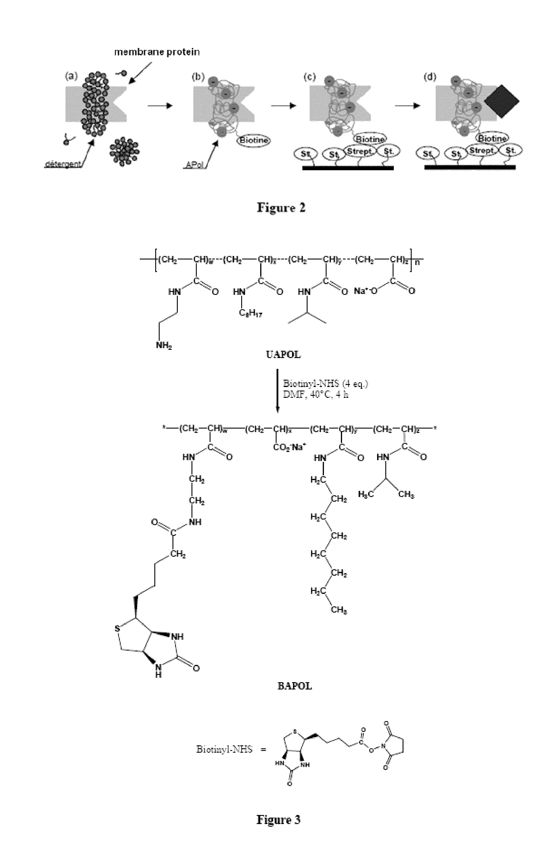
[0214]1.1 Principle

[0215]The experimental principle developed for immobilizing membrane proteins at the surface of solid supports is the following (FIG. 2): the membrane protein is solubilized in a detergent (step a) then trapped with a biotinylated amphipol and the detergent is removed (step b); the resulting complex is contacted with a support grafted with streptavidin groups with which it can be combined via a specific biotin / streptavidin interaction (step c). A protein ligand can then be circulated on the support and the ligand / protein interaction can be studied (step d).

[0216]Immobilization was experimentally monitored by a surface plasmon resonance (SPR) technique. This technique allows detection and real-time monitoring of interactions between circulating molecules and one or more immobilized molecule(s), by continuously observing for a change in surface plasmon re...

example 2

Membrane Protein Immobilization at the Surface of Streptavidin-Carrying Magnetic Beads by Means of a Biotinylated Amphipol

[0255]The inventors tested bacteriorhodopsin (BR) immobilization on magnetic beads functionalized by binding to streptavidin (SA beads).

[0256]BR was complexed with biotinylated amphipol (BAPol) according to the following method: a BR solution in octyl thioglucoside detergent (OTG) contains 1.1 g / l protein, 18 mM OTG. 17 μl of a 100 g / l BAPol solution in water are added to 300μl of protein solution (i.e. 5 g of BAPol per g of BR). After incubation for 15 min, 80 mg of Bio-beads are added and incubation is left to proceed under stirring for 3 hr at 4° C. (adsorption of OTG) followed by recovery of the solution and removal of the Bio-beads.

[0257]100 mg of SA beads were washed 3 times in NaCl-HEPES buffer (150 mM NaCl, 10 mM HEPES at pH 7.4), then excess liquid was removed; the protein solution was diluted to 13 mg / L in NaCl-HEPES buffer.

[0258]At time zero, the prote...

example 3

Synthesis of a Polyhistidine-Labelled Amphipol (HISTAPol)

[0261]The structure of biotinylated amphipol (BAPol) is described in FIG. 3.

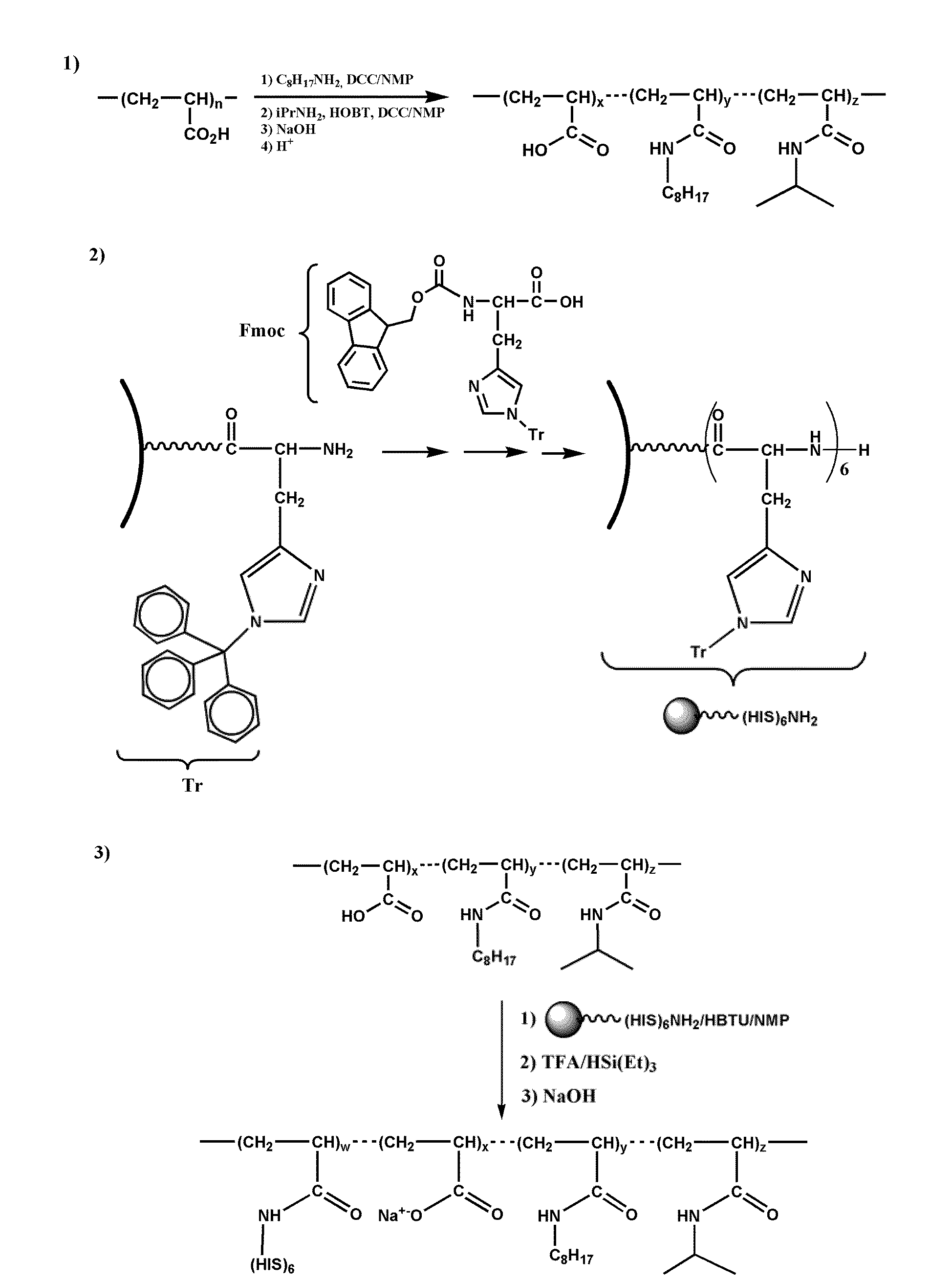
[0262]The method for synthesizing polyhistidine-labelled amphipol (HISTAPol) is derived from the method for non-functionalized amphipols (7, 16-17) and comprises three steps.

[0263]The first step comprises forming amide bonds with the carboxylic acid functionalities of a low molecular weight polyacrylic acid and appropriately selected alkylamines, such as isopropylamine and octylamine.

[0264]The second step involves work-up of a histidine hexamer. This synthesis is carried out on a solid support using an automated technique involving double coupling of the selectively protected monomer.

[0265]The last step comprises condensing the peptide N-termination onto the acid functionalities of the amphipol resulting from the first step of the synthesis. The peptide is used while still protected on its side chains and bound to the solid support. The amounts of amph...


PUM

PropertyMeasurementUnit
pHaaaaaaaaaa
timeaaaaaaaaaa
amphiphilicaaaaaaaaaa

Abstract

The invention pertains to the field of membrane protein immobilization onto supports. It relates to a product comprising a support and at least one membrane protein attached to the surface thereof, characterized in that said membrane protein is attached to said support using an amphiphilic molecule with which said membrane protein is complexed. It also relates to a process for preparing such product, as well as to various applications in the fields of diagnosis, drug design and biotechnologies. It further relates to a kit, together with a functionalized amphiphilic molecule, for preparing a product according to the invention comprising a support and an amphiphilic molecule, wherein the amphiphilic molecule and the support interact through a hydrophobic bond, an ionic bond, a specific bond or a covalent bond.

Description

FIELD OF THE INVENTION[0001]The invention pertains to the field of membrane protein immobilization onto supports. It relates to a product comprising a support and at least one membrane protein attached to the surface thereof, characterized in that said membrane protein is attached to said support using an amphiphilic molecule with which said membrane protein is complexed. It also relates to a process for preparing such product, as well as to various applications in the fields of diagnosis, drug design and biotechnologies. It further relates to a kit, together with a functionalized amphiphilic molecule, for preparing a product according to the invention comprising a support and an amphiphilic molecule, wherein the amphiphilic molecule and the support interact through a hydrophobic bond, an ionic bond, a specific bond or a covalent bond.BACKGROUND ART[0002]Integral membrane proteins represent about 30% of the coding sequences of eukaryotic genomes. Their functions, and especially thos...

Claims


Application Information

Patent Timeline
Patent Type & Authority Applications(United States)
IPC IPC(8): C12Q1/34C07K17/00C07K14/76C07K14/555C07K16/00C12N11/00G01N33/543C40B30/00C12N9/00C08F210/00C07K2/00C08B37/00C08F251/00
CPCC07K17/06C07K1/1077
Inventor POPOT, JEAN-LUCCHARVOLIN, DELPHINEGIUSTI, FABRICE
Owner CENT NAT DE LA RECHERCHE SCI
Why Eureka
Try Eureka