Method for detecting lysosomal storage diseases

a lysosomal storage and disease technology, applied in the field of lysosomal storage disease detection, can solve the problems of inaccurate screening methods to be performed before such an expensive diagnosis, and neither discloses nor suggests, and achieves the effects of convenient, sensitive method, and low cos

Inactive Publication Date: 2009-11-12
SEIKAGAKU KOGYO CO LTD +1
View PDF8 Cites 5 Cited by
  • Summary
  • Abstract
  • Description
  • Claims
  • Application Information

AI Technical Summary

Benefits of technology

[0045]Accordingly, an object of the present invention is to provide a very accurate, sensitive method for detecting lysosomal storage diseases which can be performed conveniently, quickly, and at low cost. Another object of the invention is to provide a kit for detecting lysosomal storage diseases.
[0046]The present inventors have carried out extensive research in an attempt to attain the above objects, and have found the following: in spite of conventional understanding that, in accordance with the type (classification) of mucopolysaccharidosis, only GAG of a specific species is excreted into body fluids in large amounts, in reality other species of GAGs are also excreted abundantly. Moreover, the present inventors found that GAG is also excreted into body fluids in large amounts in mucopolysaccharidoses, mucolipidoses, GM1 gangliosidoses, fucosidosis, galactosialidoses, metachromatic leukodystrophy, Niemann-Pick diseases, Tay-Sachs disease, Sandhoff disease, GM2 gangliosidoses, Krabbe disease, Fabry disease, Gaucher diseases, glycogen storage diseases and lipofuscinoses. On the basis of this finding, the inventors have achieved a highly accurate, highly sensitive, convenient, effective, inexpensive method and kit for detecting lysosomal storage diseases, thus leading to completion of the invention.
[0084]When blood is used as the body fluid, the collected blood sample may be used as is without any treatment, or serum or plasma derived from the collected blood sample may be used. Preferably, serum or plasma is employed. Alternatively, hydrophilic components may be extracted from the blood sample, after which GAG contained in the extract is measured. No particular limitation is imposed on the method for extracting hydrophilic components; for example, a droplet of a blood sample is added onto commercial filter paper, and hydrophilic components are extracted from the filter paper. Extraction of the hydrophilic components from the filter paper may be conveniently performed by soaking, in an aqueous solution, the filter paper bearing the droplet of the blood sample.
[0193]When the measurement of GAG of a single species (GAG level) is lower than that (GAG level) of healthy animals, the measurement can be correlated to “free of lysosomal storage diseases” or “low risk of lysosomal storage diseases”

Problems solved by technology

However, Japanese Patent Application Laid-Open (kokai) No. 10-153600 neither discloses nor suggests that each case of the aforementioned mucopolysaccharidoses involves, in addition to the GAG species shown in Table 1-1 being excreted, excretion into body fluid of large amounts of other species of GAGs.
Moreover, this publication neither discloses nor suggests whether measurement of GAG of a single species enables detection of all types of mucopolysaccharidoses, regardless of the classification (type) of mucopolysaccharidoses.
However, in Japan, for example, the number of newborns per year exceeds 1,000,000, and the frequency of onset of mucopolysaccharidoses, etc. is as low as one per 40,000 to 50,000, and since current diagnosis therefor, which detects deficiency or abnormality of enzymes, is cumbersome and expensive, demand exists for an accurate screening method to be performed before such an expensive diagnosis.

Method used


Image

  • Method for detecting lysosomal storage diseases
  • Method for detecting lysosomal storage diseases
  • Method for detecting lysosomal storage diseases

Examples


example 1

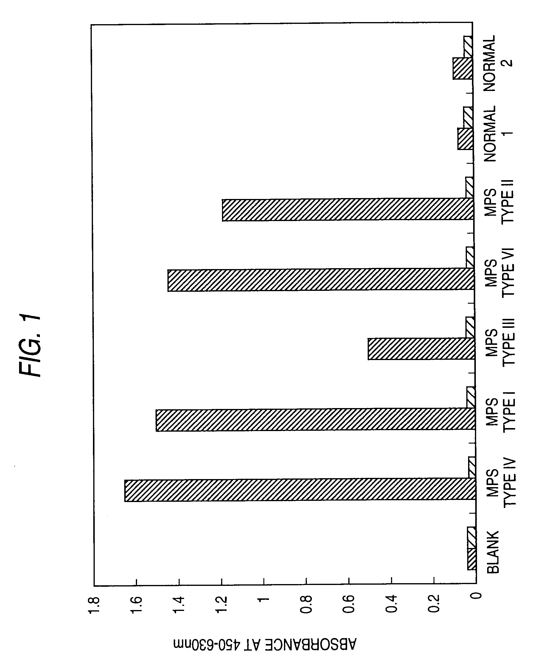
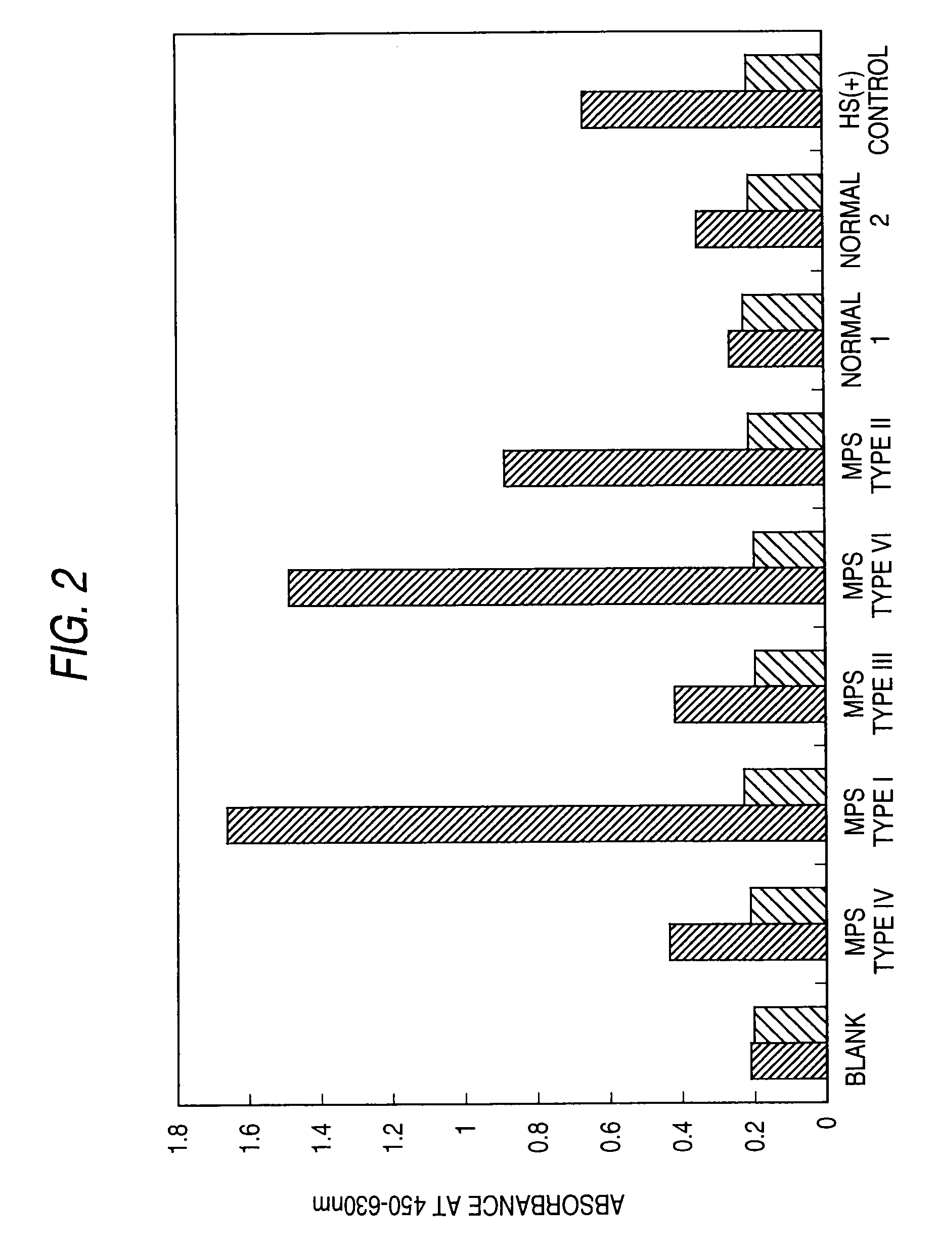
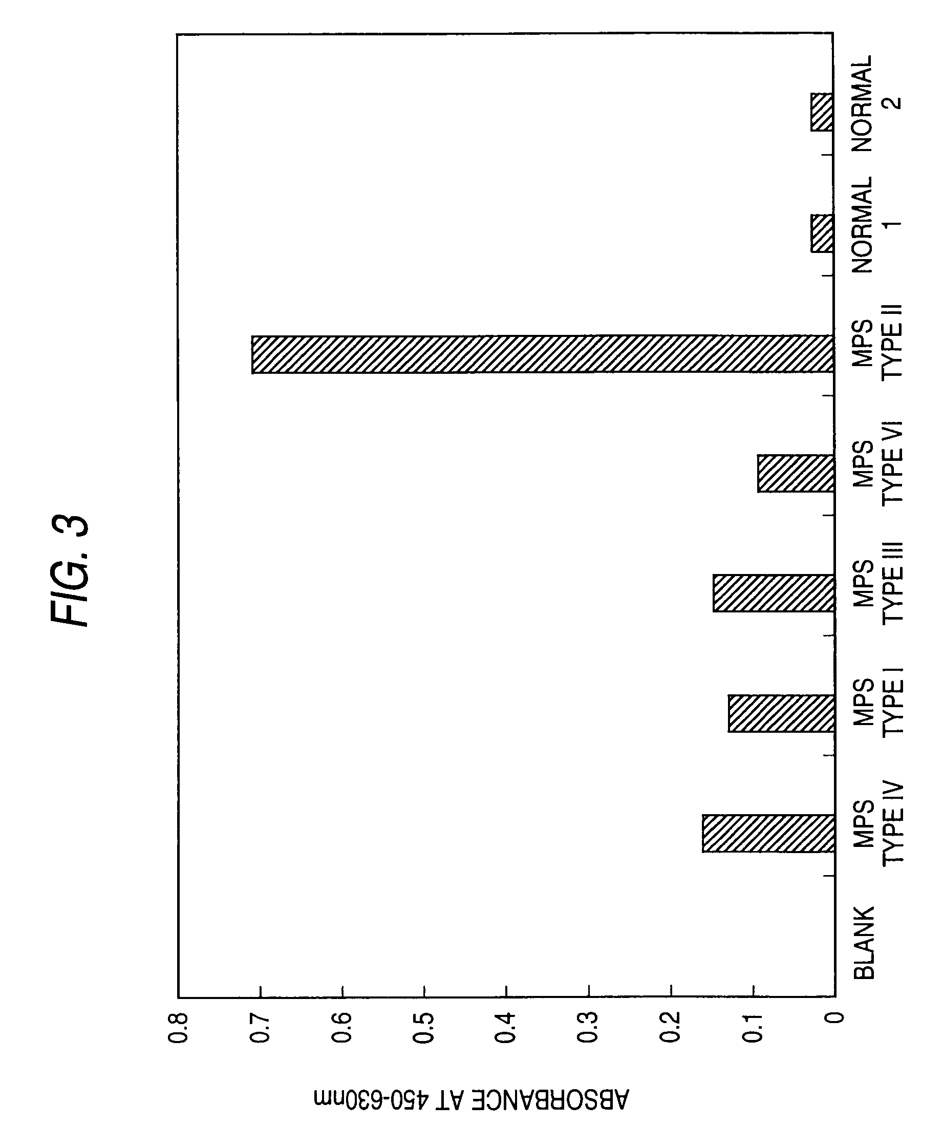
[0228]A sandwich assay was performed to detect mucopolysaccharidoses in human urine samples.

(1) The Specimens, Reagents, Etc. Employed in Example 1 were as Follows.

Specimens and Standard Samples:

[0229]The specimens were human urine samples collected from patients suffering human mucopolysaccharidosis types I, II, III, IV, or VI (1 subject each) and healthy humans (humans not suffering mucopolysaccharidosis; 2 subjects).

[0230]The standard GAGs employed were as follows:

[0231]HS (derived from bovine kidney, produced by Seikagaku Corporation):

[0232]This KS is available from Seikagaku Corporation as reagent catalogue code No. 400700, and has the following properties.

Nitrogen Content:

[0233]2.6 to 3.2% (measured by the method described in Z. Anal. Chem., 22, 366 (1883))

Sulfur Content:

[0234]5.0 to 6.0% (measured by the method described in Mikrochim. Acta., 123 (1955))

Uronic Acid Content:

[0235]28.0 to 30.0% (oxynol reaction)[0236]36.0 to 40.0% (carbazole reaction)

Glucosamine Content:

[0237]30...

example 2

[0295]An inhibition assay was performed to detect mucopolysaccharidoses in serum or urine samples from dogs or cats.

(1) Specimens, Reagents, Etc. Employed in Example 2 are as Follows.

Specimens:

[0296]The specimens employed are serum and urine samples collected from mucopolysaccharidosis type IIIB or VII model animals (dogs); mucopolysaccharidosis type I, VI, or VII model animals (cats); and healthy animals (dogs and cats not suffering mucopolysaccharidosis).

[0297]The standard GAG employed is KS derived from bovine cornea (Sigma Co.).

Antibodies:

[0298]The primary antibody employed is anti-KS antibody “5D4” (1 / 20 / 5D4; ICN Immunobiologicals). The secondary antibody employed is horseradish-peroxidase-bound anti-mouse IgG (H+L) (Pierce Co.).

Antigen-Immobilized Plate:

[0299]An antigen-immobilized plate (i.e., a plate to which KS is immobilized) was prepared as described below.

[0300]0.2 U Chondroitinase ABC (produced by Seikagaku Corporation) was added to 1 mg KS solution (bovine-cornea-deriv...

example 3

Mass-Scale Detection of Mucopolysaccharidoses

[0314]An attempt was made to detect mucopolysaccharidoses in a mass scale by the sandwich method using human urine or plasma as samples. The method is the same as the “(2) Detection of mucopolysaccharidoses through measurement of KS” in Example 1.

[0315]The results of using urine as samples (values corrected for creatinine (Cre) concentration) are shown in FIG. 5, and the results of using plasma in FIGS. 6 and 7.

[0316]In FIG. 5, abbreviations show the following results:[0317]Control: result of healthy human (n=67);[0318]All MPS IVA: result of all samples of mucopolysaccharidosis type IVA human (regardless of age, n=78);[0319]Severe: result of mucopolysaccharidosis type IVA (severe type) human (n=54);[0320]Milder: result of mucopolysaccharidosis type IVA (mild type) human (n=11);[0321]Cont 0-5: result of healthy human (from 0 to less than 5 years; n=21)[0322]IVA 0-5: result of mucopolysaccharidosis type IVA human (from 0 to less than 5 year...


PUM

PropertyMeasurementUnit
temperatureaaaaaaaaaa
concentrationaaaaaaaaaa

Abstract

A method for detecting lysosomal storage diseases including the steps of performing an assay for a single species of glycosaminoglycan contained in a specimen and correlating results of the assay with lysosomal storage diseases. A body fluid such as urine or blood can be employed as a specimen. The assay can be performed by use of a polypeptide that is capable of specifically binding to a glycosaminoglycan-containing molecule. The polypeptide may be an antibody, or a polypeptide having an antigen-binding site of an antibody.

Description

TECHNICAL FIELD[0001]The present invention relates to a method for detecting lysosomal storage diseases and to a kit therefor.BACKGROUND ART[0002]The following abbreviations are used throughout the present specification:[0003]GAG: glycosaminoglycan[0004]KS: keratan sulfate[0005]HS: heparan sulfate[0006]CS: chondroitin sulfate[0007]CS-4S: chondroitin-4-sulfate[0008]CS-6S: chondroitin-6-sulfate[0009]DS: dermatan sulfate (also called chondroitin sulfate B)[0010]GSD I: glycogen storage disease type 1[0011]GSD II: glycogen storage disease type 2 (Pompe disease)[0012]Hep: heparin[0013]HA: hyaluronic acid[0014]LIPO: lipofuscinoses[0015]MPS: mucopolysaccharidoses[0016]ML: mucolipidoses[0017]MLD: metachromatic leukodystrophy[0018]NP: Niemann-Pick diseases[0019]TS: Tay-Sachs disease[0020]Lysosomal storage diseases are diseases caused by abnormality of enzymes present in lysosomes.[0021]Mucopolysaccharidoses are kinds of lysosomal storage diseases and form a class of hereditary diseases caused...

Claims


Application Information

Patent Timeline
Patent Type & Authority Applications(United States)
IPC IPC(8): G01N33/53C12Q1/02G01N33/66G01N33/68
CPCG01N33/6893G01N2400/40G01N33/00G01N2800/10Y10S436/811G01N2800/042
Inventor OKAMURA, KAZUOMIYAURA, SHUICHITOMATSU, SHUNJI
Owner SEIKAGAKU KOGYO CO LTD
Why Eureka
Try Eureka