Ultra-sensitive detection of analytes

a technology of analytes and sensitive detection, applied in the field of screening methods, can solve the problems of increasing assay time, difficult, expensive, time-consuming, etc., and achieve the effect of expanding the flexibility, adaptability and usefulness of techniques, and facilitating detection

Inactive Publication Date: 2010-02-11
NANOSHPERE INC
View PDF10 Cites 48 Cited by
  • Summary
  • Abstract
  • Description
  • Claims
  • Application Information

AI Technical Summary

Benefits of technology

[0011]The invention overcomes many of the problems of the prior art while greatly expanding the flexibility, adaptability and usefulness of techniques directed to the amplification of a signal to facilitate detection. The present invention relates to methods, probes, compositions, and kits that utilize binding moieties, such as oligonucleotides as biochemical barcodes, for detecting at leas

Problems solved by technology

However, it is difficult, expensive, and time-consuming to simultaneously detect several protein structures under assay conditions using the aforementioned related protocols.
Although these approaches are notable advances in protein detection, they have several drawbacks: 1) limited sensitivity because of a low ratio of DNA identification sequence to detection antibody; 2) slow target binding kinetics due to the heterogeneous nature of the target capture procedure, which increases assay time and decreases assay sensitivity; 3) complex conjugation chemistries that are required to chemically link the antibody and DNA-markers; and 4) require a PCR amplification step.45 Therefore, a sensitive, and rapid method for detecting target analytes in a sample that is amenable to multiplexing and easy to implement is needed.
Current techniques cover the shift in the frequency of scattered light as a consequence of target-mediated

Method used


Image

  • Ultra-sensitive detection of analytes
  • Ultra-sensitive detection of analytes
  • Ultra-sensitive detection of analytes

Examples


example 1

Scatter Light Generated by Gold and Silver Nanoparticles in Flow Cytometry Assays

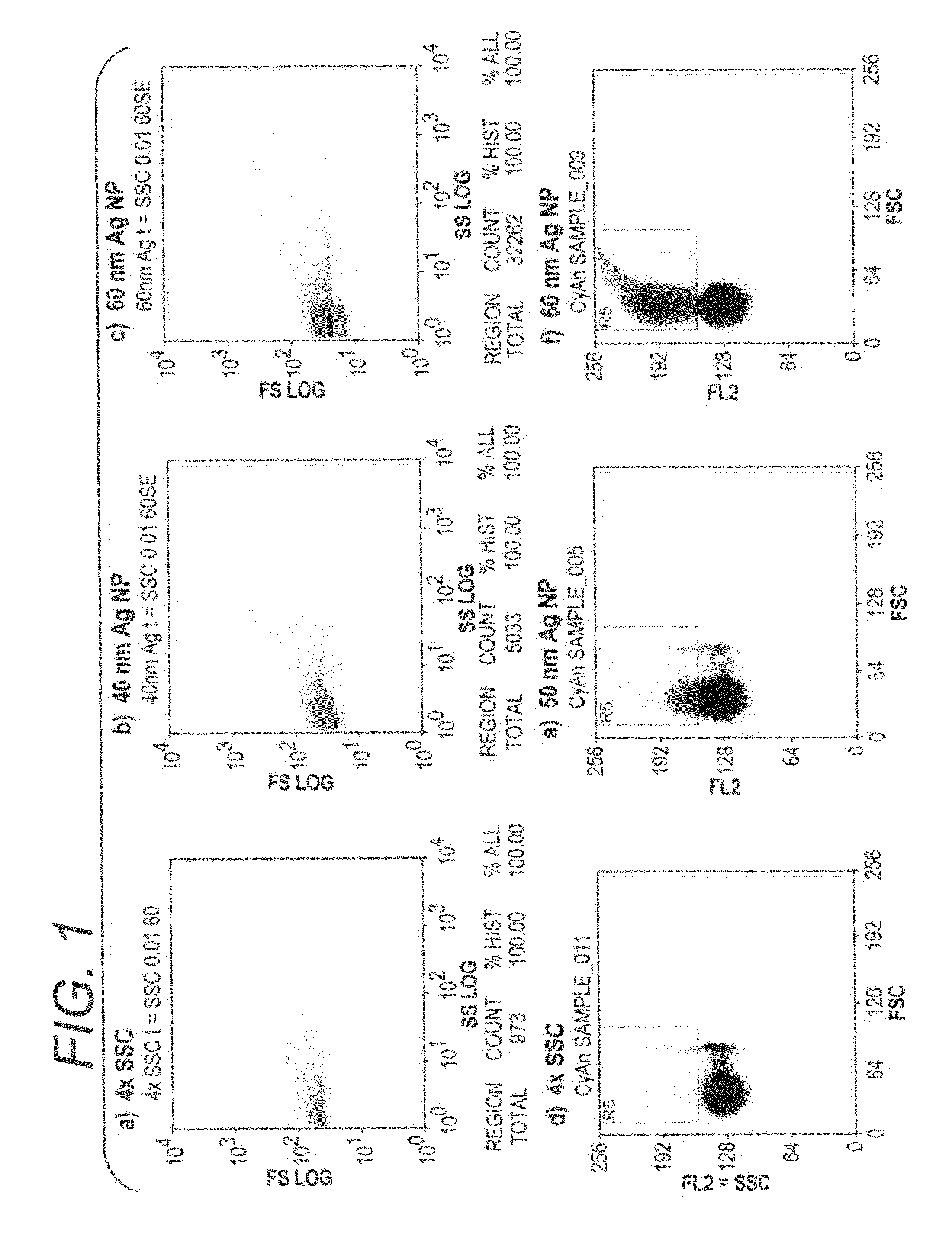
[0104]Gold and silver nanoparticles of various sizes were used to demonstrate the capability of nanoparticles to be used in flow cytometry assays. Using a Dako CytoMation 405 nm laser (Dako Denmark A / S, Glostrup, Denmark) or the Dako MoFlo 530 nm laser, forward and side scatter was adjusted to detect sub-micron particles. Gold and silver nanoparticles were obtained from BBInternational Ltd., Cardiff, UK. To demonstrate scatter light from 40 nm and 60 nm particles, 106 Ag nanoparticles in 500 uL 4×SSC (Saline Sodium Citrate) were measured by side scatter in a 60 sec analysis (FIG. 1b-c), and 106 Au nanoparticles in 500 uL 4×SSC were detected based on their red signal in a 60 second run (FIG. 1e-f), and were compared to measurement of 4×SSC alone (FIGS. 1a and 1d).

[0105]Nanoparticles of both types and sizes produced a bright and tight population, and larger nanoparticles produced more scatter. Aggregated ...

example 2

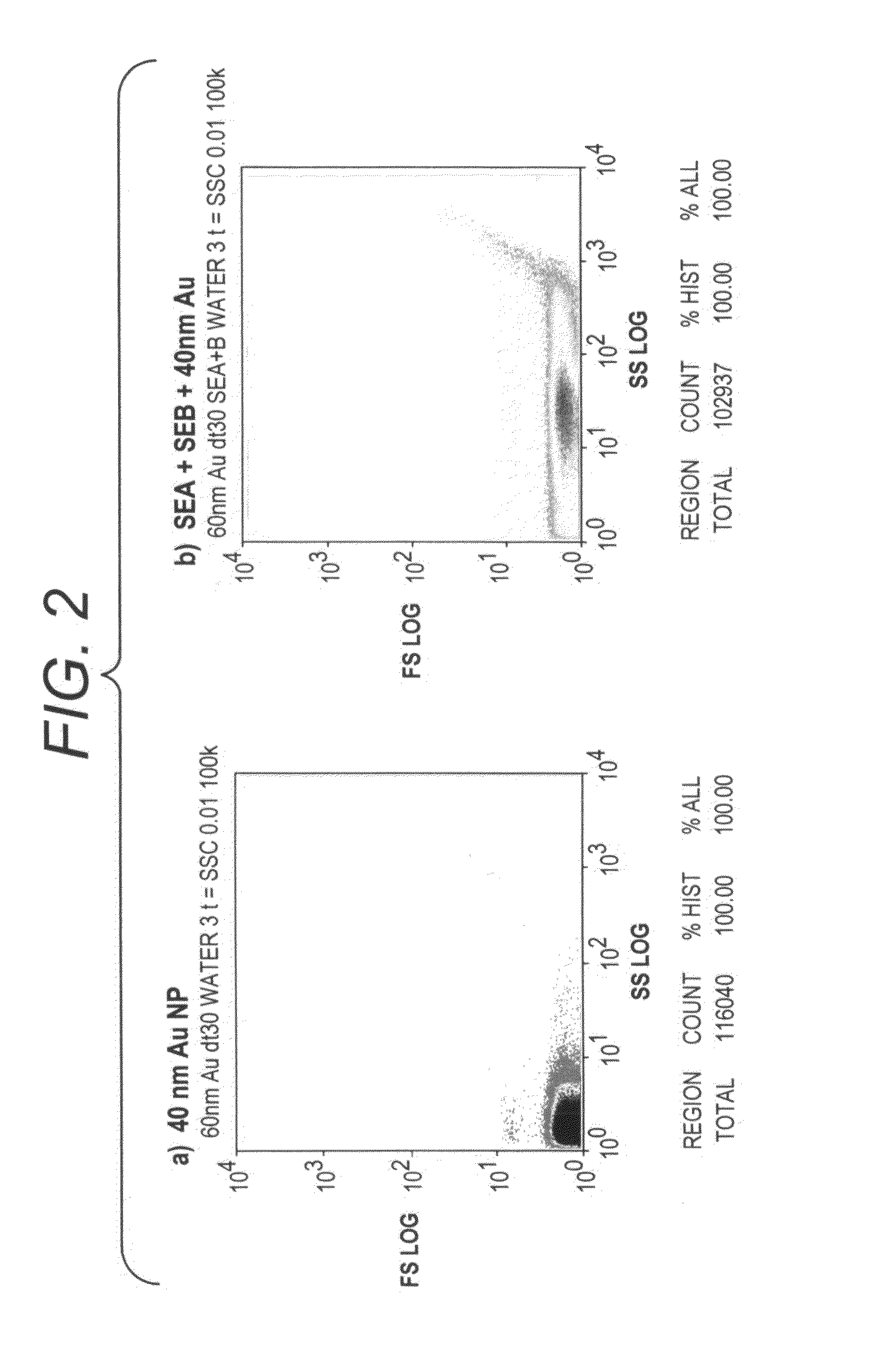
Silver Amplification Induces Broad Side Scatter Shift of Gold Nanoparticles

[0107]As shown in FIG. 2, silver staining of gold nanoparticles causes a large shift in side scatter and forward scatter, indicating a significant change in particle size and scatter properties. The experiments were conducted using 2 uL 40 nm gold nanoparticles were mixed with silver solution (2 uL Signal Enhancement A (SEA; Nanosphere, Northbrook, Ill.) and 2 uL Signal Enhancement B (SEB; These solutions are commercially available from Nanosphere, Northbrook, Ill. There are functionally equivalent commercially available Silver Enhancement reagents available (e.g. Silver Enhancement Solution A, #S-5020 and Silver Enhancement Solution B, #S-5145 Sigma-Aldrich, St. Louis, Mo.) and reacted for 5 minutes at room temperature. The reaction was stopped by diluting with 500 uL water. Scatter was detected with the CytoMation 405 nm laser. Silver-coating of gold nanoparticles caused a large shift in side scatter and a ...

example 3

Silver Particles in Solution Detectable by Flow Cytometry

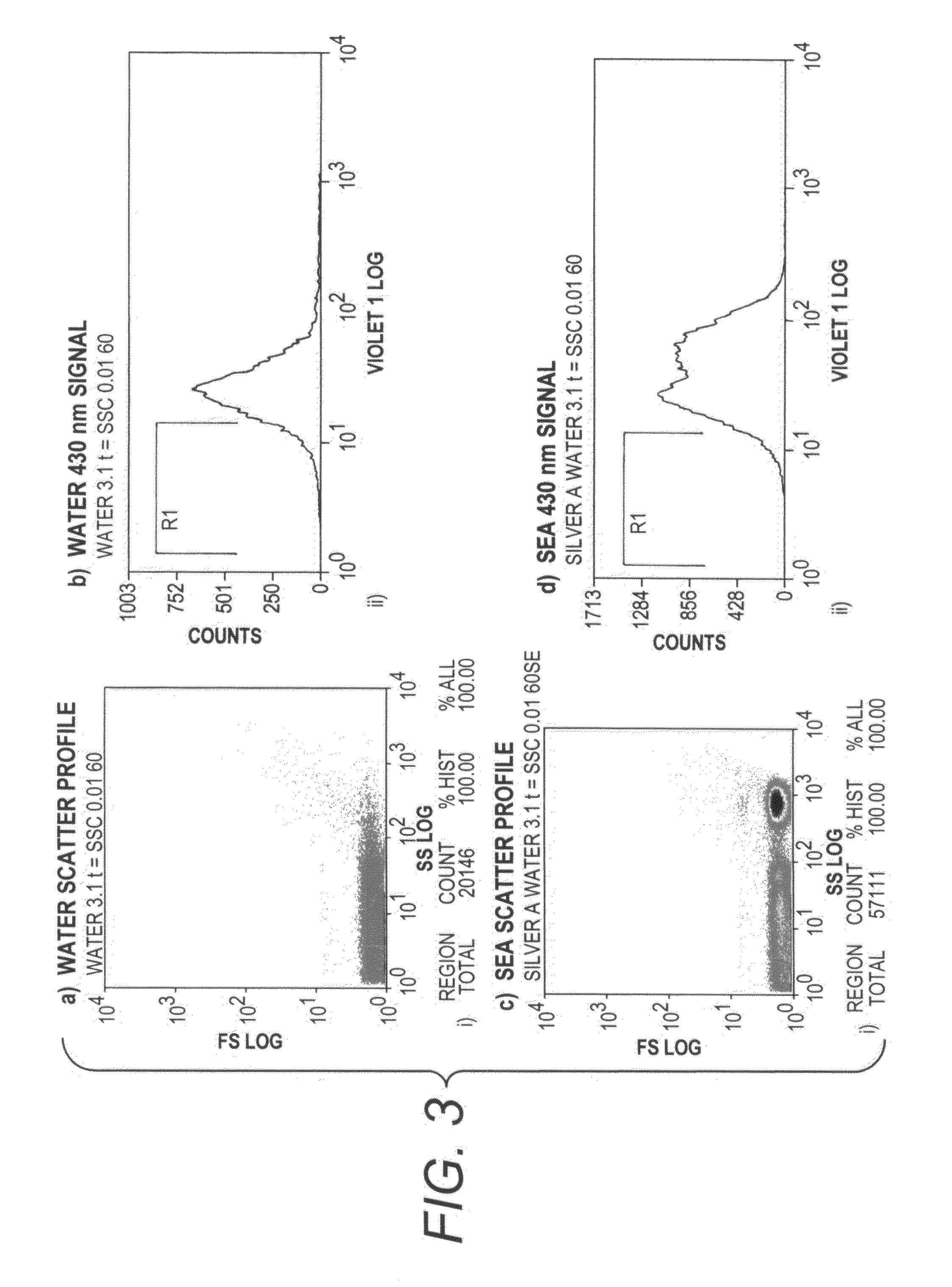
[0108]As shown in FIG. 3, Plasmon scatter light from silver particles can be seen by flow cytometry. A 100 uL aliquot of signal enhancement solution A (SEA) was transferred to a clear 1.5 mL tube. Due to opening of the box it was stored in, the SEA was briefly and randomly exposed to ambient light. Using the CytoMation 405 nm laser and 430 nm filter, silver particles induced by exposure to light were detected by side scatter (FIG. 3c). An increased 430 nm signal was also detected (FIG. 3d), indicating the plasmon scatter light from silver particles can be seen by flow cytometry.


PUM

PropertyMeasurementUnit
Diameteraaaaaaaaaa
Nanoscale particle sizeaaaaaaaaaa
Concentrationaaaaaaaaaa

Abstract

The present invention relates to screening methods, compositions, and kits for detecting for the presence or absence of one or more target analytes, e.g. biomolecules, in a sample. In particular, the present invention relates to methods that utilize nanoparticle probes in an in-solution homogeneous assay system for high-sensitivity detection of target proteins or nucleic acids based on flow analysis of single particles.

Description

[0001]This application claims the benefit of priority to U.S. Provisional Application Ser. No. 60 / 819,766, filed Jul. 10, 2006, which is incorporated by reference in its entirety.FIELD OF THE INVENTION[0002]The present invention relates to a screening method for detecting for the presence or absence of one or more target analytes, e.g., proteins, nucleic acids, or other compounds in a sample. In one application, the present invention utilizes nucleic acid reporter markers as biochemical barcodes in combination with metallic nanoparticles for detecting through measuring the shifts in resonance frequency of one or more analytes in a solution with a flow-based (flow cytometry or micro-capillary) method.BACKGROUND OF THE INVENTION[0003]The detection of analytes is important for both molecular biology research and medical applications. Diagnostic methods based on fluorescence, mass spectroscopy, gel electrophoresis, laser scanning and electrochemistry are now available for identifying a ...

Claims


Application Information

Patent Timeline
IPC IPC(8): C12Q1/68G01N33/53
CPCB82Y15/00G01N33/54306G01N33/588G01N33/587G01N33/54333
Inventor MULLER, UWE R.LEFEBVRE, PHIL
Owner NANOSHPERE INC
Why Eureka
Try Eureka