

Enzyme substrates for visualizing acidic organelles
a technology of enzyme substrates and acidic organelles, applied in the field of enzyme substrates for visualizing acidic organelles, can solve the problems of ineffective monitoring of lysosomal enzyme activity in living cells, no available therapies, and limited treatment options,
- Summary
- Abstract
- Description
- Claims
- Application Information
AI Technical Summary
Benefits of technology
Problems solved by technology
Method used
Image

Examples
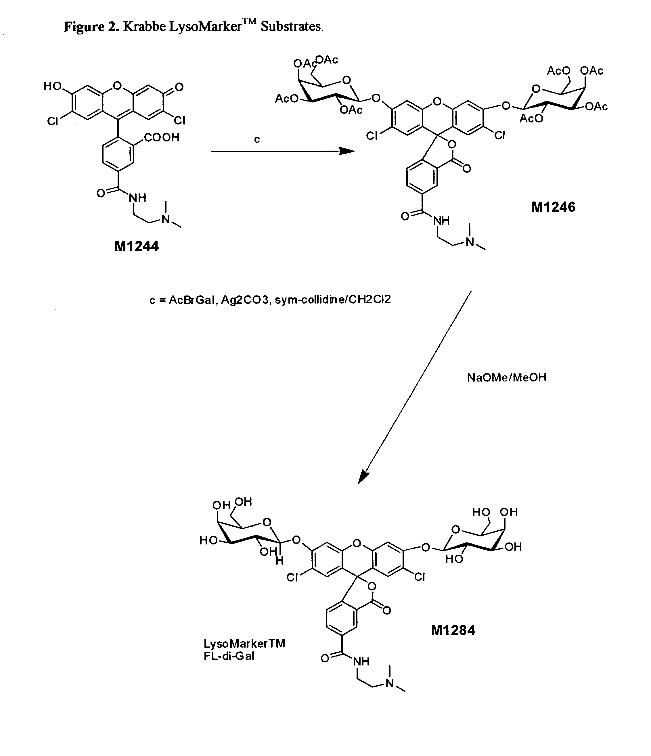
example 1
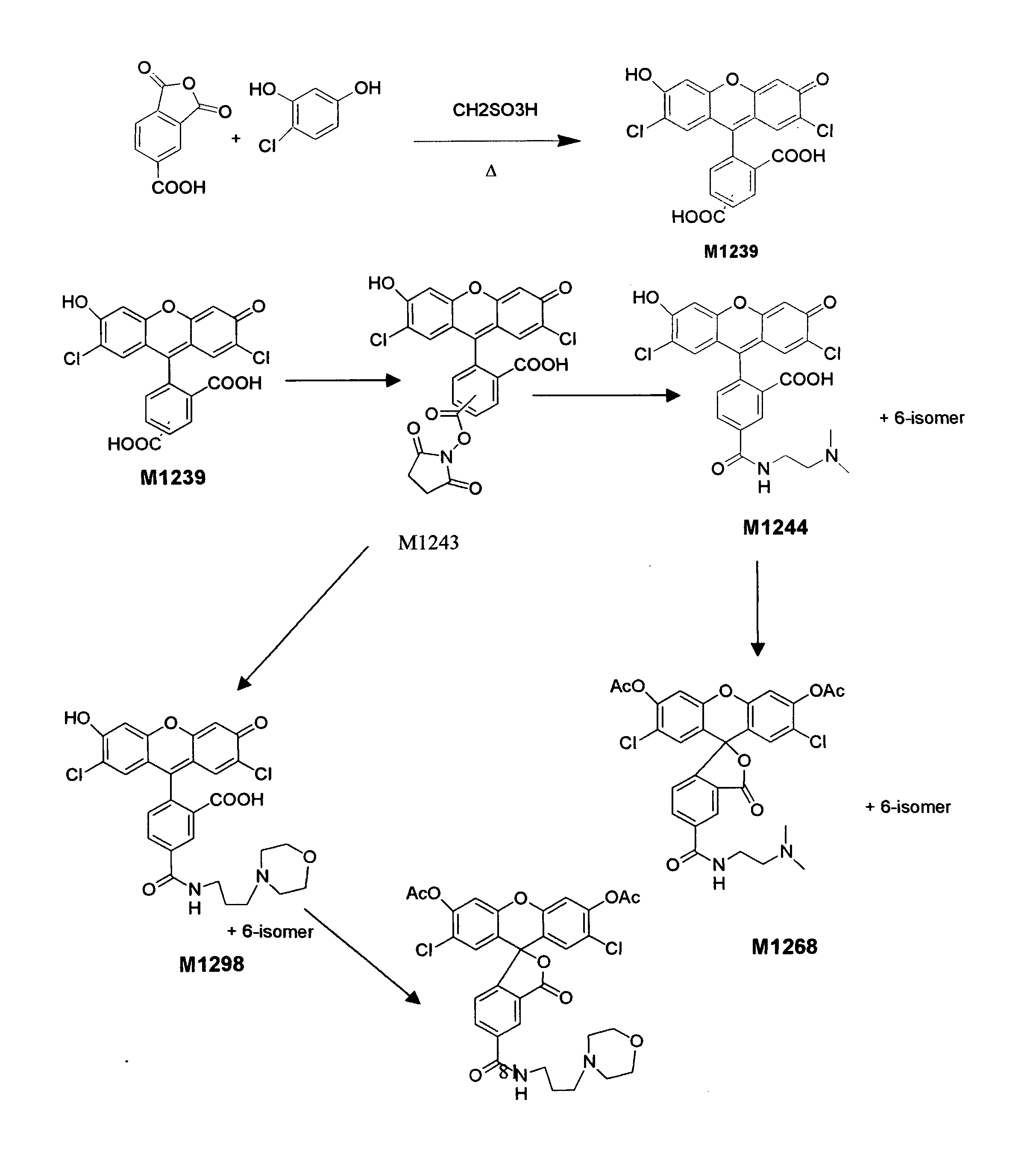
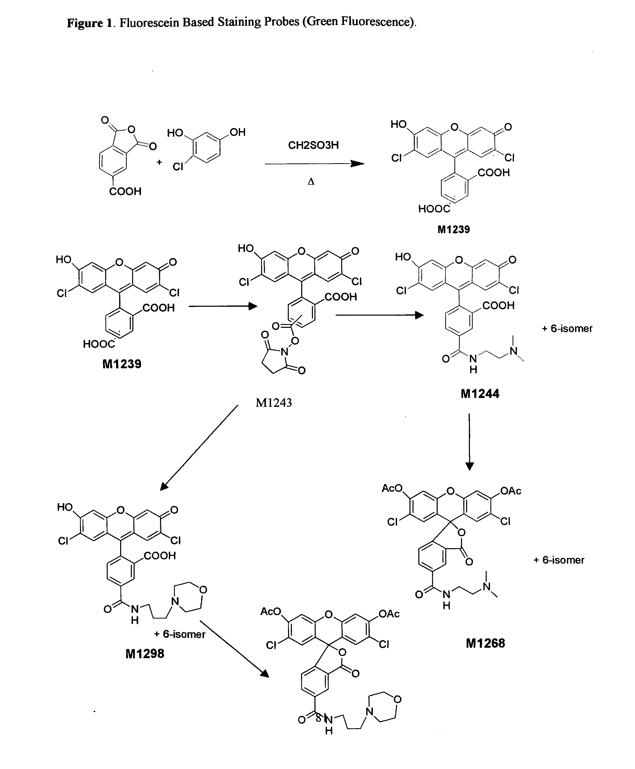
Preparation of a Lysosomal β-Galactosidase Substrate with Green Fluorescence after Enzyme Reaction
[0080]The following compound was prepared:
5(6)-(2-dimethylaminoethyl)carboxamido)-2′7′-dichlorofluorescein
[0081]To a dry 100 mL round bottom flask under anhydrous N2(g) was added N-hydroxysuccinimide (5.75 g, 50 mmol), dissolved in trifluoroacetic anhydride (20.0 mL, 144 mmol) and allowed to stir at room temperature for 1.5 h. This reaction mixture was rotary evaporated under reduced pressure and co-evaporated with dry toluene (3×20 mL) at 50° C. and dried in vacuo to give O-trifluoroacetyl-N-hydroxysuccinimide as a white amorphous crystalline solid (10.57 g, 100%).
[0082]Under anhydrous conditions, a sample of O-trifluoroacetyl-N-hydroxysuccinimide (10.57 g, 50 mmol) was dissolved in anhydrous DMF (20 mL) and 5(6)-carboxy-2′,7′-dichlorofluorescein (5.20 g, 12 mmol) and dry pyridine (10.0 mL, 130 mmol) were added. This mixture was allowed to stir at room temperature under anhydrous condi...
example 2
Preparation of an Esterase Substrate with Blue Fluorescence Emission Upon Enzyme Activity
[0086]The following compound was prepared:
[0087]To a flame dried 50 mL round-bottom flask under an atmosphere of dry N2(g) was weighed 3-(2′-dimethylaminoethylcarboxamidomethyl)-6-chloro-7-hydroxycoumarin (M1247, 99 mg, 0.3 mmole). This sample was suspended in anhydrous dichloromethane (20 mL), cooled to 0° C. (ice-bath) and acetic anhydride (1.0 mL, 10.6 mmole) and dry pyridine (1.0 mL, 12.4 mmole) added. The reaction was allowed to stir at 0° C. for 2 hours and at room-temperature overnight. The reaction mixture was diluted with dichloromethane (30 mL) and poured into ice-water (100 mL) with stirring. The organic layer was separated and washed with saturated sodium bicarbonate solution (1×100 mL) and brine solution (1×100 mL), dried over anhydrous sodium sulfate, filtered, evaporated and co-evaporated with dry toluene (2×10 mL) to give a clear glass (50 mg, 42%) homogeneous by TLC analysis (ir...
example 3
Preparation of a Lipase Substrate with Green Fluorescence Upon Enzyme Activity
[0088]The following compound was prepared:
5(6)-(2-dimethylaminoethyl)carboxamido)-2′7′-dichlorofluorescein, di-O-octanoate
[0089]5(6)-(2-dimethylaminoethyl)carboxamido)-2′7′-dichlorofluorescein (152 mg, 0.295 mmole) was suspended in anhydrous dichloromethane (10 mL) and cooled to 0° C. (ice-bath) while under anhydrous conditions under dry nitrogen gas. To this solution was added octanoyl chloride (250 uL, 1.62 mmole) and dry pyridine (1120 uL, 14.42 mmole) and the reaction mixture was placed in an ultrasonic bath for 30 min. to complete dissolution of the starting materials. This reaction mixture was allowed to react at ambient temperature under anhydrous conditions overnight. The reaction was diluted with dichloromethane (50 mL) and poured into ice-water (50 mL), extracted, and the resulting organic layer washed with ice-water (1×50 mL), saturated sodium bicarbonate solution (1×50 mL) 1 N HCl solution (1×5...
PUM
 Abstract
Description
Claims
Application Information