Cell growth method and pharmaceutical preparation for tissue repair and regeneration

Active Publication Date: 2010-10-07
SAPPORO MEDICAL UNIVERSITY
View PDF12 Cites 12 Cited by
  • Summary
  • Abstract
  • Description
  • Claims
  • Application Information

AI Technical Summary

Benefits of technology

[0063]The present invention achieves significant improvement in growth rate even using allogeneic serum (e.g., autoserum of a subject from which cells are collected) instead of foreign serum (FBS) in culture, even when an anticoagulant conventionally considered to be essential or useful for cell growth is added in a trace amount or is substantially absent. In addition, according to the present invention, cells having excellent growth efficiency (growing quickly) are obtained. These surprising effects have been totally unexpected from the conventional wisdom. The cells obtained by the culture method free from autoserum are much more excellent in safety and differentiation potency than those obtained by the conventional culture method using FBS.
[0064]A pharmaceutical preparation of the present invention has therapeutic effects even when administered in the subacute phase or later of disease, which has been assumed to be ineffective for administration. Therefore, the cells to be administered do not

Problems solved by technology

Heretofore, recovery in the functions of injured nervous tissues has been considered to be very difficult.
However, the animal cells such as FBS differ in composition from lot to lot and also have the problem of possible contamination with pathogens such as virus or prion.
However, culture in such a serum-free medium hardly produces growth equivalent to that in a serum-supplemented medium under the present circumstances.
On the other hand, in an attempt to use human serum, human adult serum is used because use of fetal serum is difficult from the ethical standpoint (see e.g., Patent Documents 6 to 8).
The disadvantage of use of the adult serum is lower growth-promoting activity than that obtained using, for example, FBS.
However

Method used


Image

  • Cell growth method and pharmaceutical preparation for tissue repair and regeneration
  • Cell growth method and pharmaceutical preparation for tissue repair and regeneration
  • Cell growth method and pharmaceutical preparation for tissue repair and regeneration

Examples


example 1

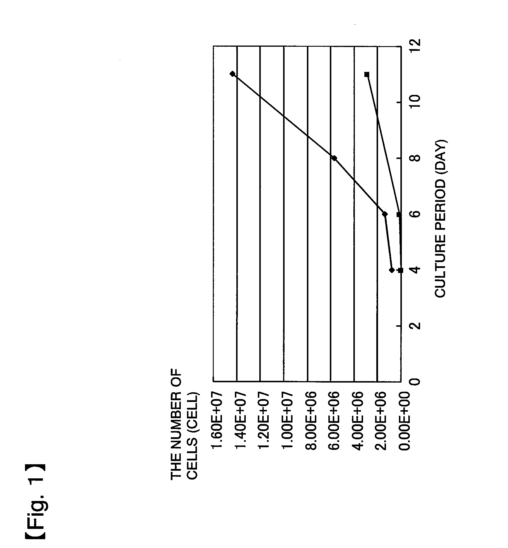
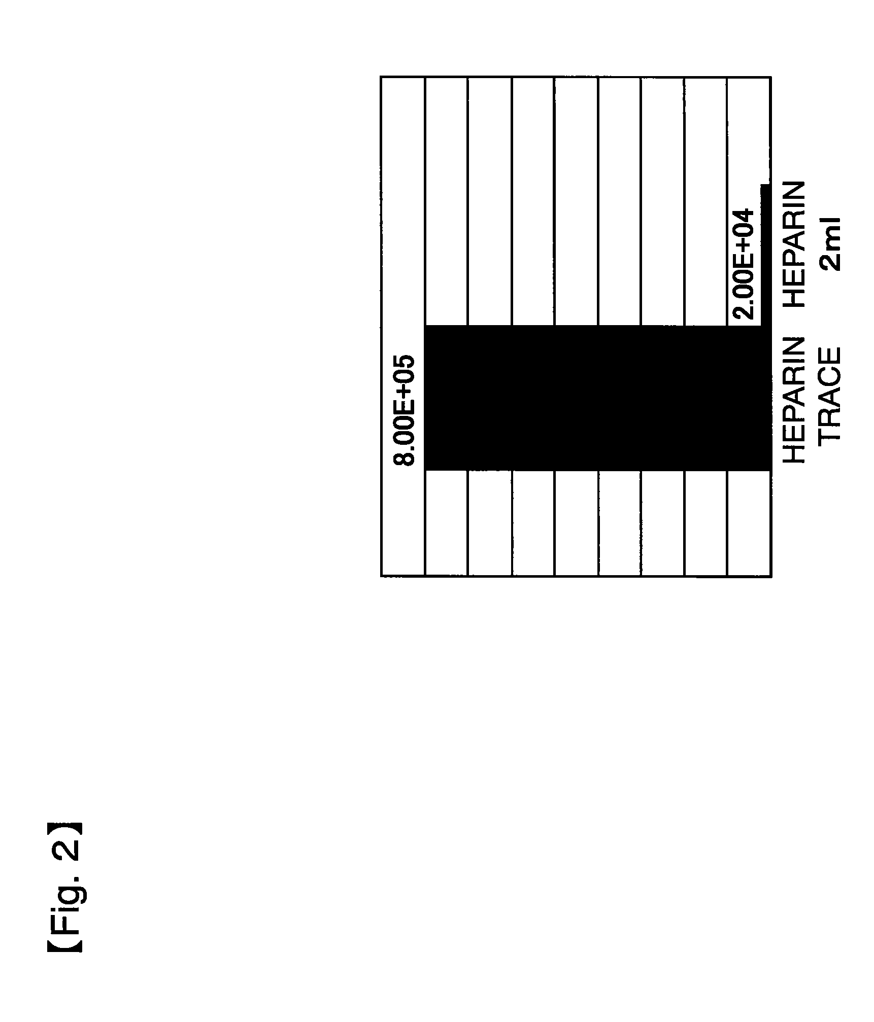
[0213]60 mL of a bone marrow fluid was collected from a brain disease patient using a blood collection tube whose inner wall was wetted in advance with trace heparin (0.1 U per mL of bone marrow fluid). The collected bone marrow fluid was added to 210 mL of a medium to bring the whole amount to 270 mL. This bone marrow fluid-containing culture solution was divided to 18 portions, each of which was then seeded in an amount of 15 mL to a dish of 150 mm in diameter (Tissue culture dish #3030-150 manufactured by IWAKI). 5 mL of the medium was added thereto to bring the whole amount to 20 mL per dish. After the seeding, these dishes are left standing in separate incubators (9 dishes per incubator), followed by culture at 37±0.5° C., in a 5% CO2 atmosphere. The bone marrow fluid was confirmed in advance by peripheral blood examination to be not infected with HIV, ATL, HB, HC, syphilis, human parvovirus B19, and the like.

[0214]The medium was prepared by adding 56.8 mL of serum derived from...

example 2

[0222]Culture was performed under the same conditions as in Example 1 except that an αMEM medium was used instead of DMEM as a standard medium. The results are shown in FIG. 6. These results demonstrated that when an αMEM medium was used, the use of human serum produces more rapid growth than obtained with FBS, by reducing the amount of heparin as well.

example 3

[Method]

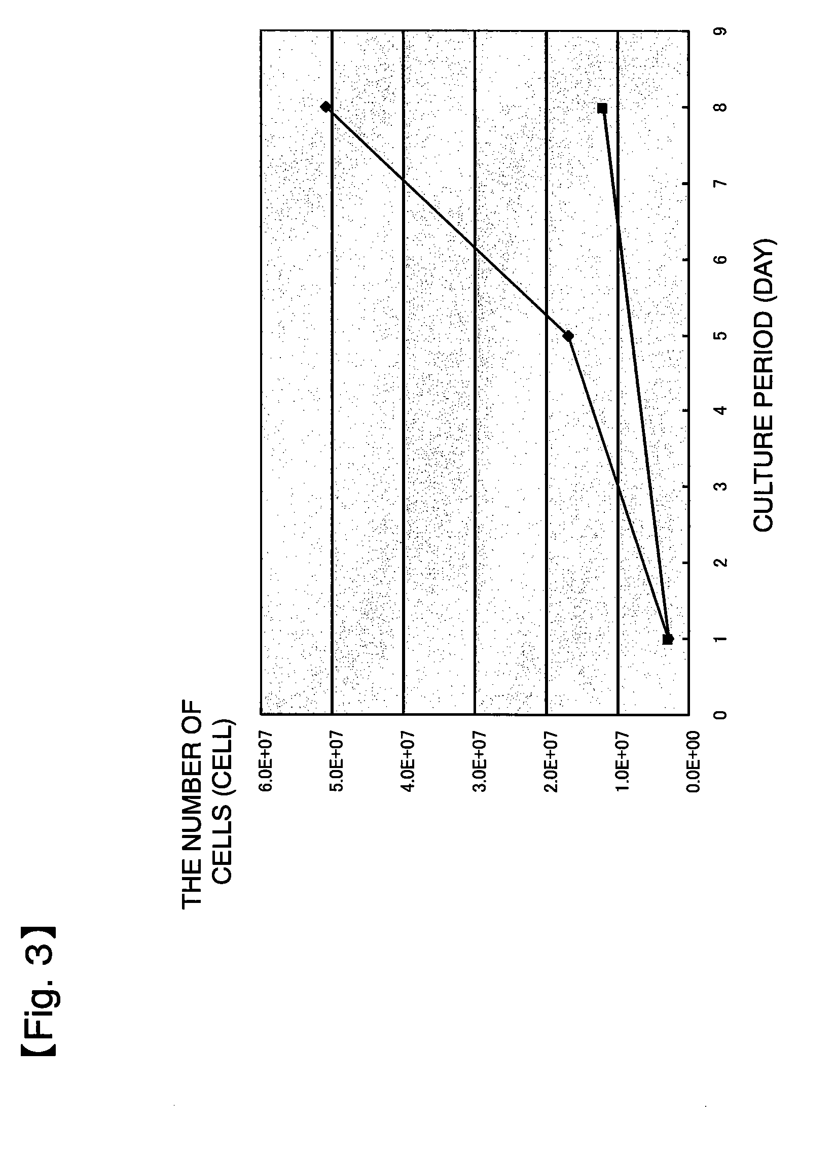
[0224]Three rat thigh bones were collected, and bone marrow cells were washed out of each thigh bone using DMEM (5 ml) and supplemented with the following agents to prepare three sample groups:[0225](i) DMEM (5 ml)[0226](ii) DMEM (5 ml)+heparin (2 μl)[0227](iii) DMEM (5 ml)+heparin (2 μl)+protamine (2 μl)

[0228]Each sample was washed with DMEM and placed in 10 ml of a culture solution (DMEM+10% FBS+1% penicillin / streptomycin+2 mM L-glutamine), which was then seeded to a 10 cm dish and cultured for 14 days. Subculture was started at the point in time when the cell density exceeded 5,500 cells / cm2.

[Results]

[0229]As shown in FIG. 8, the cells untreated with heparin had a larger initial amount and also a higher growth rate than those of the cells treated with heparin, and this was obvious on culture days 8 to 11. Moreover, the cells supplemented with protamine, a substance inhibiting heparin activity, also had a low initial amount and a low growth rate, as in the cells treated wi...


PUM


Abstract

The present invention relates to a method for growing, rapidly and massively ex vivo, cells collected from a living subject to provide a safe and effective pharmaceutical preparation for biological tissue repair/regeneration. Specifically, the present invention relates to a method for growing cells in a sample collected from a living subject by culturing the cells in a medium containing allogeneic (including autogenic) serum. Preferably the allogeneic serum has been determined as being negative for a serum tumor marker and/or an infectious factors, and the amount of the anticoagulant (e.g., heparin, a heparin derivative, or a salt thereof) added to the collected sample is less than 5 U/mL with respect to the volume of the sample or the amount of the anticoagulant in the medium at the start of culture is less than 0.5 U/mL. The present invention further relates to use of the method.

Description

TECHNICAL FIELD[0001]The present invention relates to: a method for growing, rapidly and massively ex vivo, cells collected from a living subject; cells grown by the method; a pharmaceutical preparation for biological tissue repair and regeneration comprising the cells; and a method for producing the pharmaceutical preparation. This method is suitable particularly for autotransplantation of mesenchymal stem cells and is applicable particularly to repair and regeneration of a tissue of the nervous system.BACKGROUND ART[0002]Heretofore, recovery in the functions of injured nervous tissues has been considered to be very difficult. However, neural stem cells that retain the ability to grow autonomously and pluripotency have been found in the adult brain in recent years. Based on this finding, regenerative medicine has been studied energetically on the central nervous system as well. Cell therapy for replenishing injured cells has been thought to be closest to practical use as the regene...

Claims


Application Information

Patent Timeline
IPC IPC(8): A61K35/12C12N5/0775A61P1/16A61P1/18A61P13/12A61K35/15A61K35/16A61K35/28A61P3/00A61P3/06A61P3/08A61P9/00A61P9/10A61P13/08A61P25/00A61P25/28A61P43/00C12N5/07C12N5/074C12N5/0797
CPCA61K35/28A61K2035/124C12N2501/91C12N5/0663C12N2500/84A61K35/12A61K35/16A61P1/16A61P1/18A61P13/08A61P13/12A61P25/00A61P25/28A61P3/00A61P3/06A61P3/08A61P43/00A61P9/00A61P9/10A61P3/10C12N5/0662C12N5/0018A01N1/0205A01N1/0284C12N2500/62C12N2501/90C12N5/068
Inventor HONMOU, OMASUHOUKIN, KIYOHIRO
Owner SAPPORO MEDICAL UNIVERSITY
Why Eureka
Try Eureka