Integration of very short electric pulses for minimally to noninvasive electroporation

a technology of electric pulses and electroporation, applied in the field of biomedical engineering and medical treatment of diseases and disorders, can solve the problems of reducing the amount of underlying tissue that can be treated, and achieve the effects of promoting cell death, large electric field, and high energy

Active Publication Date: 2010-10-14
VIRGINIA TECH INTPROP INC
View PDF102 Cites 224 Cited by
  • Summary
  • Abstract
  • Description
  • Claims
  • Application Information

AI Technical Summary

Benefits of technology

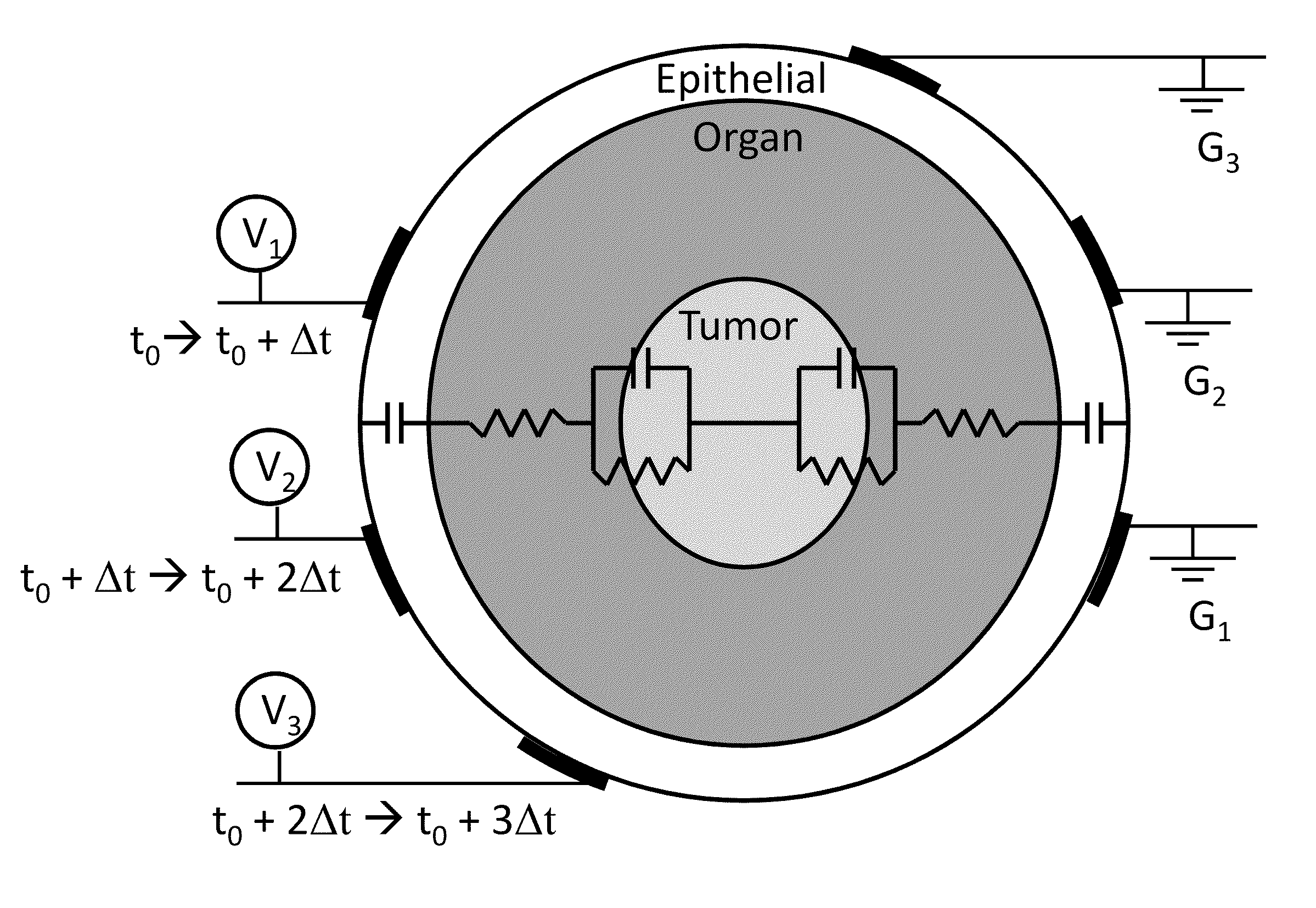
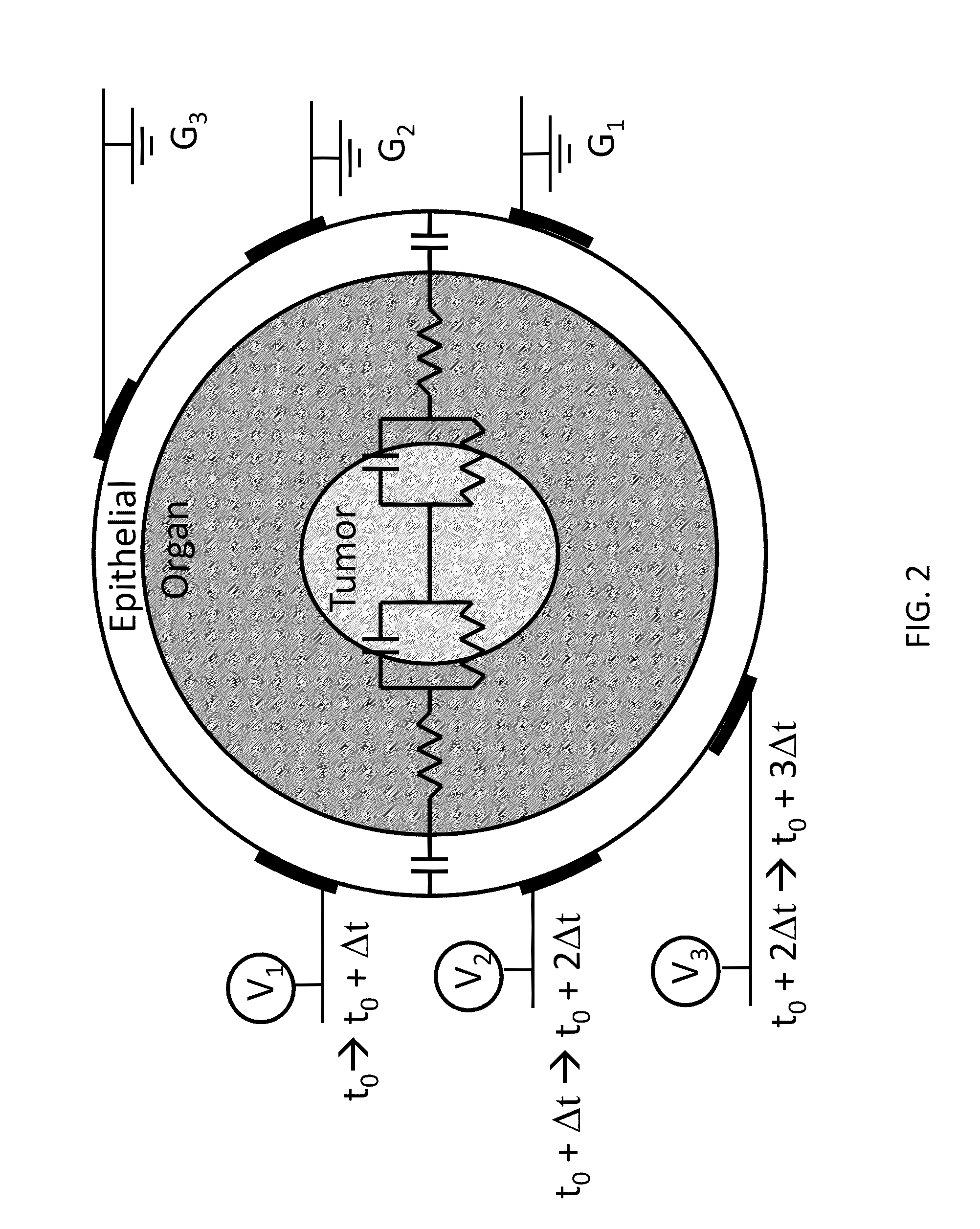
[0014]The present invention provides an advancement over tissue ablation techniques previously devised by providing methods for precisely and rapidly killing diseased, damaged, disordered, or otherwise undesirable biological tissues in situ. More specifically, the present invention provides methods comprising electric pulse therapies for ablating target cells and tissues for the treatment of diseases and disorders. Surprisingly, it has been found that the use of ultra-short pulses that have the ability to cause cell death can be effective as a treatment process for aberrant cell growths. The inventors have developed electroporation techniques using nanosecond-scale pulses as a controlled, precise way to destroy aberrant cells of a tissue or organ, without the deleterious side effect of heating the healthy cells in the vicinity of the undesirable cells. In these methods, one or more electrodes are placed within, near, or around the targeted region to deliver a series of high energy electric pulses to promote cell death. The packing of cells within a tissue is largely heterogeneous, and most organs are covered with epithelial cells joined by tight junctions to form a continuous sheet that rests on a layer of fibrous connective tissue. Further, organs can contain multiple sites of epithelial cells within underlying layers of tissue, for example, the cells forming the lining of ducts. Tight junctions are the preferred sites for electroporation when microsecond-scale pulses longer than the

Problems solved by technology

This, in turn, reduces the amount of

Method used


Image

  • Integration of very short electric pulses for minimally to noninvasive electroporation
  • Integration of very short electric pulses for minimally to noninvasive electroporation
  • Integration of very short electric pulses for minimally to noninvasive electroporation

Examples


example 1

Optimization of Voltage Parameters and Nanorod Concentrations for Selectively Killing Leukemia Cells In Vitro

[0099]Gold nanorods can be used to enhance the selectivity of pulsed electric field therapies to treat leukemia, a cancer that starts in the bone marrow and causes a large number of blood cells to be produced. Treatment of leukemia depends on the type and extent of the disease but often involves chemotherapy or radiation therapy. In autologous stem cell transplantation, stem cells from the patient's own marrow or blood are obtained and engrafted after the patient receives an intense dose of chemotherapy or radiation in an attempt to restore hematologic and immunologic function following treatment. There is the potential for disease recurrence if the engrafted stem cells contain even a single leukemic cell. Gold nanorods combined with antibody targeting techniques provide a means to selectively kill leukemic cells through the localized amplification of an applied external elec...

example 2

Computational Model for Investigating the Electric Field Distribution Within Bony Tissues

[0105]Imaging can be combined with computational modeling to provide insight into the unintuitive, highly complex interaction of electric fields with biological tissue. The electric field distribution within spongy bone can be determined to quantify treatment areas following supra-poration. Results from in vitro experimental data can be used to define electric field thresholds for killing bone marrow cells in the model. Further, image reconstructions and treatment area calculations obtained from in vivo experiments further refine the model geometry and properties.

[0106]A mathematical simulation for predicting the electric field distribution within a spongy bone structure where bone marrow resides among the trabeculae has been developed. A commercial finite element method (FEM) package, COMSOL Multiphysics (version 3.5 a), was selected because of its variety of features and functionality, as well...

example 3

Cell Death in Bony Tissue Using Pulsed Electric Fields

[0111]To demonstrate that pulsed electric fields can safely and predictably induce cell death in bony tissue, a rabbit model is used as the animal of study because techniques have been established for performing imaging and histological analysis on bony substructures in rabbits. Like humans, rabbits have a well developed haversian system, making them ideal candidates for obtaining translatable results from experiments on bone substructures. Six-month old rabbits are used because it is known that they achieve skeletal maturity at nineteen to twenty-four weeks. The IRE and supra-poration pulse generators are used in conjunction with a custom made bipolar electrode (4 mm in diameter) to deliver the electric pulses. Two IRE pulsing protocols and two supra-poration pulsing protocols are implemented for a comparison of treatment areas (Table 2). For each of the trial groups, including an untreated control group, four rabbits are utiliz...


PUM


Abstract

The present invention provides methods, devices, and systems for in vivo treatment of cell proliferative disorders. The invention can be used to treat solid tumors, such as brain tumors. The methods rely on non-thermal irreversible electroporation (IRE) or supra-poration to cause cell death in treated tumors. In embodiments, the methods comprise the integration of ultra-short electric pulses, both temporally and spatially, to achieve the desired modality of cell death.

Description

CROSS-REFERENCE TO RELATED APPLICATIONS[0001]This application relies on and claims the benefit of the filing date of U.S. Provisional Patent Application No. 61 / 167,997, filed 9 Apr. 2009, and U.S. Provisional Patent Application No. 61 / 285,618, filed 11 Dec. 2009, the entire disclosures of both of which are hereby incorporated herein in their entireties.BACKGROUND OF THE INVENTION[0002]1. Field of the Invention[0003]The present invention relates to the field of biomedical engineering and medical treatment of diseases and disorders. More specifically, the invention relates to methods for destroying aberrant cells, including tumor tissues, using a series of electrical pulses having durations on the nanosecond scale.[0004]2. Description of Related Art[0005]Treatment of abnormal cell growth in or on normal body tissues and organs can be achieved in many different ways to achieve reduced aberrant cell growth and even destruction of an aberrant cell mass. In general, treatments known in th...

Claims


Application Information

Patent Timeline
IPC IPC(8): A61B18/18A61B5/055A61B8/00A61B5/05
CPCA61B2018/0016A61N1/327A61N1/0412A61B18/1477A61B2018/00613A61N1/0424
Inventor DAVALOS, RAFAELARENA, CHRISTOPHER B.CALDWELL, JOHN
Owner VIRGINIA TECH INTPROP INC
Why Eureka
Try Eureka