Sterile Autologous, Allogenic or Xenogenic Implant and the Method of its Production

Active Publication Date: 2010-11-18
MEDICEM TECH SRO
View PDF9 Cites 7 Cited by
  • Summary
  • Abstract
  • Description
  • Claims
  • Application Information

AI Technical Summary

Benefits of technology

[0069]The inventors found and verified that the implant according to the invention can be advantageously used as a biological cover for burns, crural sores, harvesting areas and other skin defects. Dehydrated implant can be placed directly onto the bleeding or seeping wound, which will hydrate the implant in situ without a significant increase of the wound cover area and will contribute to the reduction of wound bleeding and seeping. For this use are especially suitable implants softened by suitable additives, such as glycerine. The sterile implant can be also hydrated before use in a sterile physiological solution, possibly with the addition of appropriate bactericidal agents (such as furantoin, boric acid solution or a proteinaceous silver solution in water (a silver-protein complex)), and placed onto the wound. Its great advantage is its ability to closely cover the topographical features of the wounded surface, lowers wound pain and has a hemostatic effect. Another great advantage is the fact that the healing of the whole area occurs without any change of wound dressings, which is necessary in case of other wound dressings, is often expensive and is especially traumatic for the patient (in case of severe burns it even has to be done under full anesthesia). Another advantage in comparison to other biological covers is the fact that it doesn't matter which side is in contact with the wound. The acellular dermal cover protects the wound and accelerates healing by supporting biological activity connected to healing, such as the migration and proliferation of patient's keratinocytes. Native keratinocytes will adhere to the inner surface of the implant (possibly to the fibrine created on the implant after contact with blood or plasma of the patient) and they migrate on its surface, so the implant becomes part of the skin for the period of time when active healing is taking place. As soon as healing is complete and the ep

Problems solved by technology

Excessively fast implant degradation can leave behind an inflammatory focus, which will heal with difficulty and can lead to scarring.
The infiltration of cells into the implant's structure would not be advantageous, because it could l

Method used


Image

  • Sterile Autologous, Allogenic or Xenogenic Implant and the Method of its Production
  • Sterile Autologous, Allogenic or Xenogenic Implant and the Method of its Production
  • Sterile Autologous, Allogenic or Xenogenic Implant and the Method of its Production

Examples


example 1

Porcine Skin as a Xenotransplant

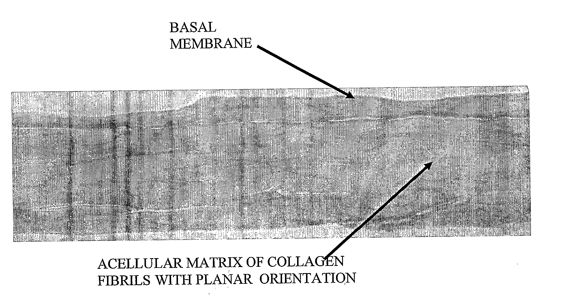
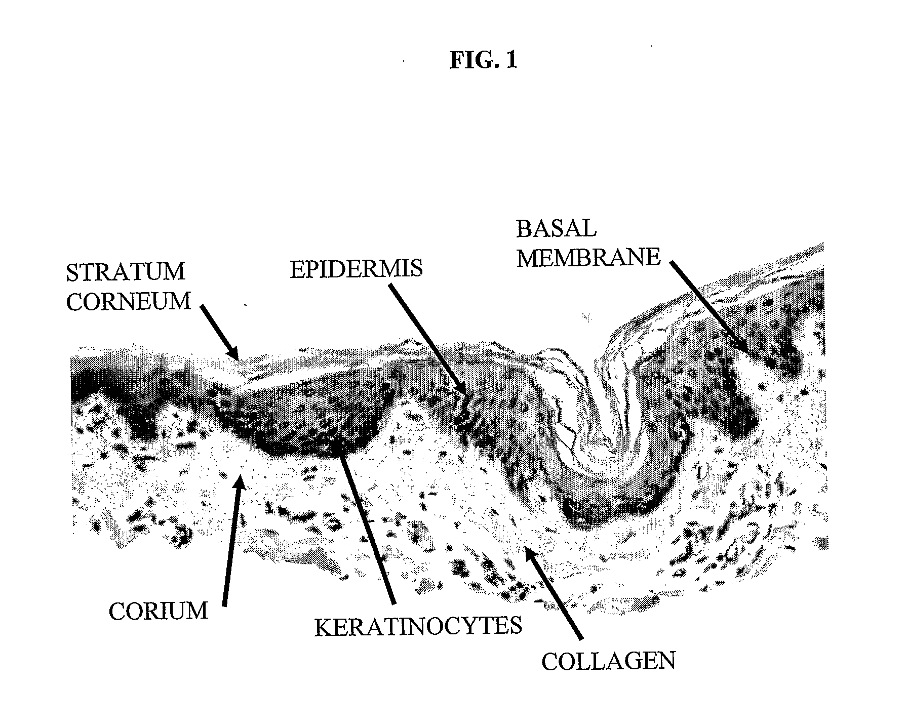
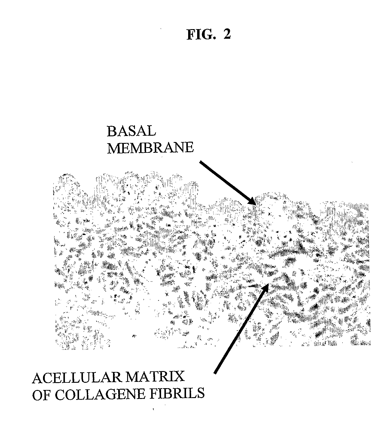
[0082]Using a dermatome, a 300-400 micron thick layer was cut from a shaved and cleaned porcine epidermis including the papillary corium layer. Histogram of the removed layer is in FIG. 1. The removed strip of porcine skin was immersed 3 times for 20 minutes at 37° C. and the for 12 hours at 4° C. in a 0.25% trypsin solution, which removed most dermal cells and separated the epidermis. The obtained epidermis was washed 6 times in demineralized water (3× for 1 hour, 1× or 12 hours, 2× for 0.5 hours) to remove remaining cells and trypsin. The FIG. 2 histogram shows, that the non-cellular structure was retained. The strip of dermis was then attached with an adhesive to a glass Petri dish and dried at room temperature to constant weight. In this state the dermis contained approx. 18% water. Thus dried acellular dermis had the same footprint area s the original hydrated dermis, but its thickness was less than half. The stretched acellular dermis was then i...

example 2

Implant Prepared According to Example 1 was Used on a 3rd Degree Burn

[0092]The damaged tissue was carefully cut off. The dehydrated transplant was removed from its sterile packaging, was adjusted to the size and shape of the treated area using scissors, was placed on the bleeding wounded area, and was sprayed with a sterile antibiotic solution. The xenotransplant softened quickly and adhered to the wounded area's surface, which stopped the bleeding. The implant retained its dehydrated state footprint, but its thickness increased upon hydration. This caused a perfect conformance to the wound. The implant was covered with a protective layer of tulle gras dressing for the first 3 days. After 8 days the implant dried out to a scab-like consistency and began to separate from the newly formed skin under the cover. New epidermis began to slowly form under the implant, as shown on FIG. 7. After healing was complete, healthy and naturally structured skin was formed, including natural pigment...

example 3

Human Cartilage as an Allotransplant

[0093]Cartilage harvested from a hip joint of a deceased donor was made free of cells by repeated soaking in a trypsin solution and in distilled water, then it was attached, using vacuum, with its concave side onto the convex side of a suitably shaped porous substrate made of fritted glass. While on this substrate, it was placed into an excess of a solution of 20 parts (by weight) methanol and 5 parts (by weight) dimethylsulfoxide (DMSO) at 35° C. for a period of 48 hours. Thus the acellular implant was dehydrated, and at the same time the collagen contained within it was partially denatured in a plane-oriented state. After this period the acellular allotransplant was removed from the solution and methanol was evaporated at room temperature using air stream. At the end of this step the transplant contained approx. 13% (by weight) DMSO, approx. 4% (by weight) water and less that 0.5% (by weight) methanol. After being removed from the porous substra...


PUM

PropertyMeasurementUnit
Temperatureaaaaaaaaaa

Abstract

The subject of the invention is a sterile, dehydrated acellular implant, which during its rehydration by water or bodily fluids displays anisotropic expansion and can act as a substrate for adhesion, migration and growth of live cells. Collagen structures of the transplant are at least partially denatured through the action of heat or organic solvents, such as lower aliphatic alcohols and ketones, which simultaneously act as preservative and sterilization agents, especially for certain types of viruses. The implant is sterilized by radiation while in an substantially dehydrated state, preferably by accelerated electrons. The transplant can be derived from various animal tissues, especially mammalian tissues, such as human or porcine tissues. The tissues suitable for the invention can be, for example, skin, placenta, pericardium, peritoneum, intestinal wall, tendon, blood vessel, etc. The implant is suitable for use in human and veterinary medicine, for instance as a temporary wound and burn cover, for the repair, substitution and regeneration of tissues, and also as a substrate for laboratory cell cultivation.

Description

FIELD OF THE INVENTION[0001]The invention relates to wound healing and tissue regeneration. Specifically it concerns a sterile dehydrated acellular implant (transplant), which during its rehydration by water or bodily fluids displays anisotropic expansion. Product of this type can be called either an implant or a transplant, depending on context and custom in various areas of expertise. In the present application, both terms are used according to the context, however, it is important to keep in mind that they are mutually interchangeable. The implant is sterilized using radiation while in an essentially dehydrated state, preferably using accelerated electrons. The implant can derive from various animal tissues, especially from mammalian tissues, such as for example human or porcine tissues, such as, for example, skin, placenta, pericardium, peritoneum, intestinal wall, tendon, blood vessel, et cetera. The implant, according the invention, is suitable for use in human and veterinary ...

Claims


Application Information

Patent Timeline
IPC IPC(8): A61L27/60A61K38/39A61P17/02
CPCA61K38/39A61L27/3633A61L27/3691A61L2430/40A61L27/3813A61L27/50A61L27/60A61L27/38A61P17/02A61P39/00A61P43/00A61L27/24A61L27/36A61L27/44A61L2430/00
Inventor DRUNECKY, TOMAS
Owner MEDICEM TECH SRO
Why Eureka
Try Eureka