
Method and apparatus for dermatological treatment and tissue reshaping

Inactive Publication Date: 2011-01-13
THE GENERAL HOSPITAL CORP
View PDF8 Cites 95 Cited by
  • Summary
  • Abstract
  • Description
  • Claims
  • Application Information

AI Technical Summary

Benefits of technology

[0023]It is therefore one of the objects of the present disclosure to provide exemplary embodiments of apparatus and method that can controllably deliver a substance to predetermined locations within the dermis or other tissue and direct electromagnetic radiation onto and / or into the substance within the tissue while substantially avoiding absorption of the radiation by surrounding tissue. It is another object of the present disclosure to provide exemplary embodiments of a needle apparatus configured to be inserted into biological tissue, where the needle apparatus can provide a visible signal that indicates whether the tip region of the needle is located within or outside of a blood vessel or other tissue structure.
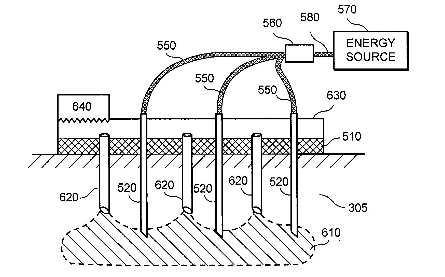
[0026]In a still further exemplary embodiment according to the present disclosure, method and apparatus can be provided that facilitate a placement of a curable cosmetic filler material in tissue and in situ curing or cross-linking of the filler material, e.g., to reduce or prevent absorption or migration of the filler material after placement. The filler material can be injected at a particular location in a tissue through the lumen of one or more needles configured to be inserted into the tissue. The electromagnetic radiation can then be delivered to the filler material using one or more needles that can be configured to penetrate the tissue to one or more desired depths where the filler material has been implanted, and which can include an optical waveguide or the like coupled to an electromagnetic radiation source. The one or more needles can be optical delivery needles that include both a lumen for material delivery and a waveguide for delivery of optical energy.
[0027]In a further exemplary embodiment of the present disclosure, method and apparatus can be provided that facilitates a photodynamic therapy (PDT) treatment of biological tissues. The tissue depth at which conventional PDT techniques can be performed may be limited by the depth to which light energy from a noninvasive external energy source can penetrate tissue overlying the treatment region. Exemplary embodiments of the present disclosure can facilitate a controlled introduction of a photosensitizer and / or photosensitizer precursor into a particular location within the tissue, and subsequent irradiation of the particular location using one or more waveguides inserted into the tissue, even if such treated tissue lies well below a tissue surface. The photosensitizer substance and the optical energy can both be delivered using one or more optical delivery needles that may include both a lumen for delivering the substance and one or more waveguides for directing electromagnetic radiation into the particular location. Undesirable or harmful interactions between the applied energy and tissue overlying the treatment region (e.g., heating, absorption, scattering, etc.) can thereby be reduced or avoided by using the exemplary optical needles and / or arrays of such optical needles described herein to direct energy into the treated tissue.
[0028]In yet another exemplary embodiment of the present disclosure, method and apparatus can be provided that facilitates determination of whether a tip of a hypodermic needle is located within or outside of a blood vessel or other particular structure prior to injecting or withdrawing a substance through the needle. A waveguide can be provided with the needle such that a distal end of the waveguide is located proximal to the needle tip. Light having one or more certain wavelengths can be provided through the waveguide to the distal end thereof Observation of the light emitted from the tip region of the needle from outside of the tissue surface can indicate whether the needle tip is inside or outside of a blood vessel. For example, this emitted light may be substantially reduced in intensity if the needle tip is within a blood vessel because of increased local absorption of the light by hemoglobin compounds within the vessel. The light can have a wavelength between about 530 nm and about 560 nm, which can exhibit a high absorption coefficient by hemoglobin compounds.

Problems solved by technology

For example, aging skin tends to lose its elasticity, leading to increased formation of wrinkles and sagging.
Other causes of undesirable wrinkles in skin include excessive weight loss and pregnancy.
These surgical approaches include facelifts, brow lifts, breast lifts, and “tummy tucks.” Such approaches can produce a number of negative side effects including, e.g., scar formation, displacement of skin from its original location relative to the underlying bone structure, and uneven tightening.
Certain treatments which use electromagnetic radiation have been developed to improve skin defects by inducing a thermal injury to the skin, which results in a complex wound healing response of the skin and / or certain biological structures located therein, such as blood vessels.
However, certain patients may experience major drawbacks after such LSR treatment, including edema, oozing, and burning discomfort during first fourteen (14) days after treatment.
These drawbacks can be unacceptable for many patients.
Indeed, LSR procedures can also be relatively painful and therefore generally may require an application of a significant amount of analgesia.
One of the limitation of LSR is that this ablative resurfacing in areas other than the face generally may have a greater risk of scarring because the recovery from skin injury within these areas is not very effective.
Although NCR techniques can assist in avoiding epidermal damage, they may have limited efficacies.
Even after multiple treatments, the clinical improvement is often below the patient's expectations.
In addition, a clinical improvement may be delayed for several months after a series of treatment procedures.
The NCR procedure can be moderately effective for wrinkle removal, and may generally be ineffective for dyschromia.
A further limitation of NCR procedures relates to the breadth of acceptable treatment parameters for safe and effective treatment of dermatological disorders.
The NCR procedures generally rely on an optimum coordination of laser energy and cooling parameters, which can result in an unwanted temperature profile within the skin leading to either no therapeutic effect or scar formation due to the overheating of a relatively large volume of the tissue.
This treatment approach can be painful, and may lead to a short-term swelling of the treated area.
In addition, because of the relatively large volume of tissue treated and the need to balance application of the RF current with the surface cooling, this RF tissue remodeling approach may likely not allow a fine control of damage patterns and subsequent skin tightening.
The current in monopolar applications generally flows through the patient's body to the remote ground, which can lead to unwanted electrical stimulation of other parts of the body.
However, low-viscosity fillers may be easily resorbed by the body and / or may flow or migrate from the initial application site.
Migration of fillers can also lead to alteration of the appearance of the filler-containing tissue, including reappearance of wrinkles, lumpiness, etc.
Fillers that are too ‘thin’ (e.g., those having a low viscosity) may not provide sufficient mechanical or rheological stability to remain in place or support surrounding tissue.
It may be difficult to inject or implant such ‘thick’ fillers into tissue using a needle, and it may also be difficult to implant them evenly to provide a natural appearance.
It can be difficult to control the extent of the crosslinking reaction, and such reactions may produce undesirable by-products.
However, it may be difficult to provide sufficient optical energy within the filler material, which may be located below the tissue surface, to initiate and / or control the extent of such crosslinking reactions.
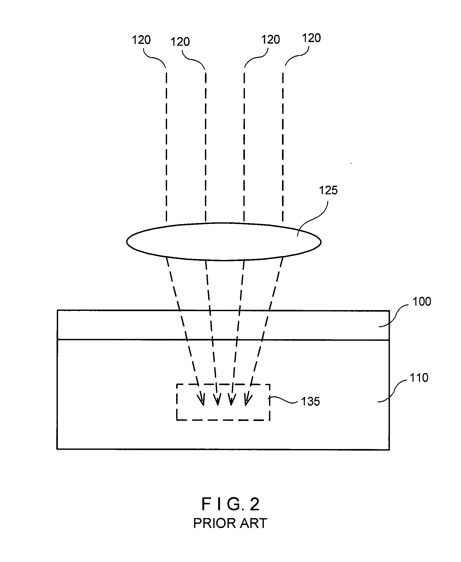
For example, tissue overlying the filler material may absorb, scatter, or otherwise interact with such energy that is directed into the tissue, which can lead to unwanted absorption, heating and / or damage of the overlying tissue and may result in a reduced amount of such energy reaching the filler material.
Further, electromagnetic energy having shorter wavelengths may be preferable for initiating crosslinking or photocuring in such materials; however, such higher-energy radiation can be more strongly absorbed by the overlying tissue and thus may not penetrate sufficiently into the subsurface filler to cure it sufficiently.
For example, introducing substances such as fillers into a blood vessel can lead to obstruction of the vessel or other undesirable effects.
It is generally difficult to identify the precise location of the tip of a hypodermic needle when it is inserted into tissue, and to ascertain whether it is within or outside of a blood vessel.
Skin may also exhibit various discolorations or other pigmentation defects which may be aesthetically undesirable.
In general, an application of EMR to skin or other tissue to treat such defects can be inefficient or lead to unwanted side effects.
Thus, it may be difficult to provide such highly-absorbed EMR to a region of tissue which lies below the surface of the skin, and there may be significant undesirable absorption of such EMR in tissue which lies above the treatment region.

Method used


Image

  • Method and apparatus for dermatological treatment and tissue reshaping
  • Method and apparatus for dermatological treatment and tissue reshaping
  • Method and apparatus for dermatological treatment and tissue reshaping

Examples


Embodiment Construction

[0023]It is therefore one of the objects of the present disclosure to provide exemplary embodiments of apparatus and method that can controllably deliver a substance to predetermined locations within the dermis or other tissue and direct electromagnetic radiation onto and / or into the substance within the tissue while substantially avoiding absorption of the radiation by surrounding tissue. It is another object of the present disclosure to provide exemplary embodiments of a needle apparatus configured to be inserted into biological tissue, where the needle apparatus can provide a visible signal that indicates whether the tip region of the needle is located within or outside of a blood vessel or other tissue structure.

[0024]These and other objects can be achieved with an exemplary embodiment of the apparatus and method according to the present disclosure, in which one or more needles are provided that may be used to inject and / or withdraw material from a particular region of a biologi...


PUM


Abstract

Exemplary method and apparatus can be provided for directing a substance and electromagnetic radiation to a particular location in a biological tissue, for example, to inject and cure a cosmetic filler in situ. The apparatus can include a needle configured to be inserted into the tissue, and a waveguide configured to direct the electromagnetic radiation, such as optical energy, to a location proximal to the needle tip. The substance can be injected into the tissue through the needle and irradiated by the optical energy. In addition, the exemplary method and apparatus can be provided for determining the location of the needle tip in a biological tissue based on characteristics of light directed through the waveguide and emitted proximal to the needle tip. For example, intensity of the emitted light can indicate whether the needle tip is located inside or outside of a blood vessel.

Description

CROSS REFERENCE TO RELATED APPLICATION[0001]The present application claims priority from U.S. Provisional Patent Application Ser. No. 61 / 185,495 filed Jun. 9, 2009, the disclosure of which is incorporated herein by reference in its entirety.FIELD OF THE DISCLOSURE[0002]The present disclosure is directed to a cosmetic method and apparatus for improving skin appearance. More specifically, the present disclosure is directed to exemplary embodiments of such method and apparatus in which electromagnetic energy, e.g. optical energy, is provided proximal to a tip of one or more needles that are inserted into a biological tissue. Such energy can be directed onto and / or into particular regions within skin or other tissue containing a curable material, e.g., a cosmetic filler, to cure such material in situ. The optical energy can optionally be used to determine whether the needle tip is located within a blood vessel when the needle is inserted into tissue, e.g., prior to injecting or withdraw...

Claims


Application Information

Patent Timeline
IPC IPC(8): A61N5/08A61M5/31A61B6/00
CPCA61B18/201A61B18/203A61B2018/00452A61M37/00A61L31/18A61L2400/06A61B2018/00458
Inventor MANSTEIN, DIETER
Owner THE GENERAL HOSPITAL CORP
Learn More