Detection method

a detection method and diagnostic moiete technology, applied in the field of detection methods, can solve the problems of insufficient, complicating assays, and far exceeding the fluorescence lifetime of terbium, and achieve the effect of reducing the cost of antigen production techniques

Inactive Publication Date: 2011-02-17
THE SEC OF STATE FOR ENVIRONMENT FOOD & RURAL AFFAIRS ACTING THROUGH THE ANIMAL HEALTH & VETERINARY LAB AGENCY
View PDF8 Cites 8 Cited by
  • Summary
  • Abstract
  • Description
  • Claims
  • Application Information

AI Technical Summary

Benefits of technology

[0037]Therefore, the method directly identifies the presence, in the sample, of a moiety as the result of exposure of a human or animal to a specific infectious organism. There is no requirement for a general immune response to have occurred. Advantageously, this allows the user of the method to detect exposure of a human or animal to a specific infectious organism at an early stage, even in t

Problems solved by technology

However the fluorescence lifetime of terbium far exceeds that of the background noise.
Previous attempts to increase the sensitivity have focused on the addition of additional reagents such as fluoride ions (see U.S. Pat. No. 5,627,074) but this has the effect of further complicating the assay, and the results have not been sufficient to ensure that the technique has found widespread use in diagnosis.
Furthermore, diagnosis of disease is relatively infrequently carried out on the basis of high throughput screening.
Even here its re-introduction remains a real threat to livestock and human health as well as the rural economy.
As such the detection of Brucella in livestock is a major issue facing any country with a livestock industry.
The economic burden of effective brucellosis surveillance, where large numbers of serum and/or milk samp

Method used


Image

  • Detection method
  • Detection method
  • Detection method

Examples


example 1

Diagnosis of Brucellosis

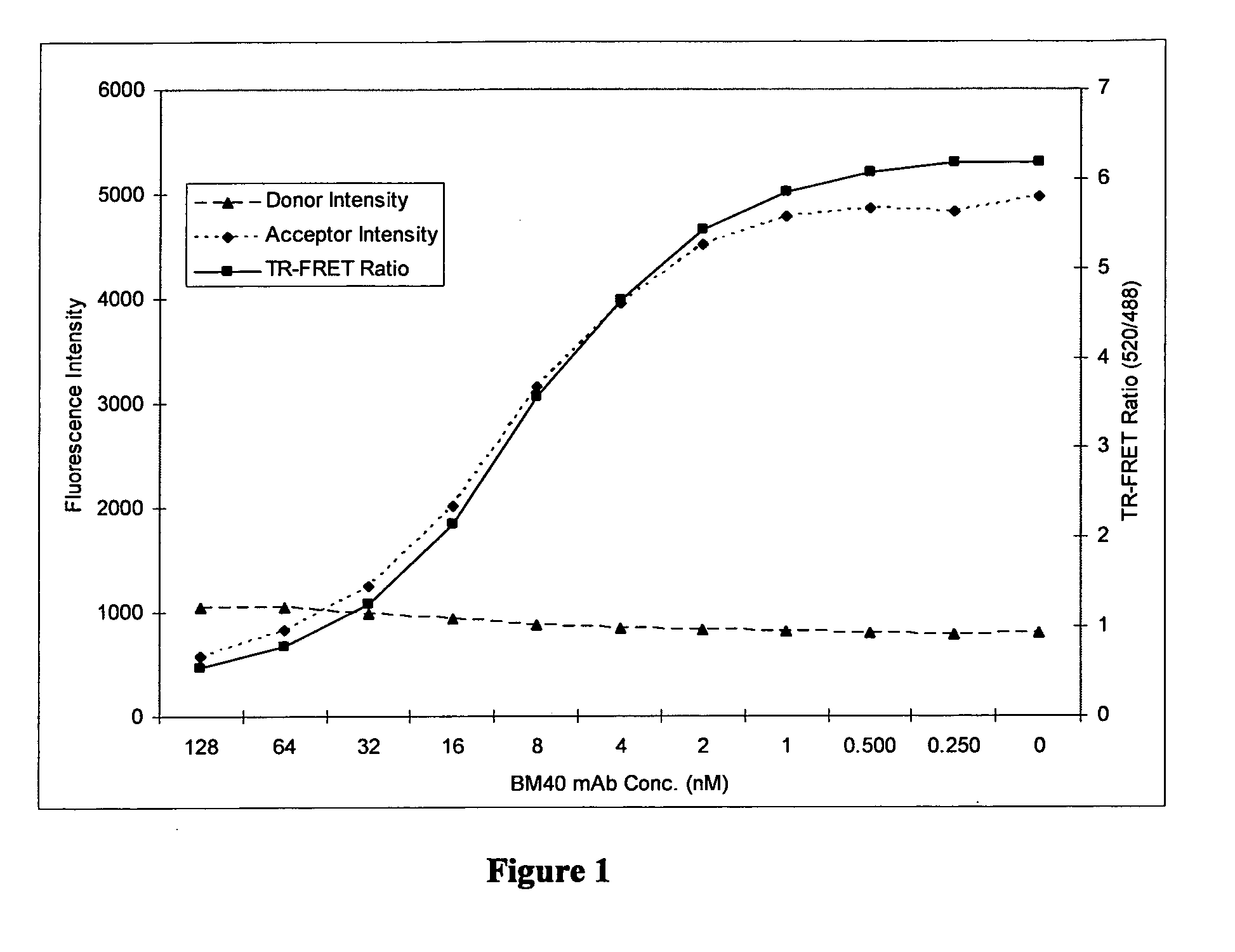
[0087]The applicants developed a TR-FRET protocol as described below. The method was used to analyse samples from Brucella infected and uninfected cattle and the results are illustrated below.

[0088]Antibody Labelling with Terbium

[0089]The BM40 mAb used was a mouse IgG1 antibody specific to Brucella ‘M’ O-antigen epitopes (Greiser-Wilke & Moenning, Ann Inst. Pasteur Microbiol. 1987 138 (5) 549-60). The supernatant from a BM40 producing B-cell hybridoma cell culture was affinity purified using a protein G column.

[0090]To label the antibody, 3 ml of BM40 was dialysed against sodium carbonate buffer (pH 9.5) for 21 hours at 4° C. using a 1-3 ml 10 kDa Molecular Weight Cut-Off (MWCO) Slide-a-lyzer (Pierce™) dialysis cassette. The BM40 mAb was recovered from the cassettes and centrifuged in 3 kDa MWCO Centricons (Millipore, Billerica, Mass.) at 4000 g for 90 minutes at +4° C. which decreased the volume to 0.7 ml. This was spectrometrically determined to be at a con...

example 2

Further Studies Relating to Diagnosis of Brucellosis

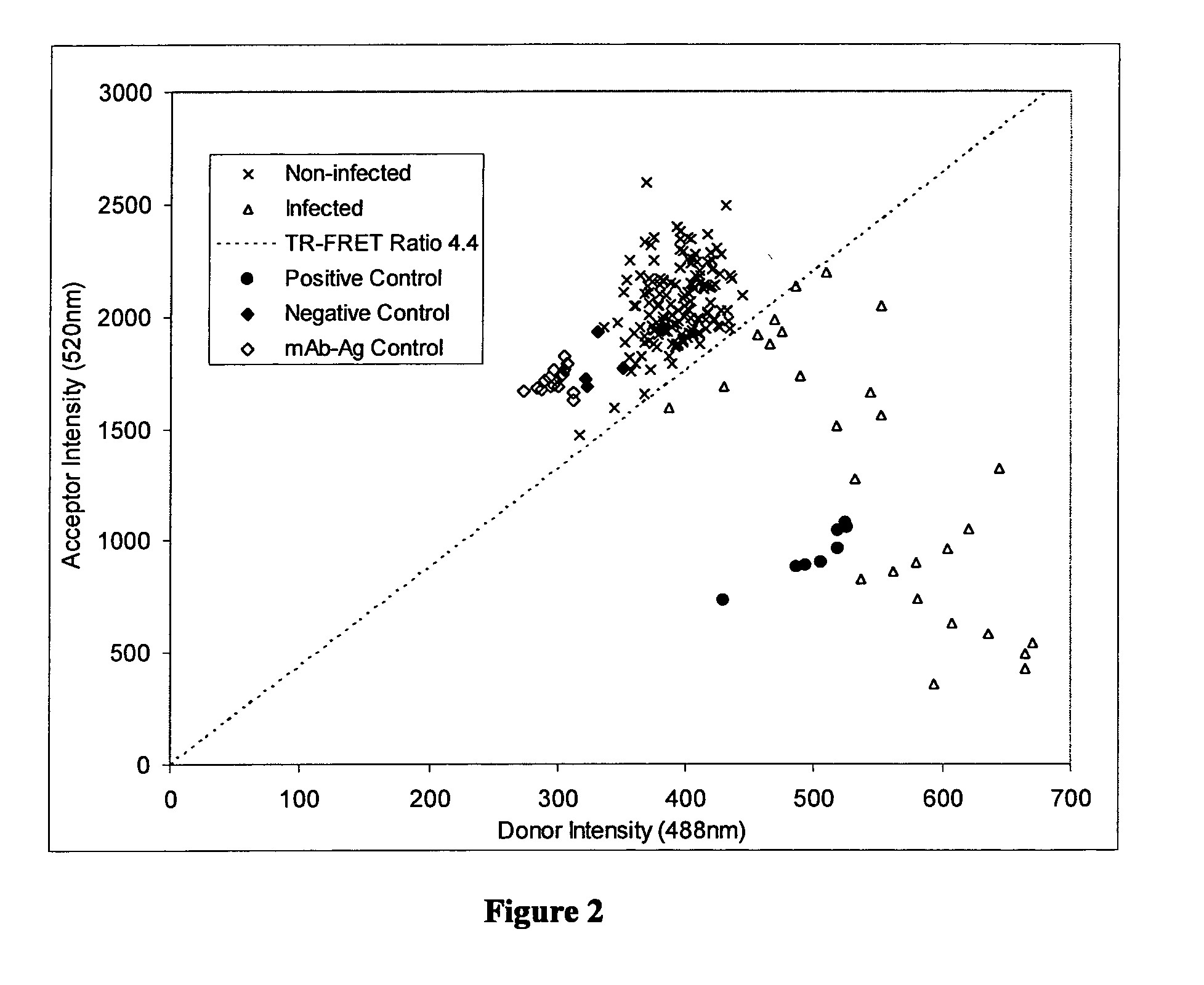
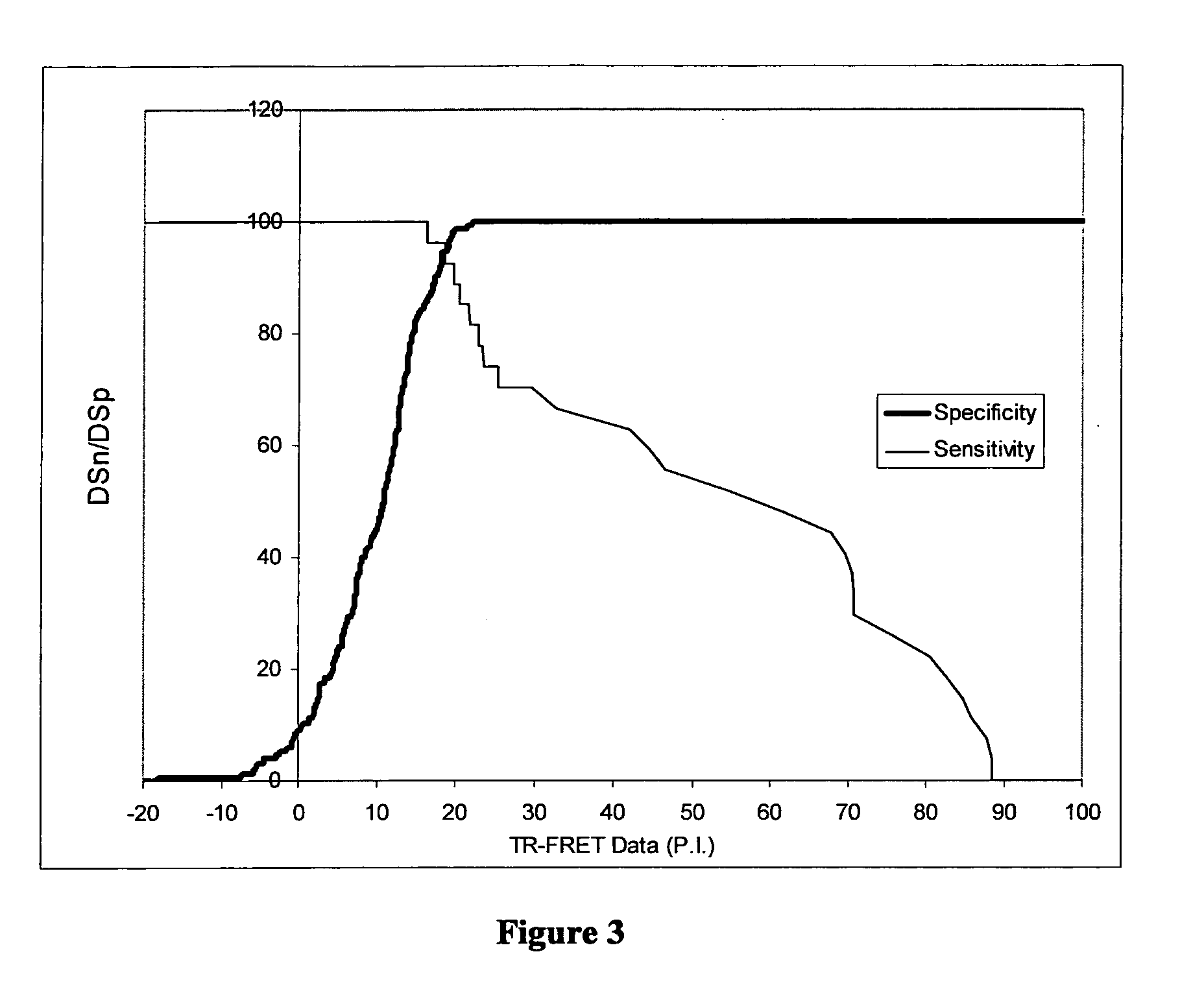
[0111]The methods described above in Example 1 were further optimised and validated as described below. The results of further studies using the optimised protocols are also described.

[0112]Test Method

[0113]The labelling of terbium to BM40 was improved by increasing the conjugation time to 6 hrs and removing excess unconjugated terbium by desalting with a 5 ml Zebra™ column (Pierce), as described above, without prior dialysis. This improved the terbium to BM40 molar ratio to more optimal levels. The production yield of Brucella sLPS-FITC was improved by desalting using a PD-10 column (GE Healthcare) following the manufacturers instructions, rather than a Zebra™ column (Pierce). Titration of these reagents against control serum (see above) demonstrated optimal reagent concentrations were 2 nM BM40-Tb and a 1 / 1750 dilution of Brucella sLPS-FITC. The optimal serum sample concentration was determined to be ⅕.

[0114]The TR-FRET assay pla...

example 3

Detection of Diagnostic Moieties for Bovine Viral Diarrhoea (BVD) by TR-FRET

[0139]The applicants developed TR-FRET protocols as described below. The method was used to analyse samples containing anti-BVD antibodies and BVD viral antigens.

[0140]Development of Competitive TR-FRET Method

[0141]The BVD TR-FRET method was developed using the following reagents in a competitive format: biotinylated recombinant BVD E2 antigen, terbium conjugated streptavidin and fluorescein conjugated anti-E2 monoclonal antibody WB214.

[0142]Production of recombinant baculovirus expressing the E2 glycoprotein for BVDV type 1a (strain C24V) was achieved by firstly cloning the region of the bovine viral diarrhoea virus genome delineated by primers BVDV C24V E2 EcoRI and BVDV C24V E2 6His XhoI (Amin Asfor PhD thesis; RVC, University of London, 2006) into the general cloning vector pGEM-T easy (Promega). The primers introduced an EcoRI site, a start codon and a Kozak consensus sequence at the 5′ terminus of the ...


PUM


Abstract

A method for detecting the presence of a diagnostic moiety indicative of exposure to an infectious organism in a biological sample taken from a human or animal, said method comprising; (a) adding to said sample a first fluorescently labelled reagent which binds said diagnostic moiety, and a second fluorescently labelled reagent which either binds said diagnostic moiety in addition to said first fluorescently labelled reagent, or which binds the first fluorescently labelled reagent or a complex comprising the first fluorescently labelled reagent in competition to the said diagnostic moiety, wherein a label on one of the first or second fluorescently labelled reagent acts as a fluorescent energy donor compound and wherein the other of the first or second fluorescently labelled reagent acts as a fluorescent energy acceptor compound which is able to accept fluorescent energy from said donor compound; (b) exciting the fluorescent energy donor compound by illuminating with light of a wavelength which is absorbed by said fluorescent energy donor compound; (c) measuring fluorescent signal emitted by said fluorescent energy acceptor compound as a result of its absorption of the fluorescent energy from the donor compound after a time delay; and (d) relating the results to the presence or absence of diagnostic moiety in said sample.

Description

FIELD OF INVENTION [0001]The present invention relates to a method for the detection of diagnostic moieties, in particular to methods based upon Time Resolved Fluorescent Energy Transfer (TR-FRET) technology to measure the proximity of moieties such as antibodies or antigens in biological samples, which is useful in the diagnosis and screening for diseases caused by infectious organisms such as brucellosis and the virus which causes Bovine Viral Diarrhoea (BVD Virus), as well as kits useful in the method.BACKGROUND [0002]FRET technology has been known for many years. In FRET, a donor fluorophore is excited by light, and if a suitable acceptor is in close proximity, the excited state energy from the donor can be transferred to the acceptor. For the acceptor to be suitable it must have an excitation wavelength that overlaps with the emission wavelength of the donor. The energy transfer leads to a decrease in the donor's emission intensity and an increase in the acceptor's emission int...

Claims


Application Information

Patent Timeline
IPC IPC(8): C12Q1/70G01N33/569G01N33/566
CPCG01N33/542G01N2800/26G01N2333/005
Inventor MCGIVEN, JOHN
Owner THE SEC OF STATE FOR ENVIRONMENT FOOD & RURAL AFFAIRS ACTING THROUGH THE ANIMAL HEALTH & VETERINARY LAB AGENCY
Why Eureka
Try Eureka