Compositions and methods for making and using laminin nanofibers

a technology of laminin nanofibers and nanofibers, which is applied in the field of compositions, can solve the problems of prohibitive three-dimensional scaffolding and high cost of laminin and reconstituted basement membrane manufacture, and achieve novel biomimetic effects, long shelf life, and greater tensile strength

Active Publication Date: 2011-09-29
UNIV OF VIRGINIA ALUMNI PATENTS FOUND
View PDF0 Cites 58 Cited by
  • Summary
  • Abstract
  • Description
  • Claims
  • Application Information

AI Technical Summary

Benefits of technology

[0012]The present invention provides large cost savings over other techniques. The cost of laminin and reconstituted basement membrane (RBM) manufacture is extremely high and approaches being prohibitive in its usage as a three-dimensional scaffold, such as nanofiber meshes. The present invention provides a mixture of at least one synthetic polymer and at least one extracellular matrix protein, thus decreasing the costs for preparing the mixed nanofiber of the invention because less protein is needed. In one aspect, the synthetic polymer is PCL. In one aspect, the extracellular matrix protein is laminin. In one aspect, the laminin is laminin I.
[0013]It is also desirable to increase the tensile strength of laminin-rich materials and reduce their stickiness to electrodes and molds during manufacture. To achieve these improvements, methods are disclosed herein to fabricate co-spun laminin and synthetic biodegradable polymer materials that can be fabricated into nanofiber meshes, films, and microspheres among a broad variety of possible biomedical applications. The synthetic polymer provides a better base for encapsulation of drugs and growth factors, a mechanically stable substrate with defined decay rates, and a platform for manipulating fiber morphology and geometry, while serving as a low cost filler to reduce the amount of laminin necessary. The applications for this protein-polymer electrospun mixture will dramatically increase as the cost of manufacture decreases and the repeatability increases. The electrospun nanofibers can be applied in culture, device fabrication for tissue engineered constructs, conduits, or even as a bandage, or component thereof, or other topical application.
[0014]Laminin, which contains many biologically active moieties (including a moiety having a sequence consisting of an isoleucine, a lysine, a valine, an alanine, and another valine), may especially benefit from orientation at the individual molecule level in a nanofiber mesh. Providing the biologically active molecule laminin within the framework of intraluminal nanofiber scaffold may improve axonal guidance and support during regeneration.
[0016]The present invention further provides compositions and methods for aligning the laminin-polymer nanofiber mixtures of the invention. In addition to the physical benefit of providing an aligned substrate for directional outgrowth, there exist two further potential benefits of aligned nanofibers for peripheral nerve regeneration, both occurring as a result of the electric field changes created by the insulting gap. First, high field forces at the edges of the gap exert stronger forces on individual polymer fibers, causing fibers to stretch across the gap, decreasing their resulting diameter. The ideal conditions for alignment and stretching have been explored by our collaborators in a recent publication [10]. This stretching effect decreases the lower bound of mean fiber diameters below 100 nm, yielding fiber diameters mimetic of the natural basement membrane, which has feature sizes in the range of 75-150 nm [11]. Second, fiber alignment across two electrodes separated by an insulating gap results in molecular level orientation of individual polymer molecules within the fiber [12].
[0024]The nanofibers comprising a mixture of laminin and at least one synthetic polymer prepared by the methods of the invention should have a very long shelf life when stored with desiccation. They have far greater tensile strength than matrigel gels. The nanoscale fibers are similar to the fibers seen by cells encountering laminin in real basement membranes, thus they may be expected to demonstrate novel biomimetic effects. The materials fabricated by this process may, for example, be used as an anhydrous coating of scaffold biomaterials for tissue engineering, as well as substrate for ex vivo cultivation of both specialized tissue cells and stem cells. The latter could be a tremendous aid to basic science research as differentiation and phenotype expression of cells on biomimetic laminin scaffolds may be more representative of in vivo behavior.
[0043]In one embodiment, the nanofibers form a mesh. Meshes may, for example, be applied directly to a site of injury. Additionally, the nanofibers may be applied to or in a substrate or vehicle and then delivered or administered. For example, a nanofiber mesh of the invention may be applied to the lumen of a conduit, which can be used when reapposing a severed nerve and the nanofibers will help stimulate axonal regeneration and healing of the injured nerve.

Problems solved by technology

The cost of laminin and reconstituted basement membrane (RBM) manufacture is extremely high and approaches being prohibitive in its usage as a three-dimensional scaffold, such as nanofiber meshes.

Method used


Image

  • Compositions and methods for making and using laminin nanofibers
  • Compositions and methods for making and using laminin nanofibers
  • Compositions and methods for making and using laminin nanofibers

Examples


embodiments

[0187]The present invention provides compositions and methods for mimicking three dimensional scaffolding as found in vivo to better mimic how cells grow and differentiate and to aid in, inter alia, attachment and proliferation of cells, cell differentiation, wound repair, regeneration of injured tissue, etc. In one embodiment, the injured tissue is nerve tissue.

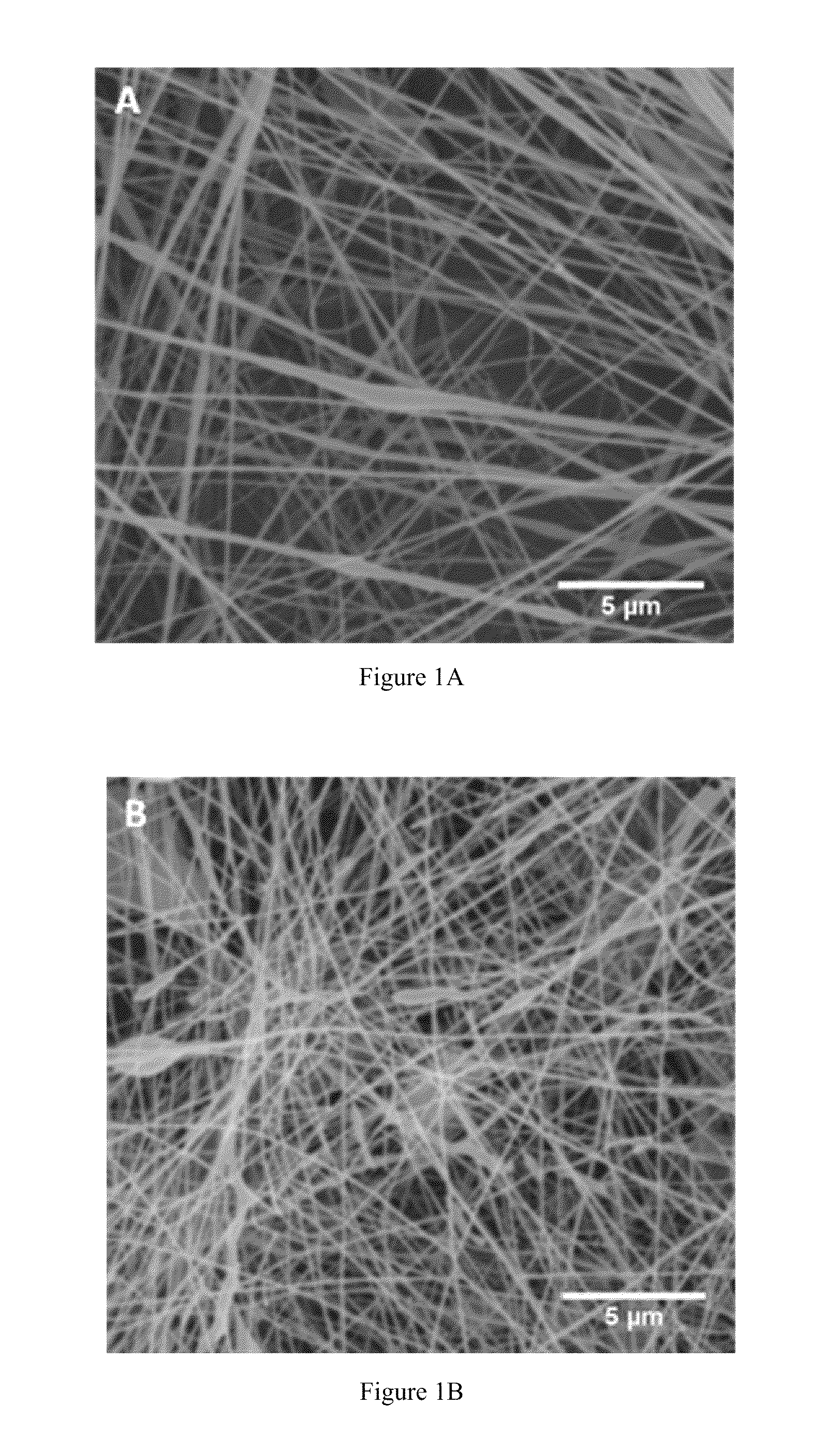
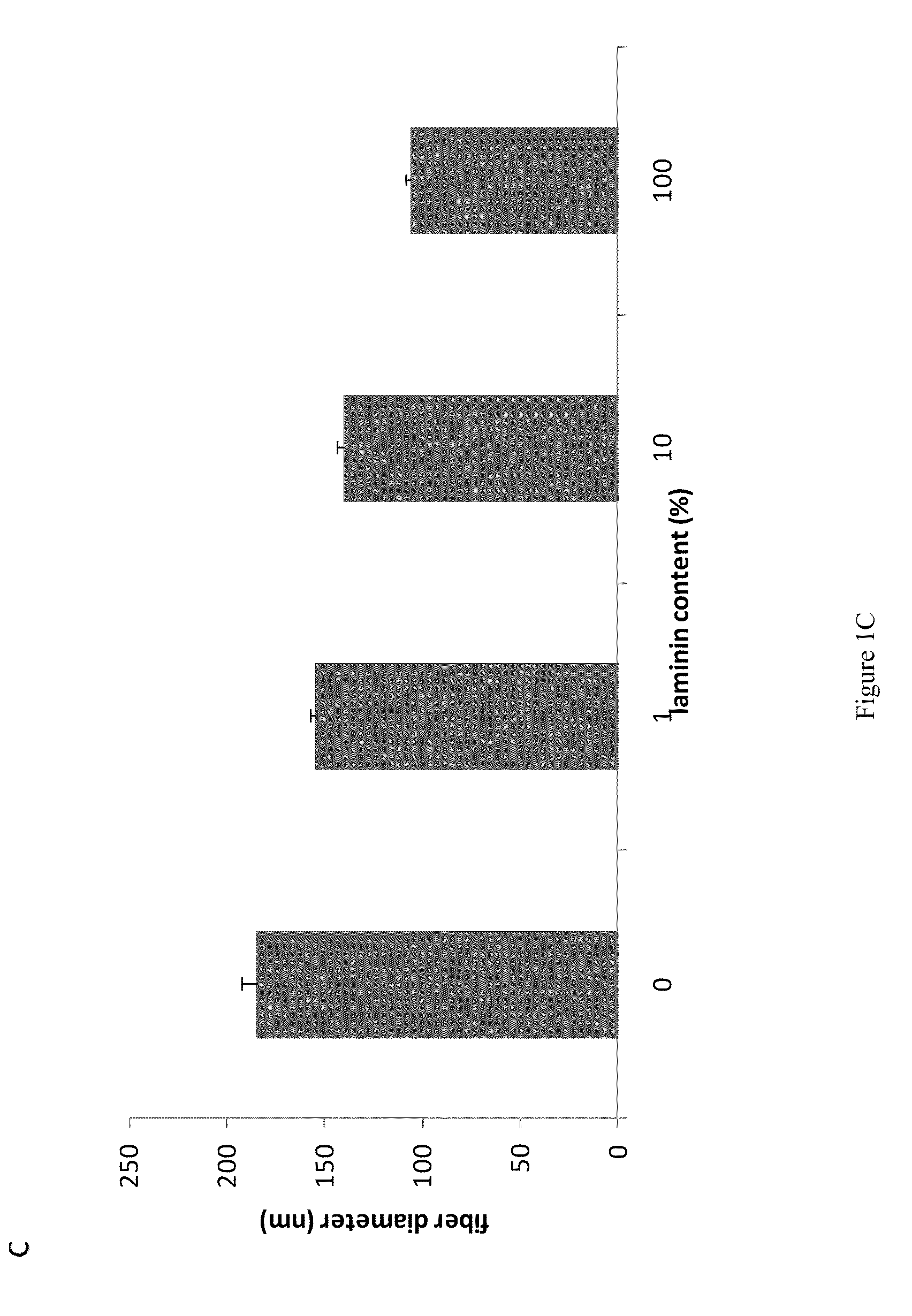
[0188]The present invention provides methodologies and parameters for fabrication of the hybrid biomaterial by blending pure laminin or complex extracts of tissues containing laminin with biopolymers such as polycaprolactone (PCL), polylactic / polyglycolic acid copolymer (PLGA) or Polydioxanone (PDO) in, for example, HFP, TFA, or TFE, fabrication of substrates and scaffolds and devices from the hybrid biomaterial in forms such as films, nanofibers by electrospinning or microspheres, and the biological or biomedical use of the material or devices derived from it.

[0189]Previous work prepared and used nanofibers that were homoge...

example 1

Materials and Methods

[0309]The solvent, 1,1,1,3,3,3-hexafluoro-2-propanol (HFP) was purchased from Sigma (St Louis, Mo.). All cell culture reagents were purchased from Fisher Scientific (Pittsburgh, Pa.).

[0310]Laminin Isolation

[0311]Laminin I was purified from the EHS tumor according to previously established methods. The final laminin solution was subjected to 2 rounds of precipitation with 45% ammonium sulfate to remove most growth factors present. Purity of laminin was evaluated by SDS-PAGE and Western analysis with affinity purified antibodies to type IV collagen, entactin / nidogen and perlecan, the major contaminants of such preparations. Purity was determined to be greater than 99% laminin (w / v). Laminin was stored at −80° C.

[0312]Laminin Electrospinning

[0313]For the parametric study, a series of process parameters was chosen within ranges shown to be successful in creating submicron or nanoscale fibers of other ECM proteins such as collagens [13] and fibrinogen. Laminin was di...

example 2

Laminin Nanofiber Mesh Substrates for Stem Cell Growth and Differentiation as Recited in U.S. application Ser. No. 12 / 598,776.

[0347]Methods—Embryonic Stem Cell Culture: D3 and ES-E14TG2a murine embryonic stem cells were cultured on STO or CF1 mouse embryonic fibroblast feeder layers, fed daily and sub-cultured every 2 or 3 days. The media used was DMEM+15% ES-qualified FBS supplemented with L-glutamine, non essential amino acids, pyruvate, 2-mercaptoethanol, and leukemia inhibitory factor (Chemicon). All tissue culture reagents were from GIBCO except as noted.

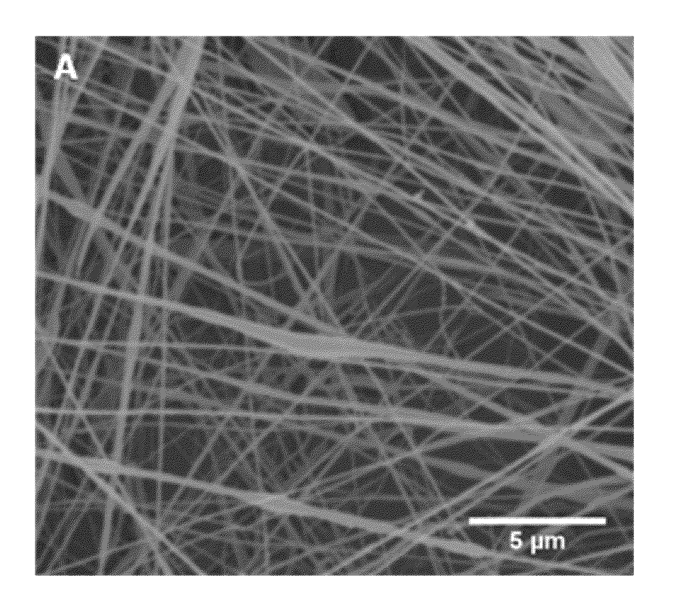
[0348]Fabricated meshes of laminin I nanofibers (LNFs) with fiber size (10-150 nM dia.), geometry, and porosity of authentic basement membranes were fabricated using electrospinning methods. Unlike previously described NFs synthesized from protein polymers, meshes of LNFs retain their structural features when wetted and do not require fixation by chemical cross-linking, which often destroys biological activity. Embryonic stem c...


PUM

PropertyMeasurementUnit
diametersaaaaaaaaaa
diametersaaaaaaaaaa
gap distanceaaaaaaaaaa

Abstract

The present invention provides methodologies and parameters for fabrication of the hybrid biomaterial by blending pure laminin or complex extracts of tissues containing laminin with biopolymers such as polycaprolactone (PCL), polylactic / polyglycolic acid copolymer (PLGA) or Polydioxanone (PDO) in fluoroalcohols (HFP, TFA), fabrication of substrates and scaffolds and devices from the hybrid biomaterial in forms such as films, nanofibers by electrospinning or microspheres, and the biological or biomedical use of the material or devices derived from it.

Description

CROSS REFERENCE TO RELATED APPLICATIONS[0001]This application is entitled to priority pursuant to 35 U.S.C. §119(e) to U.S. provisional patent application No. 61 / 312,695, filed on Mar. 11, 2010, and is a continuation-in-part of U.S. patent application Ser. No. 12 / 598,776, filed on Nov. 4, 2009, which is entitled to priority to International App. No. PCT / US2008 / 062935, which is entitled to priority to U.S. provisional patent application No. 60 / 927,584, filed on May 4, 2007, the entireties of which are incorporated by reference herein.STATEMENT REGARDING FEDERALLY SPONSORED RESEARCH OR DEVELOPMENT[0002]This invention was supported by Grant No. DE-010369-08 awarded by the National Institutes of Health and Grant No. 736002 awarded by the National Science Foundation. The United States Government therefore has certain rights in the invention.FIELD OF THE INVENTION[0003]The present invention relates to compositions, methods, and apparatuses for preparing and using electrospun protein-polym...

Claims


Application Information

Patent Timeline
Patent Type & Authority Applications(United States)
IPC IPC(8): C12N5/079C12N5/071C12N5/073C12N5/074C12N5/0775C12N5/0793C12N5/09C12N5/077B29C47/00C08L89/00B82Y5/00
CPCB82Y5/00C07K14/78C08L89/00C12N5/0619C12N2533/00C08L67/02C08L67/06C08L67/04C08L31/04A61L2430/32A61L2400/12A61L27/26
Inventor OGLE, ROY CLINTONBOTCHWEY, III, EDWARD A.NEAL, REBEKAH A.
Owner UNIV OF VIRGINIA ALUMNI PATENTS FOUND
Why Eureka
Try Eureka