
Aptamer-Based Device For Detection Of Cancer Markers And Methods Of Use

a technology of cancer markers and aptamer strips, applied in the field of aptamer-based strips, can solve the problems of inability to detect cancer markers in the native environment, both methods are sensitive to the solution environment, and both fluorescence-based methods have significant limitations in analyzing proteins in their native environmen

Inactive Publication Date: 2012-04-12
MISSOURI STATE UNIVERSITY
View PDF0 Cites 16 Cited by
  • Summary
  • Abstract
  • Description
  • Claims
  • Application Information

AI Technical Summary

Benefits of technology

The patent text describes a system and method for detecting and quantifying biomolecules in a sample with improved sensitivity and selectivity. The system uses aptamers, which are oligomeric or polymeric nucleic acids that can bind to specific molecules with high accuracy. The method involves contacting the sample with a polynucleotide molecule that can bind to the target molecule, allowing it to bind to the aptamer. The amount of target molecule bound to the aptamer is then quantified. The system can be used at the point-of-care location and is highly sensitive and selective. The patent also describes chemically modifying the aptamers for enhanced performance and introducing reporter molecules for detection. The system employs a lateral flow format and a single membrane, making it more efficient and easier to use. Overall, the patent provides a novel and effective tool for detecting and measuring biomolecules in a sample.

Problems solved by technology

All these signal-transduction techniques have their individual strength and weakness.
For instance, although FRET- or fluorescence-quenching-based probes can quantify target concentrations with changes in fluorescence intensity, both methods are sensitive to the solution environment.
More importantly, because of interference from background signals, both fluorescence-based methods have significant limitations in analyzing proteins in their native environments.
For example, when a quenching-based FRET molecular probe is used for protein studies, the probe always has some incomplete quenching, resulting in a significant probe background.
Moreover, in a native biological environment, there are many potential sources for false positive signals of the molecular probe for protein analysis.
These problems decrease the sensitivity and specificity of currently available assays.
Despite extensive research and development in bio-analysis, effective solutions to these problems remain limited.

Method used


Image

  • Aptamer-Based Device For Detection Of Cancer Markers And Methods Of Use
  • Aptamer-Based Device For Detection Of Cancer Markers And Methods Of Use
  • Aptamer-Based Device For Detection Of Cancer Markers And Methods Of Use

Examples


example 1

Preparation of Strip as the Platform for the Binding of Aptamer-Conjugated Nanoparticles to Marker Proteins

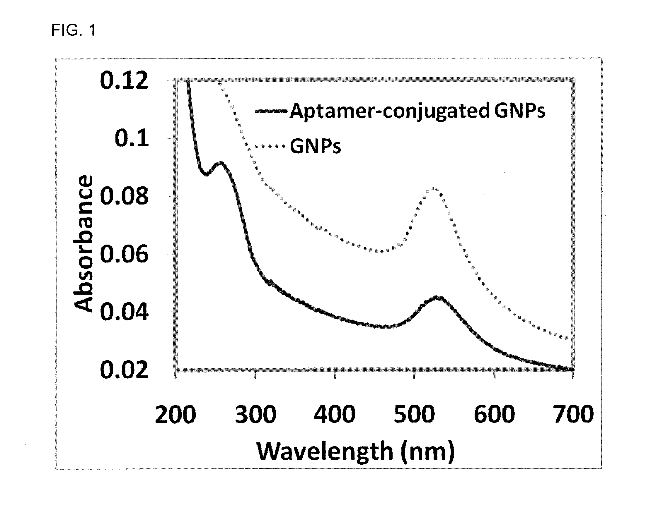
[0053]The well known lateral flow format was utilized to fabricate the strip. Unlike traditional lateral flow strips used in antibody-based assay, this strip combined traditional lateral flow kit design with contemporary fluorescent nanoparticle labels and aptamer assays. Moreover, unlike traditional lateral flow strips that incorporate several membranes, the instant strip used only one membrane that was made of a large-pore single layer hydrophilic material that was non-protein binding in nature. This material fulfilled all of the required functionalities of the components of the traditional lateral flow device, namely, as a sample pad, a conjugate pad, a membrane, and an adsorbent pad. Because the material used to build the membrane did not bind to proteins, a different strategy called “laying down boulders in the stream” was used to construct the test and control lines. Brie...

example 2

Binding of Aptamer-Conjugated Nanoparticles to Marker Proteins

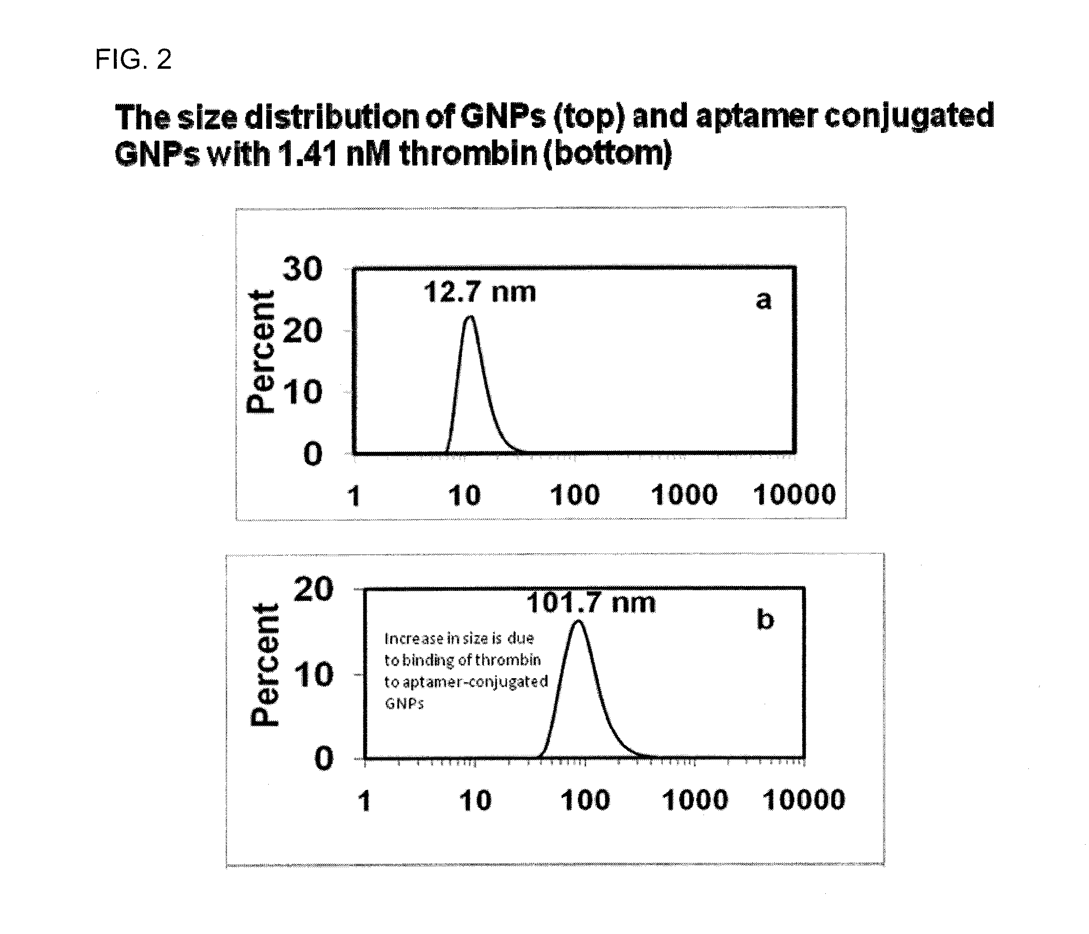
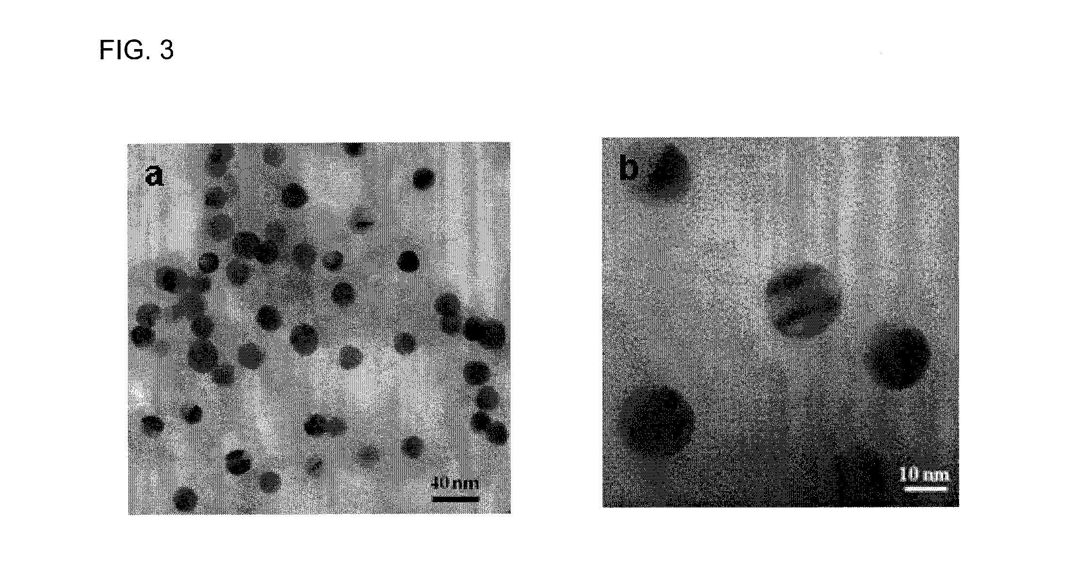
[0054]Dithiothreitol (DTT), thrombin from bovine plasma, and bovine serum albumin (BSA) were purchased from Sigma Aldrich (St Louis Mo.). Lysozyme was purchased from Sigma-Aldrich (St. Louis, Mo.). Thiolated thrombin with sequence 5′-SH-(CH2)6-TTTTTTTTTTGGTTGGTGTGGTTGG-3′ and thiolated lysozyme aptamer, 5′- / 5thioMC6-D / ATC TAC GAA TTC ATC AGG GCT AAA GAG TGC AGA GTT ACT TAG-3′ were purchased from Integrated DNA Technologies (Coralville, Iowa). Hydrogen tetrachloroaurate (III) trihydrate (HAuCl4.3H2O) was obtained from Fisher Scientific (Fair Lawn, N.J.). Sodium citrate was obtained from Spectrum Chemical Corp (New Brunswick, N.J.). Gel filtration columns (NAP-5) was purchased from GE Healthcare Bio-Sciences Corp (Piscataway, N.J.). DLS Spectroscopy was performed on a Malvern Nanozetasizer (ZS90). The DLS spectrometer was operated at 25° C. with the detector angle at 90°, incident laser wavelength of 633 nm and 4 mW laser p...

example 3

Aptamer Constructs for Selective Target Detection

[0069]In order to detect proteins in a sample using a lateral strip, an oligonucleotide construct was designed and synthesized which incorporated two known aptamer sequences that bind thrombin and ATP, respectively. As shown in the FIG. 6, the oligonucleotide had both a thrombin binding region and an ATP binding region, and these two binding regions were separated by an inter-aptamer region of several nucleotides in length. In addition, the sequences at the 5′ and 3′ end of the oligonucleotide construct were complementary to each other such that the oligonucleotide would fold into a hairpin structure as shown in FIG. 6.

[0070]In construction A, two reporters, fluorescein and Cy3, were conjugated to the 5′ terminus and to the inter-aptamer region, respectively. Fluorescein and Cy3 were known to be a fluorescent FRET pair. Changes in FRET were monitored after separate or sequential addition of thrombin and ATP targets to construct A. FRE...


PUM

PropertyMeasurementUnit
ODaaaaaaaaaa
diameteraaaaaaaaaa
sizeaaaaaaaaaa

Abstract

Systems and methods are provided for detecting and quantitating one or more compounds or molecules in a sample. An aptamer-based point-of-care device (such as a strip) is described for rapid detection of target molecules such as the cancer marker p-glycoprotein (Pgp). Fluorescent molecules or gold nanoparticles may be used to detect the binding between a target molecule and the aptamer. By way of example, fluorescence resonance energy transfer (FRET) or Dynamic Light Scattering (DLS) may be used for detecting the physical and / or chemical changes caused by the binding of the aptamers to the target molecules.

Description

RELATED APPLICATIONS[0001]This application claims priority to U.S. Patent application 61 / 388,389 filed Sep. 30, 2010, the entire content of which is hereby incorporated by reference into this application.BACKGROUND[0002]1. Field of the Invention[0003]The disclosure relates to new systems and methods for detecting and quantifying one or more compounds in a sample. More particularly, the disclosure relates to the use of aptamer-based strips for the detection of proteins or other compounds in a patient sample.[0004]2. Description of Related Art[0005]Various biological molecules, such as nucleic acids, proteins, lipids, and other chemicals, play important roles in the structure and function of many life forms. Many different systems have been used to detect the presence of a particular biological molecule in a complex sample. For example, antibodies have been used to detect the presence of a protein in blood samples, and more recently, DNA microarrays have been developed to identify pol...

Claims


Application Information

Patent Timeline
Patent Type & Authority Applications(United States)
IPC IPC(8): G01N33/53G01N21/64C07H21/00G01N21/49C12Q1/68B82Y15/00
CPCB82Y15/00G01N33/5308
Inventor WANEKAYA, ADAMDELONG, ROBERTDATTA, BHASKAR
Owner MISSOURI STATE UNIVERSITY
Learn More