
Cellular electric stimulation mediated by piezoelectric nanotubes

a piezoelectric nanotube and cell technology, applied in the field of piezoelectric nanovectors, can solve the problems of patient's death, long-term disability, and high invasiveness of current electrical stimulation procedures, and achieve the effects of maximizing the effect of effective electrical stimulation, reducing the invasiveness of present-day procedures, and eliminating or drastically reducing adverse problems and side effects

Inactive Publication Date: 2012-05-17
SCUOLA SUPERIORE DI STUDI UNIVERSITARI E DI PERFEZIONAMENTO SANTANNA +1
View PDF0 Cites 13 Cited by
  • Summary
  • Abstract
  • Description
  • Claims
  • Application Information

AI Technical Summary

Benefits of technology

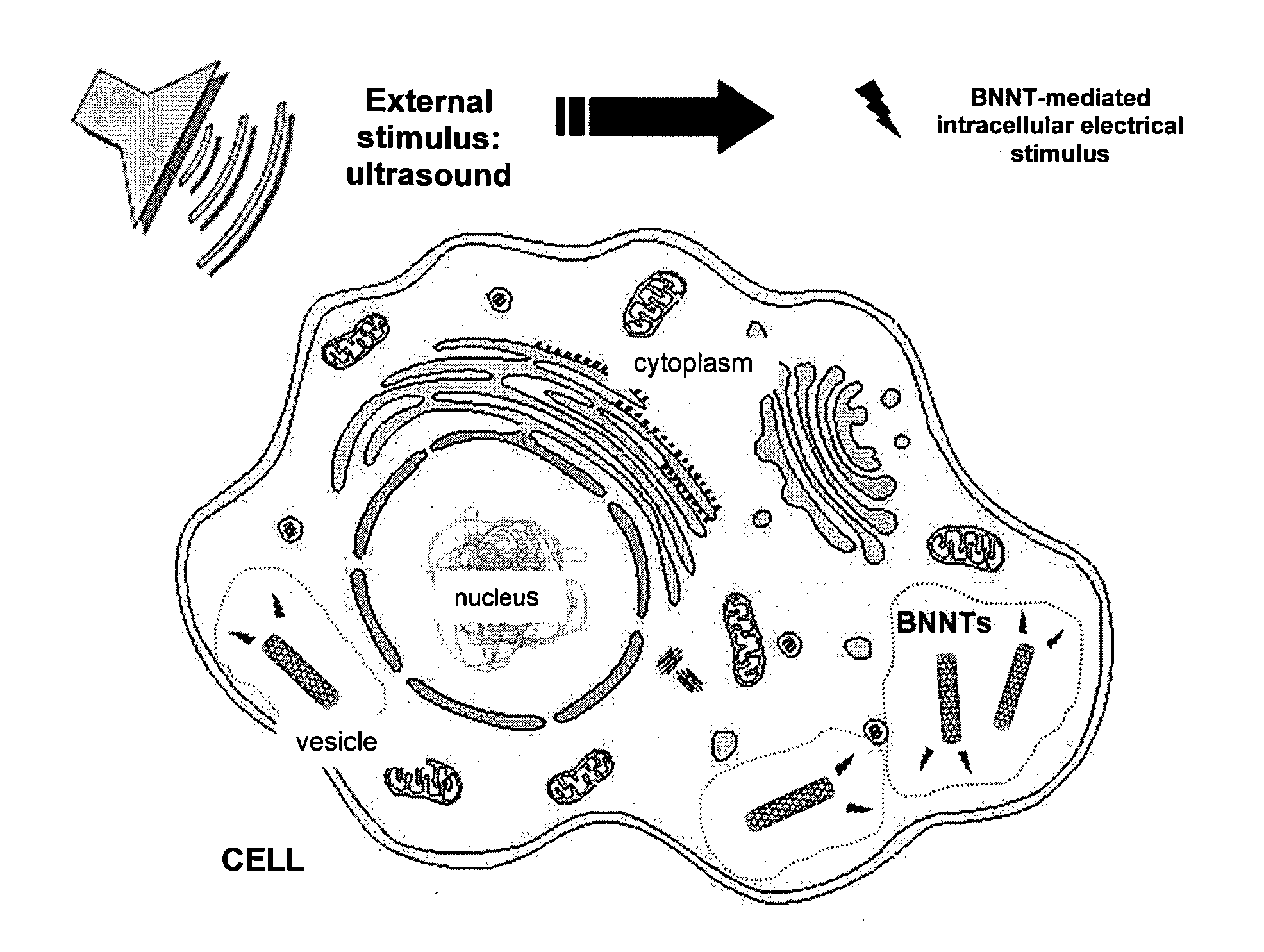
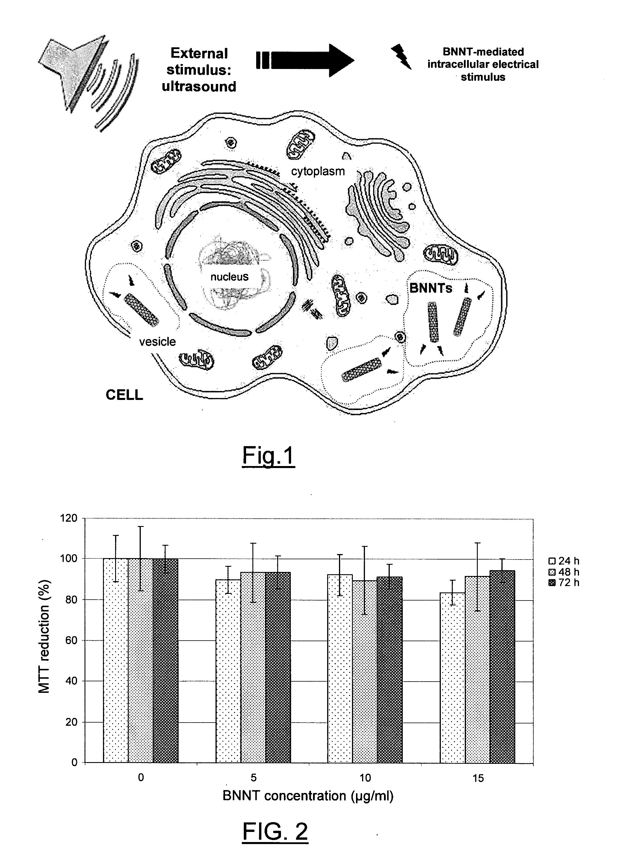
[0015]The present invention is based on the surprising discovery that piezoelectric nanotransducers can be effectively employed in a completely non-invasive treatment of electrotherapy, in which the electrical stimulus generated by the nanotransducers is caused through a (wireless-type) stress external to the patient's body, by ultrasonic waves of appropriate power. Therefore, the present invention is based on the experimental demonstration that not only piezoelectric nanotransducers can be stimulated by an ultrasonic field generated externally to the system in which the same have been localized, but the electrical stimulus produced by the nanotransducer localized inside the target cell is sufficiently high to cause an effective electrical stimulation in a real cell system, in vitro or in vivo.
[0026]The solution proposed by the invention offers the advantage of inducing an effective electrical stimulation maximizing the benefits of electrical cell stimulation, but eliminating or drastically reducing adverse problems and side effects caused by present-day clinical technologies. The proposed method totally reduces the invasiveness of present-day procedures for electrical stimulation of tissues in vivo and remarkably simplifies any form of electrical stimulation in vitro. With regard to in vitro cell stimulation, the proposed solution abolishes the electrical circuits for stimulation, electrical connections or other devices connected to the cultures, thereby facilitating the system for the improvement of cell growth conditions. The nanotransducers may be dispersed in the culture medium (CM) or embedded into support structures for cell growth (polymeric scaffolds, adhesion substrates, etc.) and then stimulated through ultrasonic fields. Moreover, both in in vitro and in vivo applications, the powers involved can be modulated casewise in order to better adapt them to different needs.

Problems solved by technology

However, current procedures for performing electrical stimulation are highly invasive.
All of these episodes can lead to long-term disabilities and, in the worst cases, to patient's death.
Moreover, oft-times infections caused by devices, not responding to antibiotic treatments, lead to a definitive removal of the electrodes.
Owing to all these complications, it is easy to understand how to date nervous stimulation, though effective and promising, is restricted to the sole treatment of advanced stages of the pathologies, when every other pharmacological therapy proves totally ineffective.
However, the technique entails the same high-invasivity problems already found in the case of nervous stimulation.

Method used


Image

  • Cellular electric stimulation mediated by piezoelectric nanotubes
  • Cellular electric stimulation mediated by piezoelectric nanotubes
  • Cellular electric stimulation mediated by piezoelectric nanotubes

Examples


example 1

Human Osteoblasts (HOBs) Isolation and Expansion

[0089]Trabecular bone samples, removed from the femoral head of a patient undergoing femoral joint replacement surgery, were used after obtaining informed consent. Samples were sectioned, under sterile conditions, into smaller pieces. Thereafter, bone fragments were placed in a sterile saline supplemented with antibiotics and antimycotics and washed several times in order to remove fat, marrow, tissue residuals and blood cells. Isolation was performed in accordance to the established method (Di Silvio et al. Human cell culture. London (UK): Kluwer Academic Publishers; 2001. p. 221-241). Cell migration from native tissue was observed within 1-2 weeks, leading to formation of an osteoid layer in the neighbourhood of the explant. Cells were cultured in a culture medium (CM) containing: DMEM low glucose (Sigma-Aldrich, Milan, I), 10% FCS (Invitrogen), 10% L-glutamine (Sigma-Aldrich), HEPES (Sigma-Aldrich), non-essential amino acids (Sigma-...

example 2

BNNTs Preparation and Conjugation

[0090]BNNTs supplied by Australian National University, Canberra, Australia, were produced by using ball-milling and annealing method (Chen Y et al. (1999) Chemical Physics Letter 299, p. 260-264; Yu J et al. (2005) Chemistry of Materials 17, p. 5172-5176). Details relating to sample purity and composition (provided by the supplier) were: yield >80%, boron nitride >97 wt %, metallic catalysts (Fe and Cr) derived from the milling process ˜1.5 wt % and adsorbed O2 ˜1.5 wt %.

[0091]The polymer used for the aqueous suspension and dispersion of BNNTs was poly-L-lysine (PLL) obtained from Fluka (81339), molecular weight 70,000-150,000. All experiments were carried out in phosphate buffered solution (PBS) as described previously (Ciofani G. et al. (2008) Biotechnol. Bioeng. 101, p. 850-858). Briefly, samples of BNNT powder in a 0.1% PLL solution were ultrasonicated for 12 h with a Branson sonicator 2510 (Bransonic). The output power of the sonicator was set ...

example 3

MTT Assay

[0096]To evaluate cell viability, MTT (3-(4,5-dimethylthiazole-2-yl)-2,5-diphenyl tetrazolium bromide, M-2128 from Sigma) cell proliferation assays were carried out after 24, 48, and 72 h of incubation with PLL-BNNT modified media, which contained a final concentration of BNNTs equal to 5, 10 and 15 μg / ml. After trypsinization and cell counting with a Bürker chamber, HOBs were plated in 96-well plates. Once the adhesion was verified (after about 6 h from the seeding), cells were incubated with MTT 0.5 mg / ml for 2 h. Then, 100 μl of dimethyl sulfoxide (DMSO, Sigma) were added in each well and absorbance at 550 nm was measured with a VERSAMax microplate reader (Molecular Devices). A reference test (cells cultured in the absence of BNNTs) was carried out as control.


PUM

PropertyMeasurementUnit
Adhesion strengthaaaaaaaaaa
Electric propertiesaaaaaaaaaa
Piezoelectricityaaaaaaaaaa

Abstract

Piezoelectric nanotransducers for use in an in vivo treatment of cell stimulation through electrical stimulation are described. The nanotransducers are localized in a target site, and an electrical stimulus is induced in the same site through external stimulation of the nanotransducers by ultrasonic waves.

Description

TECHNICAL FIELD OF THE INVENTION[0001]The invention relates to a method for inducing non-invasive electrical cell stimulation, both in vitro and in vivo, by use of piezoelectric nanovectors. Specifically, these are boron nitride nanotubes (BNNTs) capable of converting a specific non-invasive external stimulus (ultrasonic waves) into electrical inputs able to stimulate cells.STATE OF THE PRIOR ART[0002]Electrotherapy[0003]Electrical cell stimulation finds numberless applications in the biomedical field, such as deep brain stimulation, gastric stimulation following gastroparesis, cardiac stimulation, muscle stimulation, etc. In particular, in neurological disorders electrical brain stimulation is often the sole form of therapy. It has long been demonstrated that appropriate electrical stimulations induce a positive response in cultured cells with regard to proliferation, metabolism or production of specific substances. Supronowicz and collaborators demonstrate that electrical stimulat...

Claims


Application Information

Patent Timeline
IPC IPC(8): A61N1/36A61K9/00C12N5/071C12N13/00A61K33/22A61K9/50B82Y5/00
CPCA61N1/205B82Y5/00A61N1/326A61P17/02A61P25/04A61P43/00
Inventor CIOFANI, GIANNIRAFFA, VITTORIADANTI, SERENAMENCIASSI, ARIANNADARIO, PAOLOPETRINI, MARIOCUSCHIERI, ALFRED
Owner SCUOLA SUPERIORE DI STUDI UNIVERSITARI E DI PERFEZIONAMENTO SANTANNA
Learn More