Method for culturing stem cells

a stem cell and culture method technology, applied in the field of biological technology, can solve the problems of es cell mutation, affecting the maintenance of normal karyotype, and difficulty in obtaining pure es cells for biochemical and molecular biological analysis, and achieves the effect of reducing the culture cost of stem cells, simple and safe, and effectively solving contamination problems

Active Publication Date: 2012-05-17
SHANGHAI ICELL BIOTECH +1
View PDF0 Cites 7 Cited by
  • Summary
  • Abstract
  • Description
  • Claims
  • Application Information

AI Technical Summary

Benefits of technology

[0022]The stem cell culture method according to the present invention does not require the treatment of the feeder layer cells to lose the division ability, and is thus simple and safe, thereby effectively solving the problem of contamination caused by animal-derived ingredients in culture of human stem cells at present, greatly reducing the culture cost of the stem cells, and providing a safe, effective, and inexpensive stem cell culture method for the industrialization of the stem cells in the future.

Problems solved by technology

However, the disadvantages in use of the MEFs to culture the ES cells are that (1) in culturing and expanding the human ES cells, the MEFs may spread animal pathogens to the human ES cells through the culture medium; and (2) a large amount of MEFs need to be produced, since the MEFs have a limited life cycle, cannot be passaged in vitro for a long period of time, and have an ability of generating the proliferation factor and the differentiation inhibitor that is diminished or even lost with the extension of the passage time; (3) it is difficult to obtain pure ES cells for biochemical and molecular biological analysis; and (4) the release of the chromosome from dead MEFs may cause the mutation of the ES cells and affects the maintenance of normal karyotype.
However, the culture system based on human feeder layer still requires the feeder layer cells and the ES cells to grow at the same time, and the human tissues cannot meet the demand for in-vitro culture and expansion of the ES cells due to the difficulty exiting in obtaining of the human tissues.
However, the treatment through which the feeder layer cells lose the division ability, for example, irradiation with ray or treatment with mitomycin C, increases not only the operation steps, but also potential risks, because mitomycin C is a DNA inhibitor, which may cause the chromosome deformity of the ES cells.
However, the growth factors and the culture medium containing the same are very expensive, thereby increase the research and application cost.

Method used


Image

  • Method for culturing stem cells
  • Method for culturing stem cells
  • Method for culturing stem cells

Examples


embodiment 1

Preparation of Human Amniotic Epithelial Cell Feeder Layer

[0052]Isolation and culture of human amniotic epithelia cells (HAECs): the amnion was peeled off from the placenta (which was negative for various virus indication, including types A, B, C, and E hepatitis, HIV, and syphilis) obtained after parturition through cesarean section, the amniotic epithelial cells were isolated, washed with PBS to remove the blood cells, digested with 0.25% trypsin at 37° C. for 30 min, beaten, added with RPMI1640 containing 10% of human umbilical cord serum to quench the digest, filtered with a sieve of 100 meshes, and centrifuged at 1,500 rpm. The supernatant was discarded, and the cells were inoculated at 1×10⁴ / cm2 in a 10 cm petri dish, and cultured in a cell incubator at 37° C. with 5% CO2 to 80% confluence.

embodiment 2

Preparation of Mouse Embryonic Fibroblast (MEF) Feeder Layer

[0053]Isolation and culture of mouse embryonic fibroblasts (MEFs): embryos of mice during pregnancy at day 12.5 were removed of the viscera, head, and tail, and washed two times with PBS. The tissue was scissored, digested with 0.25% trypsin for 30 min, beaten uniformly with a burette, and centrifuged at 1000 rpm for 5 min The supernatant was discarded, and the cell pellets were beaten uniformly, added with DMEM culture medium (containing 10% FBS), cultured in an incubator at 37° C. with 5% CO2, and treated with mitomycin C after 2-3 passages.

embodiment 3

Culture of Human Embryonic Stem Cells

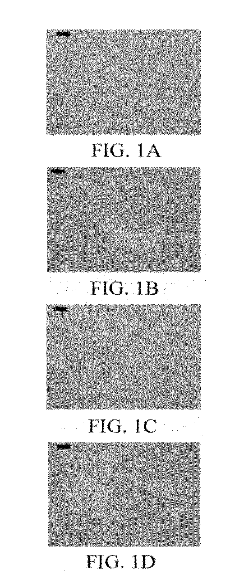
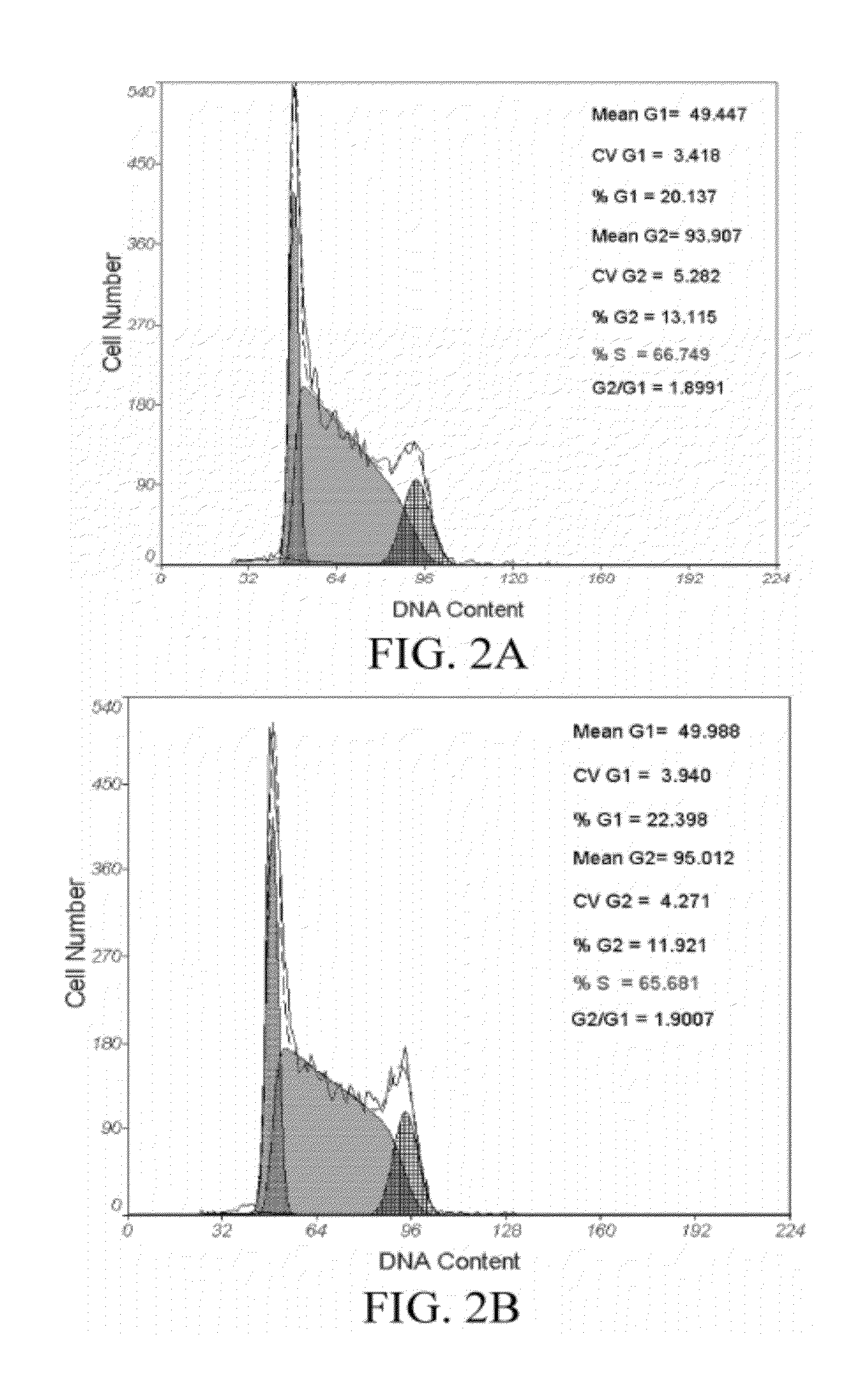
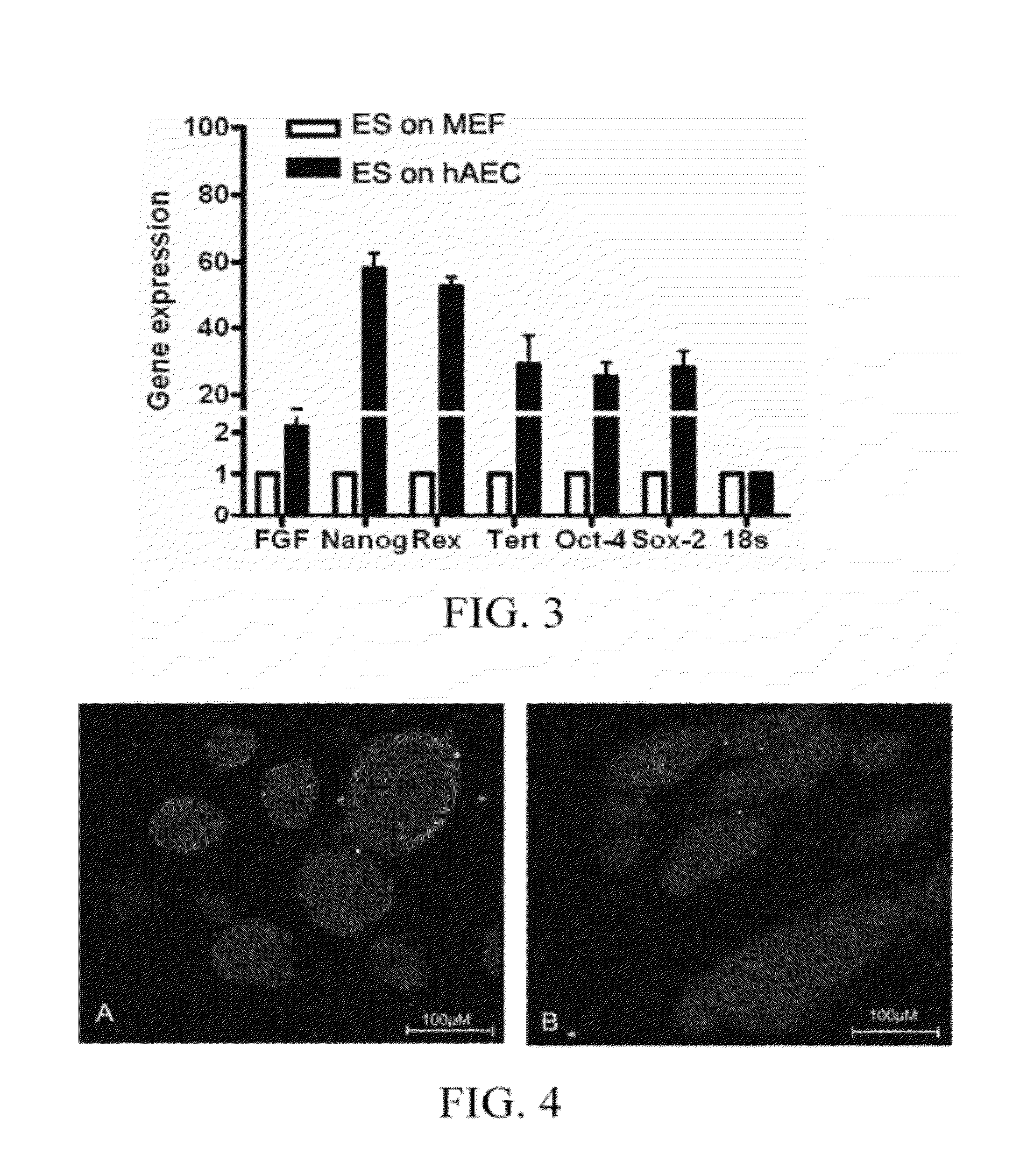
[0054]The human embryonic stem cell line hHES1 was respectively cultured on the two feeder layers obtained in Embodiments 1 and 2 [Wu C F, Tsung H C, Zhang W J, et al. Reprod Biomed Online. 2005 December; 11(6):733-9]. KO-DMEM medium supplemented with 10 ng / ml bFGF, 5% human umbilical cord serum, and 12 ng / ml hLIF was used, HES cell clones at day 3 day of culture were used for molecular biological test, and qRealtime-PCR, immunofluorescence IF, and Western blotting were used for detecting the gene expression differences of Nanog, Oct4 and other markers; and flow cytometry (FCM) was used for detecting the cell cycle difference of HES. hHES1 after 20 passages was analyzed for chromosome karyotype by G banding. 1×10⁶ HES cells was collected and intramuscularly injected respectively into the back legs of the SCID mice of 4-8 weeks. After about 7-8 weeks, it was found though touch that a tumor was formed, the mice were sacrificed, a paraffin section w...


PUM

PropertyMeasurementUnit
concentrationaaaaaaaaaa

Abstract

In the field of biological technology, a stem cell culture method is provided. The method includes preparing an amniotic epithelial cell feeder layer that is not treated to lose the division ability; and seeding the stem cells onto the amniotic epithelial cell feeder layer, and culturing in a culture medium. The stem cell culture method according to the present invention does not require the treatment of the feeder layer cells to lose the division ability, and is thus simple and safe, thereby effectively solving the problem of contamination caused by animal-derived ingredients in culture of human stem cells at present, greatly reducing the culture cost of the stem cells, and providing a safe, effective, and inexpensive stem cell culture method for the industrialization of the stem cells in the future.

Description

BACKGROUND[0001]1. Technical Field[0002]The present invention relates to the filed of biological technology, and more particularly to a stem cell culture method.[0003]2. Related Art[0004]Stem cells are a class of self-renewing pluripotent cells, which are able to be differentiated into many different functional cells under certain conditions. The stem cells may be classified by two methods. In a first method, the stem cells are classified into embryonic stem cells (ES cells) and somatic stem cells according to the development stage of the stem cells. In a second method, the stem cells are classified into totipotent stem cells (TSCs), pluripotent stem cells, and unipotent stem cells according to the development potential of the stem cells.[0005]ES cells are mainly derived from inner cell mass (ICM) of a blastocyst and early embryonic cells before a fertilized ovum is developed into a morula. Human ES cells have 2 distinguished features, one is the high in-vitro self-renewing ability,...

Claims


Application Information

Patent Timeline
Patent Type & Authority Applications(United States)
IPC IPC(8): C12N5/071C12N5/0735
CPCC12N5/0606C12N2502/025C12N2501/235C12N2501/115C12N5/0607
Inventor LAI, DONGMEICHENG, WEIWEIGUO, LIHELIU, TIANJINJIANG, LIZHENHUANG, QIN
Owner SHANGHAI ICELL BIOTECH
Why Eureka
Try Eureka