Miniaturized electroporation-ready microwell aray for high-throughput genomic screening

a microwell and genome technology, applied in the field of miniaturized electroporation-ready microwell aray for high-throughput genomic screening, can solve the problems of limited application of capital expenses and the cost of reagents necessary to perform such large screens, hampered this screening technology, and difficult functional annotation of mammalian genomes, etc., to achieve high-efficiency parallel introduction of exogenous molecules

Inactive Publication Date: 2012-09-13
THE SCRIPPS RES INST
View PDF1 Cites 6 Cited by
  • Summary
  • Abstract
  • Description
  • Claims
  • Application Information

AI Technical Summary

Benefits of technology

[0007]The invention relates to the introduction of exogenous molecules into cells. In some embodiments, the described materials and methods provide tools for high-throughput cell-based screens of exogenous genetic material. The invention is also directed to microwell arrays on an electroporation-ready substrate and procedures to achieve highly efficient parallel introduction of exogenous molecules into human cell lines and primary mouse macrophages. The microwells confine cells and offer multiple advantages during imaging and phenotype analysis. The invention is further directed to a method to load the described microwell arrays with libraries of nucleic acids using a standard microarrayer. These tools of the invention form the basis of a miniaturized high-throughput functional genomics screening platform to carry out genome-size screens in a variety of mammalian cells.

Problems solved by technology

However, the capital expenses and the cost of reagents necessary to perform such large screens have limited application of this technology.
While this work has demonstrated the feasibility of screening in very small formats, the use of chemical transfection reagents (effective only in a subset of cell lines and not on primary cells) and the lack of defined borders between cells grown in adjacent microspots containing different genetic material (to prevent cell migration and to aid spot location recognition during imaging and phenotype deconvolution) have hampered this screening technology.
Thus, functional annotation of the mammalian genome has proven particularly difficult.
Elegans, Drosophila and other lower organisms, where large genetic screens can be carried out with relative ease to discern gene purpose, tools to delineate gene function, signaling pathways and genetic networks in mammalian cells have been very limited.
These approaches share three critical weaknesses that drastically limit application of this technology.
Second, cell microarrays lack physical barriers to contain cells transfected with one nucleic acid from migrating and mixing with cells transfected with another nucleic acid.
Migration of cells between spots can lead to inter-spot contamination, confounding phenotypic analysis and hindering time-lapse studies (where spots are visited multiple times over the course of the assay).
Third, because the cells grow in a lawn without reference points, it is difficult to identify with certainty the microscale regions corresponding to individual spots during automated image analysis, and minor errors in imaging (due to tolerances in the microscope stage or during microarraying) can lead to incorrect phenotypic annotations.

Method used


Image

  • Miniaturized electroporation-ready microwell aray for high-throughput genomic screening
  • Miniaturized electroporation-ready microwell aray for high-throughput genomic screening
  • Miniaturized electroporation-ready microwell aray for high-throughput genomic screening

Examples


example 1

Bottom Electrode Microwell Array

[0042]Example 1 begins with a substrate including a conductive material bonded on top of a base material. In an example, the base material is non-opaque (e.g., transparent or translucent). The base material can be either flexible or rigid and either conductive or non-conductive. For example, the base material can include a glass including borosilicate, quartz, soda-lime, and porous or non-porous glass. In other examples, the based material can include a plastic (e.g., a polymer) including Poly-methyl-methacrylate (PMMA), Polystyrene, Polycarbonate, and Polypropylene. In another example, the base material is opaque and can be a silicon, metal, ceramic and the like. In examples where the base material is conductive, the base material can include, for example, a metal, polysilicon, doped silicon, metal alloys, and composite materials containing conductive particles.

[0043]The conductive material can be bonded to the base material and can provide a bottom ...

example 2

Side Electrode Microwell Array

[0050]Example 2 begins with a substrate including a base material. In an example, the base material is non-opaque (e.g., transparent or translucent). The base material can be either flexible or rigid and is non-conductive. For example, the base material can include a glass including borosilicate, quartz, soda-lime, and porous or non-porous glass. In other examples, the based material can include a plastic (e.g., a polymer) including Poly-methyl-methacrylate (PMMA), Polystyrene, Polycarbonate, and Polypropylene. In another example, the base material is opaque and can be a silicon, ceramic and the like.

[0051]In an example, an electrode layer is formed on the substrate. In some examples, the electrode layer provides substantially uniform electric charge across the substrate. Accordingly, the electrode layer is composed of a conductive material and is used to distribute the electric charge across the substrate.

[0052]In an example, the electrode layer is com...

example

Highly Parallel Introduction of Nucleic Acids into Mammalian Cells Grown in Microwell Arrays

Materials and Methods

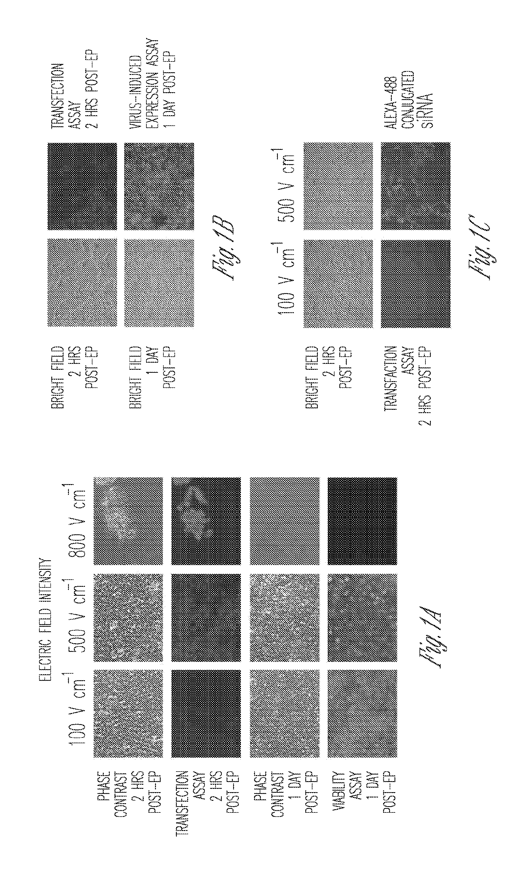
Optimization of Electroporation Parameters for HEK 293T Cells

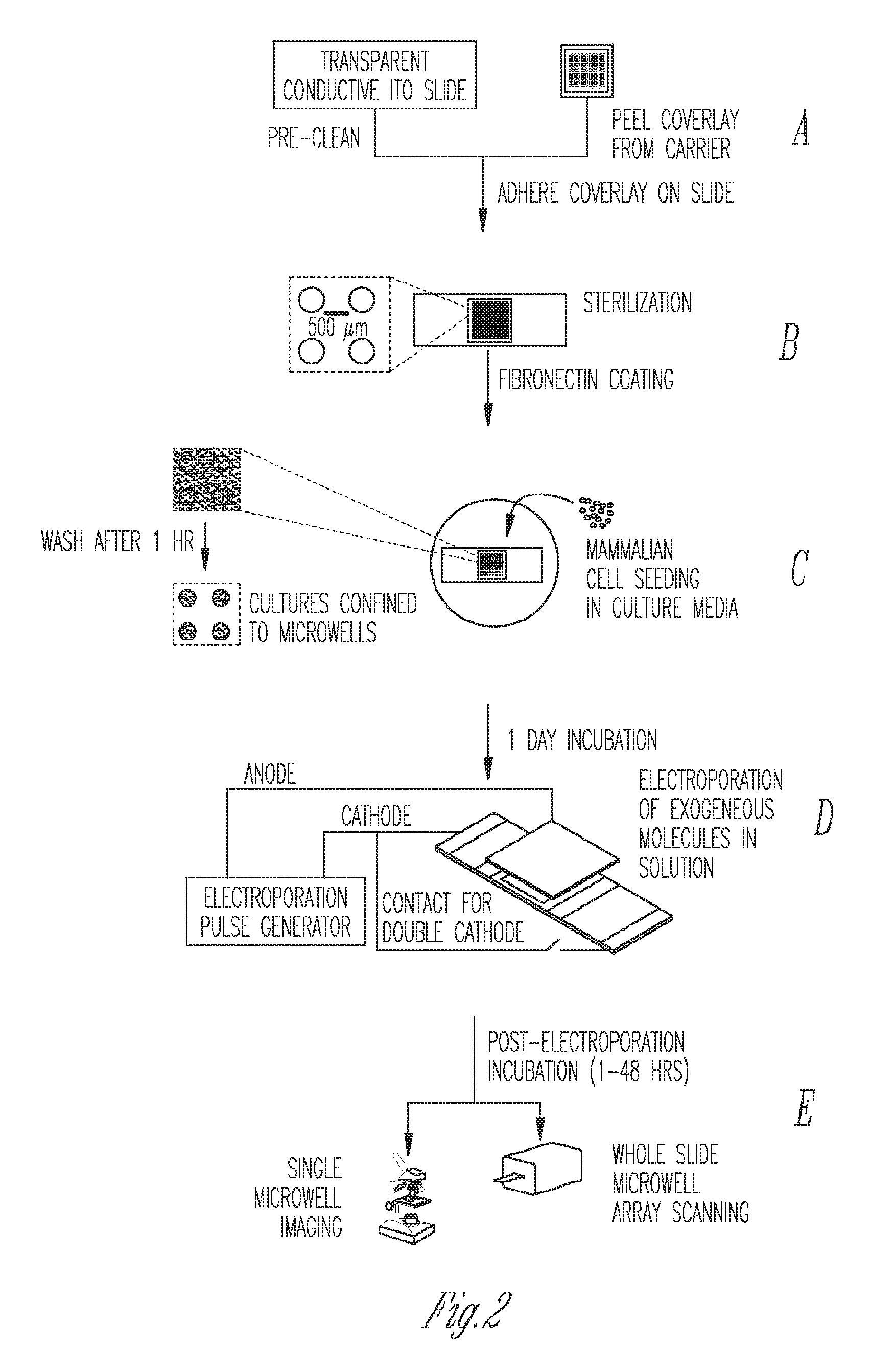
[0099]Optimization was conducted on diced Indium-Tin Oxide (ITO) coated glass (unpolished, surface resistivity 4-8 Ωsq−1, Delta Technologies, MN) pieces as shown in FIG. 8A, on a custom-built electroporation setup (FIG. 8B). Briefly, pieces 1 cm×2.5 cm were diced from single microscope slides, rinsed in de-ionized water and dried under a nitrogen stream. Thereafter, 100 μl of 10 μg m−1 fibronectin from human plasma (Sigma) was pipetted on the top half of the piece and allowed to coat for 2 hrs. After aspiration and washing with PBS, 2−3×10⁴ HEK293T cells in 100 μl of media (DMEM, 10% FBS and antibiotics) were added to the same spot. The cells were allowed to adhere to the spot for 1 hr in the incubator at 37° C. and 5% CO2, prior to washing with PBS and flooding with media. After 24 hrs of incubation, media was ...


PUM

PropertyMeasurementUnit
dispense volumeaaaaaaaaaa
thicknessaaaaaaaaaa
diameteraaaaaaaaaa

Abstract

Methods of introducing exogenous molecules into cells including cell lines and primary cells are provided. Additionally, miniaturized electroporation-ready microwell arrays are provided. These tools provide a miniaturized high-throughput functional genomics screening platform to carry out genome-size screens in a variety of cell types.

Description

CROSS-REFERENCE TO RELATED APPLICATION[0001]This application claims the benefit of U.S. Provisional Patent Application Ser. No. 61 / 251,086, filed Oct. 13, 2009, entitled “MINIATURIZED ELECTROPORATION-READY MICROWELL ARRAY FOR HIGH-THROUGHPUT GENOMIC SCREENING,” the entirety of which is incorporated herein by reference.BACKGROUND[0002]High-throughput cell-based screens of genome-size collections of cDNAs and siRNAs have become a powerful tool to annotate the mammalian genome, enabling the discovery of novel genes associated with normal cellular processes and pathogenic states, and the unraveling of genetic networks and signaling pathways in a systems biology approach. However, the capital expenses and the cost of reagents necessary to perform such large screens have limited application of this technology.[0003]Efforts to miniaturize the screening process have centered on the development of cellular microarrays created on microscope slides that use chemical means to introduce exogenou...

Claims


Application Information

Patent Timeline
Patent Type & Authority Applications(United States)
IPC IPC(8): C12N13/00G03F7/20C12M1/42B82Y5/00
CPCB01L3/0268B01L3/5085C12N15/87C12M23/16C12M35/02C12M23/12Y02A90/10
Inventor SAEZ, ENRIQUEJAIN, TILAK
Owner THE SCRIPPS RES INST
Why Eureka
Try Eureka