Biomaterial composite composition and method of use

a biomaterial and composite technology, applied in the direction of peptide/protein ingredients, packaged goods, foodstuffs, etc., can solve the problems of disc degeneration, difficult repair of damaged disc as functional living tissue, and unsatisfactory reconstruction results of these interventions

Active Publication Date: 2012-11-08
UNIVERSITY OF MEMPHIS RESEARCH FOUNDATION
View PDF0 Cites 21 Cited by
  • Summary
  • Abstract
  • Description
  • Claims
  • Application Information

AI Technical Summary

Benefits of technology

[0037]FIG. 13 shows linearized ΔCT measures for iNOS with Rn18s taken as a housekeeping gene plotted against exposure time is shown. Given the average relative increase in iNOS by the

Problems solved by technology

The reconstruction results from these interventions are often less than ideal.
Generally, repair of damaged disc as functional living tissue is difficult.
The cause of disc degeneration has been speculated to be a resultant of poor disc nutrient supply and waste removal.
This lack of transport to and from the disc likely impedes present regenerative treatment systems in the repair of damaged discs.
These occurrences were ranked fourth and ninth in prevalence, excluding pregnancy and childbirth related instances, and resulted in a financial burden of $20.2 billion (inflation-adjusted hospital costs).
Joint prostheses have an obvious advantage over fusion due to at least a partially preserved functionality, but both clinical treatments are plagued with exorbitant costs, native tissue morbidity, and extensive rehab/recovery, and they may lead to unforeseen complications and traumatic revision surgery.
Although the pains and costs associated with the treatment of diarthrodial joints such as hips and knees is evident through the long road of pain management

Method used


Image

  • Biomaterial composite composition and method of use
  • Biomaterial composite composition and method of use
  • Biomaterial composite composition and method of use

Examples


example 1

[0069]Rat mesenchymal stem cells from two donors which had been cryopreserved upon reaching 80% confluence of primary cultures where combined at a 1:1 ratio and seeded at 10̂4 cells / cm̂2 in T-flasks. Cells were passaged at 80% of this first passage and used for experimentation upon again reaching 80%. At the beginning of each experiment MSCs from two donors were combined and expanded for one passage in monolayer and were entering their second passage. Cells were grown in 10% (v / v) FBS-supplemented DMEM-HG containing penicillin, streptomycin, and amphotericin under normal culture conditions (37° C.; 5% (v / v) CO2; 100% RH).

[0070]Experimental Composition 1 and Composition 2 (Table 1) were formed in 3.6 ml and 3.3 ml sterile quantities, respectively. Calcium salts were loaded in to 3 cc syringes which were sterilized with gamma irradiation (˜2.5 Mrad). Concentrated human platelet releasate (hCPR) and water were sterilized through 0.2 μm filtration. Alginate solutions at twice the final ...

example 2

[0089]Table 1 details the biomaterial composite formulations and preparation of the formulations F1+ and F2+ which were created through syringe mixing. A syringe containing sodium alginate (Sigma; St. Louis, Mo.) solution at twice the final concentration was coupled to a second containing 125-150 μm β-tricalcium phosphate (β-TCP) granules (Plasma Biotal, Ltd.; Derbyshire, United Kingdom), calcium sulfate dihydrate (USG Corp.; Chicago, Ill.), human concentrated platelet releasate (hCPR), and, for F2+, sodium citrate (Fisher; Waltham, Mass.). Components were mixed by twenty passes between the coupled syringes, which also initiated an ionic polymerization setting reaction. This mixture can be easily delivered through injection via a 16-gauge hypodermic needle prior to curing. Additionally, material controls with deionized water replacing hCPR were created and designated as F1− and F2−. hCPR was prepared under an Institutional Review Board-approved protocol at the University of Memphis ...

example 3

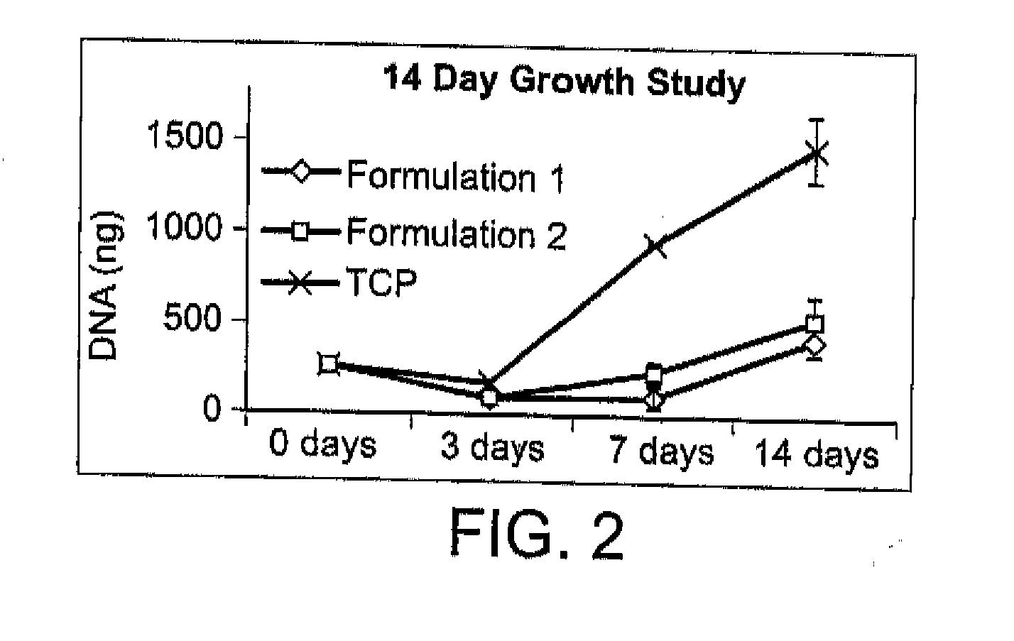
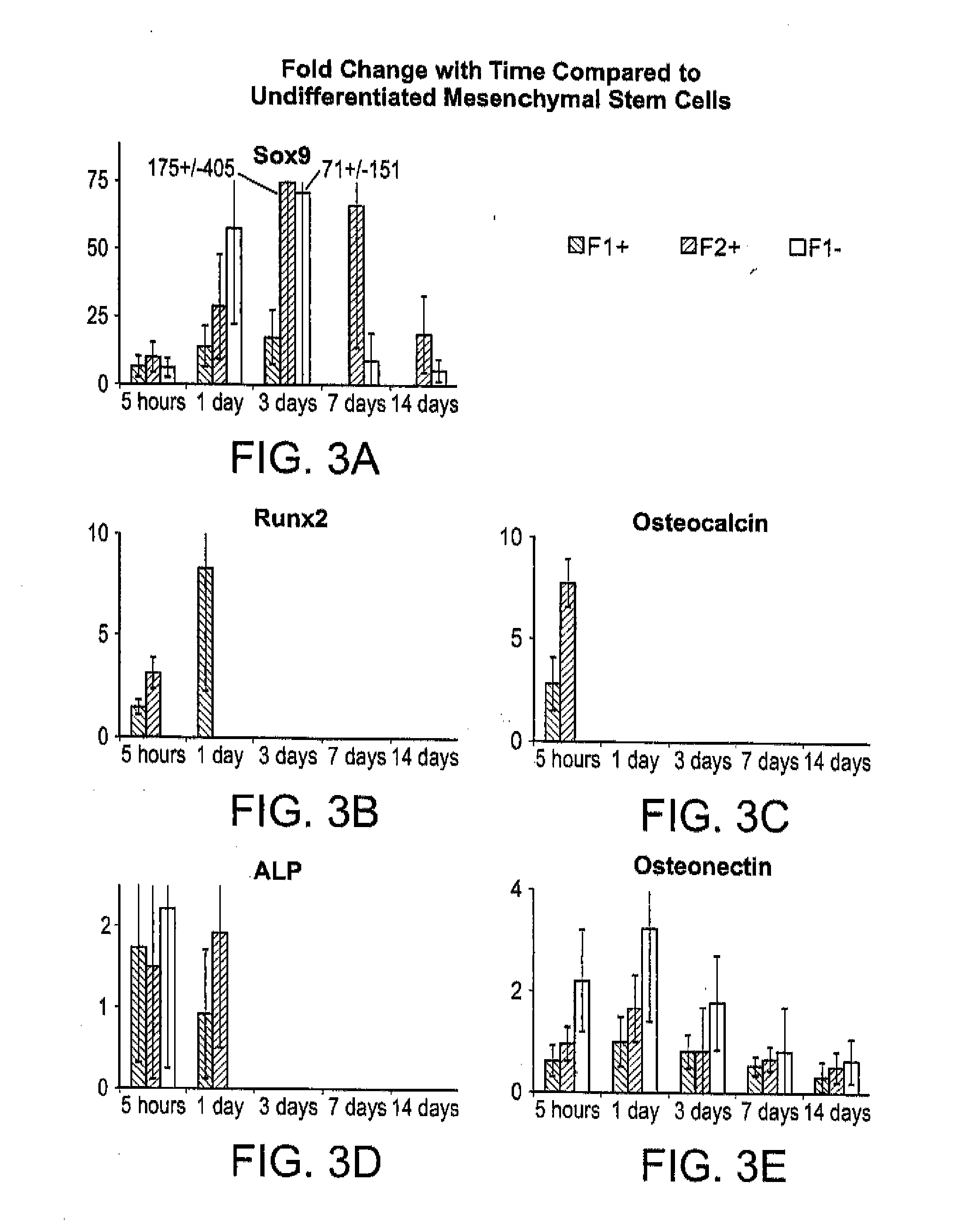
[0110]There exists a clinical need for consistent bone remodeling within problematic sites such as those of fracture nonunion, avascular necrosis, or irregular bone formations. To address such needs, a biomaterial composite composition is provided to induce early inflammatory responses after implantation and to provide later osteoconductive scaffolding for bone regeneration. Biomaterial composite-induced inflammation parallels the early stage of hematoma-induced fracture repair and allows scaffold-promoted remodeling of osseous tissue to a health state. Initiation of the wound healing cascade by two human concentrated platelet releasate-containing alginate / β-tricalcium phosphate biocomposites has been studies in vitro using the TIB-71™ RAW264.7 mouse monocyte cell line.

[0111]This in vitro example relates to tunable interactions between the platelet releasates and the biomaterial composites for enhancing hematoma-like fracture repair. Additionally, minimally invasive delivery for in ...


PUM

PropertyMeasurementUnit
Lengthaaaaaaaaaa
Fractionaaaaaaaaaa
Percent by massaaaaaaaaaa

Abstract

This invention relates to a process to facilitate osteochondral bone remodeling in a subject by inducing regeneration of this bone to a healthy, vascularized state capable of supporting the underlying hyaline cartilage of articular joints and spinal discs, both biomechanically and metabolically and to deliver a bioactive agent. This process involves the steps of: administering an effective amount of an injectable in situ curing biomaterial composite to a site. The biomaterial composite product is prepared by a process involving the steps: admixing an alginate solution with a nonporous aggregate of β-tricalcium phosphate, in a sufficient amount to initiate polymerization of the alginate solution, to form a hydrogel having from between 10 to 20 percent by volume of β-tricalcium phosphate.

Description

RELATED APPLICATIONS[0001]This application claims the benefit of US provisional patent application filed May 6, 2011 Ser. No. 61,483,446 under 35 USC §119(e) (hereby incorporated by reference).STATEMENT REGARDING FEDERALLY SPONSORED RESEARCH OR DEVELOPMENT[0002]NoneREFERENCE TO SEQUENCE LISTING, A TABLE, OR A COMPUTER PROGRAM LISTING COMPACT DISC APPENDIX[0003]Sequentially uploaded as a file.FIELD OF THE INVENTION[0004]The present invention relates to an injectable biomaterial composite, a method to make the biomaterial composite, and a method to use this biomaterial composite in medical applications.BACKGROUND OF THE INVENTION[0005]Bone damage and damage to associated tissues from less severe injury can typically heal naturally. Other damage from more severe injuries to bone and related tissues caused by extreme trauma and complications associated with disease or genetic conditions, often require surgery, tissue grafts, or artificial implants in order to reconstruct these tissue in...

Claims


Application Information

Patent Timeline
IPC IPC(8): A61K47/36A61K35/14A61K9/00A61P19/00A61P29/00B65D85/00A61K38/18A61K35/19
CPCA61K38/18A61K35/19A61K9/06A61K9/0024A61K38/1858A61P19/00A61P29/00
Inventor MCCANLESS, JONATHAN DEMPSEYHAGGARD, WARREN OLLVER
Owner UNIVERSITY OF MEMPHIS RESEARCH FOUNDATION
Why Eureka
Try Eureka