Multi-Modality Ultrasound and Radio Frequency System for Imaging Tissue

a radio frequency system and ultrasound technology, applied in the field of soft tissue imaging, can solve the problems of unreliable mammography results, unsuitable for cancer detection in young women, unnecessary biopsies and expense, etc., and achieve the effects of superior contrast, signal-to-noise improvement, and superior penetration and resolution capabilities of ultrasound

Inactive Publication Date: 2012-11-22
ULTRAWAVE LABS
View PDF4 Cites 13 Cited by
  • Summary
  • Abstract
  • Description
  • Claims
  • Application Information

AI Technical Summary

Benefits of technology

[0021]The use of two focused ultrasound beams with their focal points intersecting at a target inclusion buried in a larger mass of tissue provides the means to image submillimeter-size inclusions deep inside the tissues or inside the body. The ultrasound energy through its interaction with the nonlinearity of the inclusion induces low frequency vibration in the target inclusions which results in measurable displacement of the inclusion and causes a Doppler shift in the reflected radio frequency signals. The dielectric properties of the inclusion provide the means for contrast of the inclusion with respect to its surroundings and provide a superior contrast compared to other methods resulting in an approximately 30 to 40 dB signal-to-noise improvement. While vibro-acoustography used alone as an imaging modality takes advantage of low-frequency ultrasound harmonic components from multiple high-frequency ultrasound wave inputs to excite the target tissue, it relies on differences in elastic properties of tissue structures such as tumors, lesions, ablations, etc. relative to the surrounding tissue as the means to contrast the structure with its surroundings. Likewise microwave-only imaging modalities require solving the inverse problem which can produce imprecise results and is limited in resolution to a fraction of the wavelength used for measurement. This invention combines the superior penetration and resolution capabilities of ultrasound with the high contrast and discrimination of microwave imaging.
[0022]In this invention it should be noted that while the radio frequency energy and the ultrasound signals are applied concurrently on the target tissues in true dual modality system where both modalities work simultaneously, the ultrasound waves are used to vibrate the target tissues while the radio frequency energy provides the detection and imaging functionality. In an alternative embodiment, ultrasound transceivers can be used so that images can be generated by the radio frequency energy as well as the ultrasound waves. The vibration of the target tissue at the intersection of the two ultrasound beams results in Doppler sidebands around the radio frequency carrier frequency and separated from the carrier by a frequency equal to the difference in frequency of the two ultrasound signals. The ability to focus the ultrasound beams at points of intersection at a desired tissue depth dramatically improves the dynamic range of this imaging scheme and enables detecting and imaging sub-millimeter size targets and achieves high levels of resolution at a desired tissue depth while also achieving superior contrast.

Problems solved by technology

However, mammography results in too many false negatives and false positives which delays the detection of early cancer, or results in unnecessary biopsies and expense.
Additionally, mammography results are unreliable in the case of dense or fibrocystic breast tissues.
This makes mammography unsuitable for cancer detection in young women with familial history and dense tissues and it further exposes these women to the danger of frequent exposure to ionizing radiation.
However, the cost of MRI is prohibitive and the procedure is too long and results in discomfort and anxiety for many patients.
While ultrasound images achieve an excellent resolution and reasonable depth of penetration, the resulting images have relatively poor contrast.
However, since the wavelength of microwaves in soft tissue is on order of a few centimeters, the spatial resolution of this method is limited.
As a result, imaging methods that rely on microwaves alone are disadvantaged by the necessity to trade off low-frequency penetration against high-frequency contrast.
This proved to be quite challenging using any single imaging modality because, used alone, each has significant limitations.
It is very difficult to obtain a small spot size with a focused beam of microwave energy even if an array is used.
Such large arrays would be too large and impractical to address parts of human or animal anatomy.
Such precision timing requires a very complex computer system and extensive microwave and digital hardware to implement.
This simple integration using independent electrical signals from the ultrasound and microwave modalities, as proposed by the Rosner et al. reference, does not take advantage of the physical interaction of the ultrasound and microwave modalities.
Therefore, this method of combining the image results from microwave and from ultrasound possesses the same disadvantages of each subsystem when used independently.
It is obvious to those skilled in the art that the proposed system fails to concurrently achieve the desired combination of high penetration, high resolution, and high contrast.
Additionally, in the case of the Rosner et al. reference, as a result of the microwave wavelength, the detection is the result of large area excitation which inherently results in low resolution of microwaves which make the Rosner et al. reference's teaching capable of only detecting targets with centimeter dimensions.
Also, the proposed teaching fails to address the problem inherent in short range radar system where it is impossible to turn off the transmitter during the reflected signal detection causing tremendous degradation to the signal-to-noise ratio and the overall imaging scheme.
Conversely, other parts of human or animal anatomy that lack significant protrusion from the body could not be imaged.
It is well known to those skilled in the art that the magnitude of the difference in elastic constants for these two types of tissue is relatively small, typically on the order of 1 to 10% percent, thus resulting in a much poorer contrast compared to methods that use the difference in dielectric contrast which, as pointed out by Li et al. is on the order of 400%.
However, there is no a priori reason that the standing waves will induce significant tissue motion at the precise tumor location.
The acoustic losses of the breast-loaded cylinder will broaden the acoustic resonance, thus decreasing the amplitude of the standing waves and making precise determination of the frequency shift difficult.
Frequencies high enough to be unaffected by the predetermined mechanically rigid shape as might be the case for millimeter wave radiation are highly absorbed by the surrounding tissues and thus cannot be used.
Further, coupling the microwave energy into the predetermined mechanically rigid shape could be difficult if the predetermined mechanically rigid shape is anywhere near an electromagnetic resonant frequency.
The small low frequency signal that the Parker et al. teaching requires to be extracted may be overwhelmed by the 1 / f noise of the microwave oscillator that provides the microwave signal.
Such noise is inherent in all oscillators and cannot be arbitrarily reduced or eliminated.
In addition to the loss of contrast mentioned earlier, these factors may place further limits on the minimum size of tumors that may be detected with the Parker et al. reference methodology.
(January 2010), means for precisely and expeditiously solving the inverse problem where internal properties of an inhomogeneous medium determined by means of external measurements in conjunction with computationally-based estimation of the internal fields and properties of the medium has proved to be prone to error.
Additionally, the Meaney et al. reference because of its reliance on microwave only, suffers from the need to trade off low-frequency to achieve penetration against high-frequency to achieve high resolution.
While there may some marginal benefit from the teachings of two overlapping images or creation of a three dimensional image, the Dines et al. methodology has disadvantages inherent in each of the two methods applied separately.
Among these shortcomings are the inability to find a medium to achieve good ultrasound and microwave impedance matching into the breast tissue while uniformly heating the breast tissue due the variability in the breast size, shape and presence of inhomogeneity in the breast tissue.
The skin layer covering the breast tissue presents another major challenge due the reflection of energy at the skin and the tank fluid and the skin and the breast tissue boundaries.
This introduces significant signal clutter and interferes with the propagation path of thermoacoustic waves resulting in errors when performing the inverse calculation thus negatively impacting the image quality.
However, none of the methods proposed can get around (1) the loss of contrast due to inhomogeneities obscuring the tumors located behind them, (2) shadowing by an inhomogeneity located in front of a tumor, or (3) determining ways to overcome the effect of attenuation as the microwave propagates through the breast tissue or to precisely account for the time of flight of the reflected thermoacoustic signals.
These challenges become even greater when imaging a cystic or dense tissue.
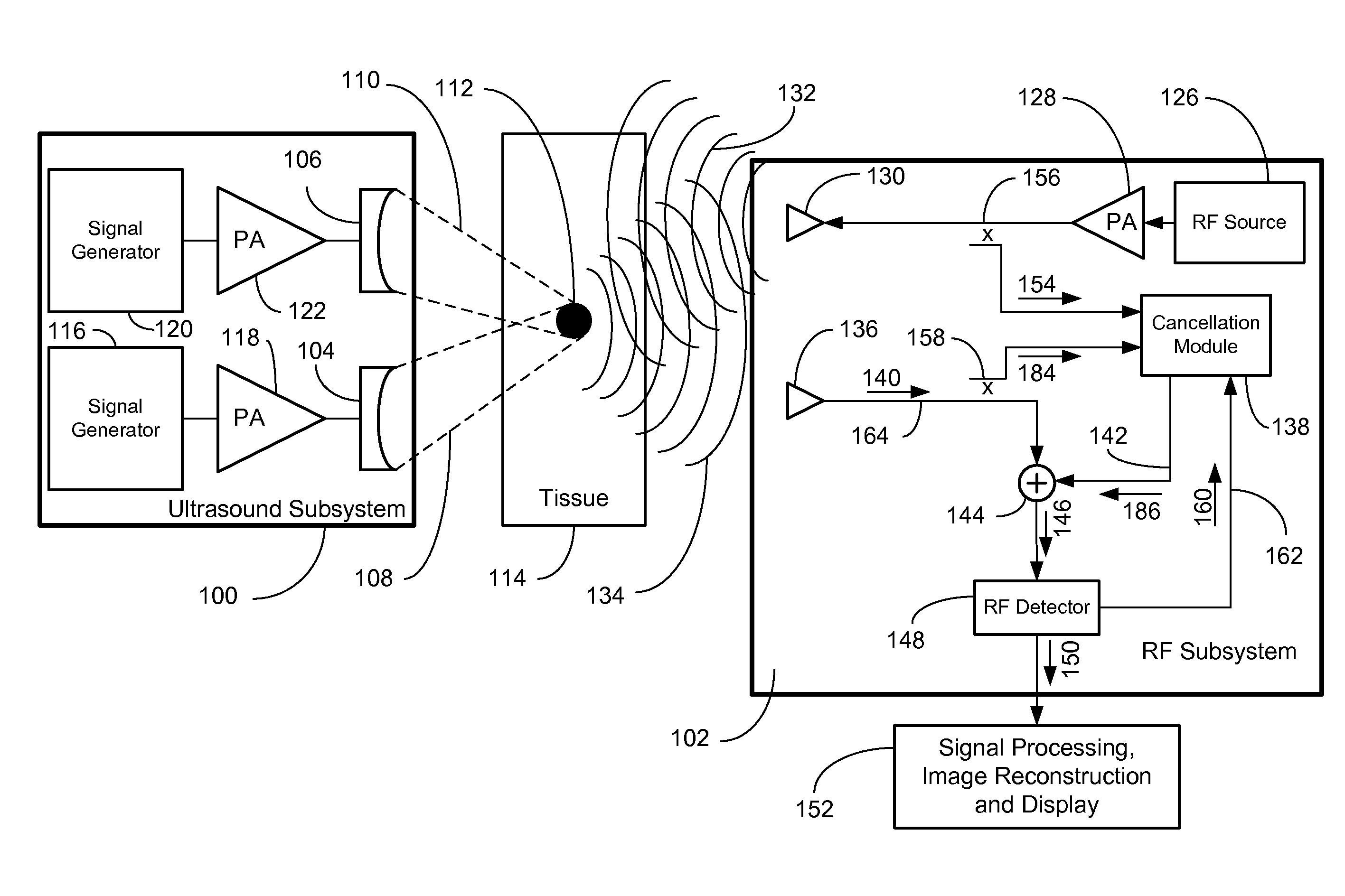
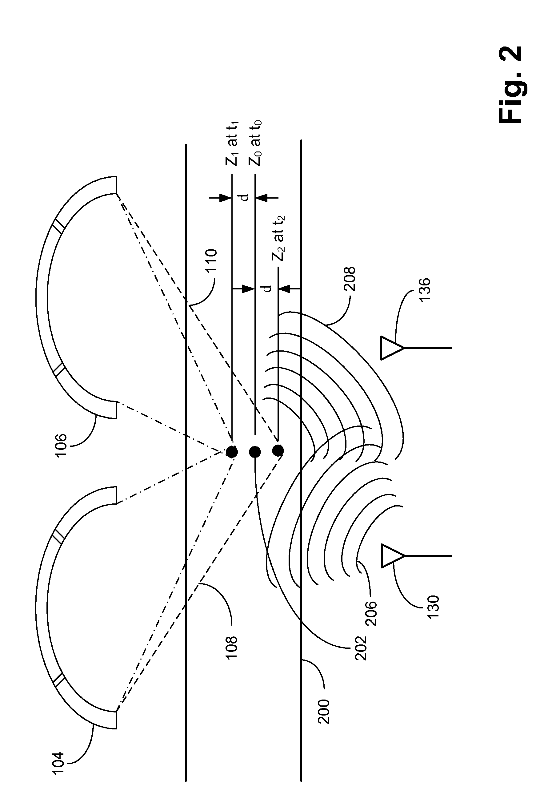
The ultrasound energy through its interaction with the nonlinearity of the inclusion induces low frequency vibration in the target inclusions which results in measurable displacement of the inclusion and causes a Doppler shift in the reflected radio frequency signals.
Likewise microwave-only imaging modalities require solving the inverse problem which can produce imprecise results and is limited in resolution to a fraction of the wavelength used for measurement.

Method used


Image

  • Multi-Modality Ultrasound and Radio Frequency System for Imaging Tissue
  • Multi-Modality Ultrasound and Radio Frequency System for Imaging Tissue
  • Multi-Modality Ultrasound and Radio Frequency System for Imaging Tissue

Examples


Embodiment Construction

[0036]FIG. 1 is a block diagram illustrating the dual-modality imaging system where the two modalities are operating at the same time. The system comprises an ultrasound subsystem 100 and a radio frequency subsystem 102. The ultrasound subsystem 100 employs at least two ultrasound transmitter 104 and 106 capable of generating two focused ultrasound beams 108 and 110 aimed at a focal point 112 where the intersecting ultrasonic beams are focused within the tissue 114 under observation. The ultrasound transmitter 104 is driven by a first signal generator 116 that passes its energy through a power amplifier 118 thus generating a signal at frequency f1. The ultrasound transmitter 106 is driven by a second signal generator 120 which passes its energy through a power amplifier 122 thus generating a signal at frequency f2. Ideally a small difference in frequency (hertz to kilohertz) between frequency f1 and frequency f2 is created so that a desired beat frequency Δf equals (f1−f2) is genera...


PUM


Abstract

This invention provides a dual-modality system for performing characterization and imaging of tissue, tumors, structures, lesions, and ablations under investigation. Specifically, the invention couples ultrasound technology comprising at least two focused ultrasound beams for vibrating target tissues located at the focal point of the ultrasound beams intersection with a radio frequency system for measuring the response of the target tissues. The ultrasound system vibrates the target tissues while the reflected radio frequency energy is transmitted into the target tissues. When reflected, the main carrier tone of the reflected radio frequency energy is cancelled and analysis is performed on the remaining sideband frequencies.

Description

CLAIM OF PRIORITY[0001]Applicant claims priority to U.S. Ser. No. 12 / 151,355 titled “Multi-modality System for Imaging in Dense Compressive Media” filed on May 6, 2008 and is incorporated by reference.BACKGROUND OF THE INVENTION[0002]1. Field of Invention[0003]The field of the invention is soft tissue imaging for deep tissue and tissues within the body using radio frequency energy and vibro-acoustography in a dual modality imaging system where the two modalities operate simultaneously.[0004]2. Related Art[0005]A number of imaging technologies are available to detect cancer in soft and compressed tissues within the human body. For breast cancer, X-ray mammography is currently the only FDA approved technology for breast cancer screening. However, mammography results in too many false negatives and false positives which delays the detection of early cancer, or results in unnecessary biopsies and expense. Additionally, mammography results are unreliable in the case of dense or fibrocyst...

Claims


Application Information

Patent Timeline
Patent Type & Authority Applications(United States)
IPC IPC(8): A61B6/00A61N7/00
CPCA61B5/4312A61B5/0048A61B5/05A61B8/485A61B8/4416A61B8/4477A61B8/085A61B5/0051A61B5/0035
Inventor ISMAIL, ALY M.CARR, FRANCISKWOK, SAI
Owner ULTRAWAVE LABS
Why Eureka
Try Eureka