Scaffold for articular cartilage regeneration and method for manufacturing same

Inactive Publication Date: 2013-04-04
TE BIOS
View PDF8 Cites 12 Cited by
  • Summary
  • Abstract
  • Description
  • Claims
  • Application Information

AI Technical Summary

Benefits of technology

[0014]The present inventors have studied a scaffold for the regeneration of articular cartilage, applicable to both the superficial zone and the middle zone of articular cartilage, which culminated in finding that when incorporated with multiwalled carbon nanotubes, a 3D collagen type II-based hydrogel can be improved in mechanical properties and can be used as a scaffold for culturing human mesenchymal stem cells, or chondrocytes or osteocytes that are differentiated from human mesenchymal stem cells, with applicability to the middle zon

Problems solved by technology

Once it is damaged, cartilage, a connective tissue found predominantly in the joints of vertebrates, is hardly apt to regenerate in the body.
Persons with damaged articular cartilage can do only limited daily activities because of serious pain they endure.
Chronically damaged articular cartilage may be further aggravated and develop into degenerative arthritis, which acts as a serious barrier to physical or v

Method used


Image

  • Scaffold for articular cartilage regeneration and method for manufacturing same
  • Scaffold for articular cartilage regeneration and method for manufacturing same
  • Scaffold for articular cartilage regeneration and method for manufacturing same

Examples


example 2

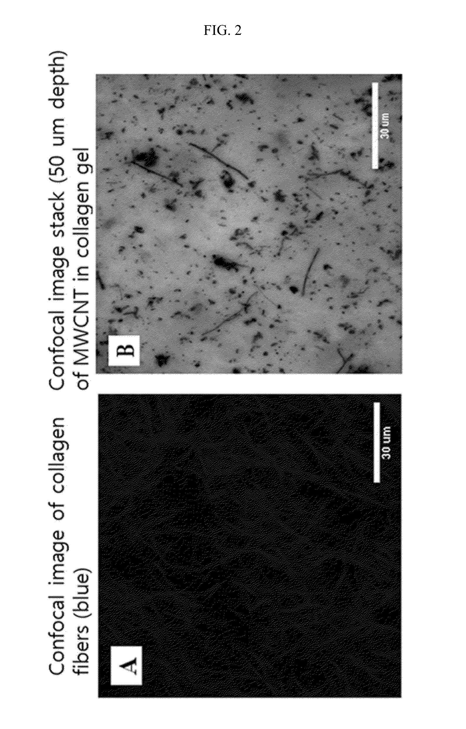
Preparation of Collagen Gel Composed of Multiwalled Carbon Nanotube-Incorporated 3-D Collagen Type II-Based Hydrogel Seeded with Human Mesenchymal Stem Cells

[0065]1. Multiwalled Carbon Nanotubes (MWCNT)-PBS Solution

[0066]To a mixture of 15 mL of sulfuric acid and 5 mL of nitric acid, 50 mg of MWCNT [240˜500 nm in outer diameter, 5˜40 ∥m in length, 95+% in purity, manufactured by catalytic chemical vapor deposition (CVD), Nanostructured and Amorphous Materials Inc.] was added, and the solution was ultrasonicated for 1 hr at 50° C. in an ultrasonication water bath, and neutralized with ammonium hydroxide. The MWCNT was collected as pellets by centrifugation for 10 min at 5,000 rpm, and the supernatant was removed. The pellets were washed four times with sterile water, ultrasonicated for 15 min, and centrifuged. After removal of the supernatant containing residual solvents, and undesired amorphous carbon, the MWCNT was resuspended and dispersed in 4 mL of phosphate buffered saline (PB...

example 3

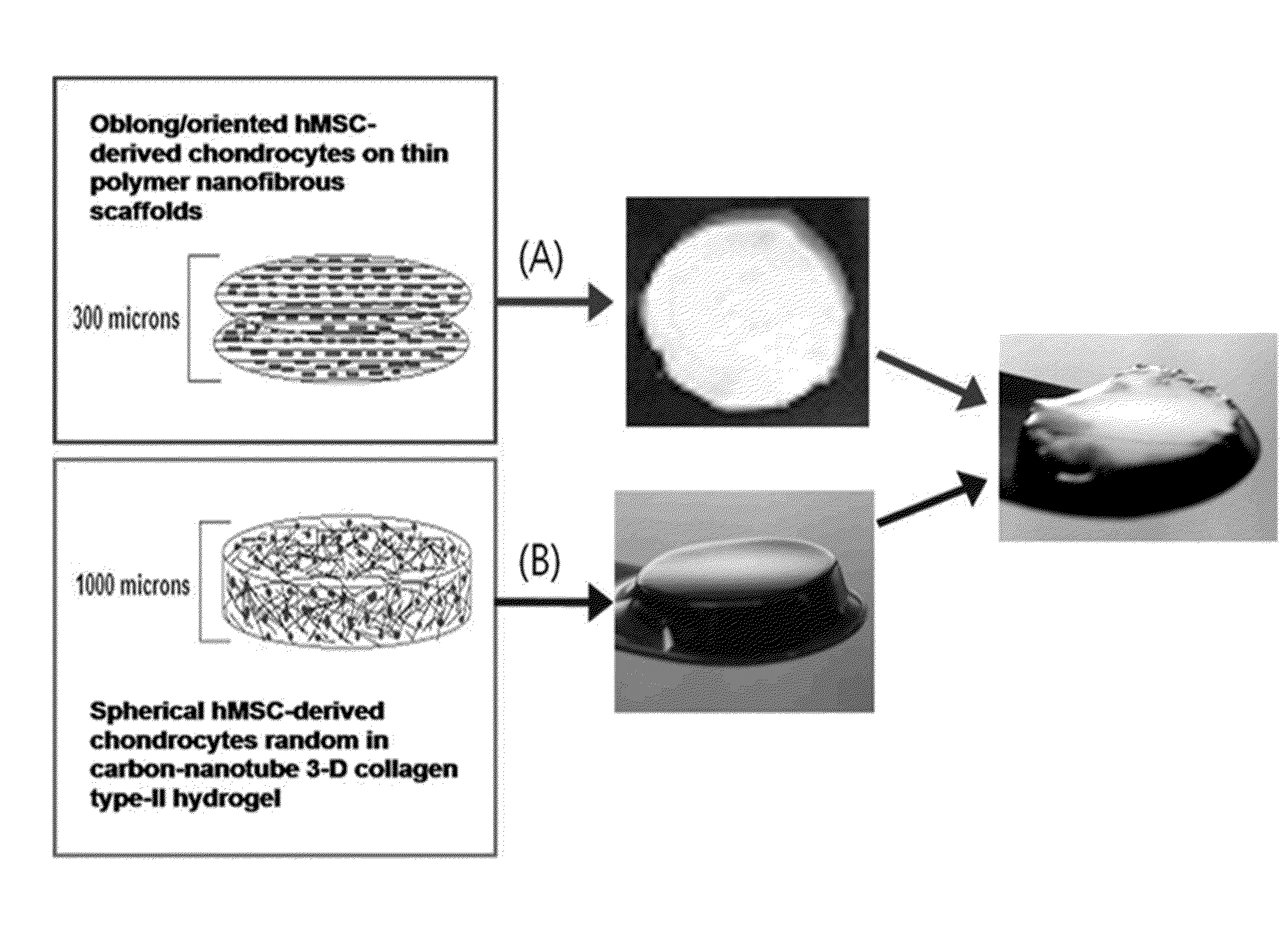
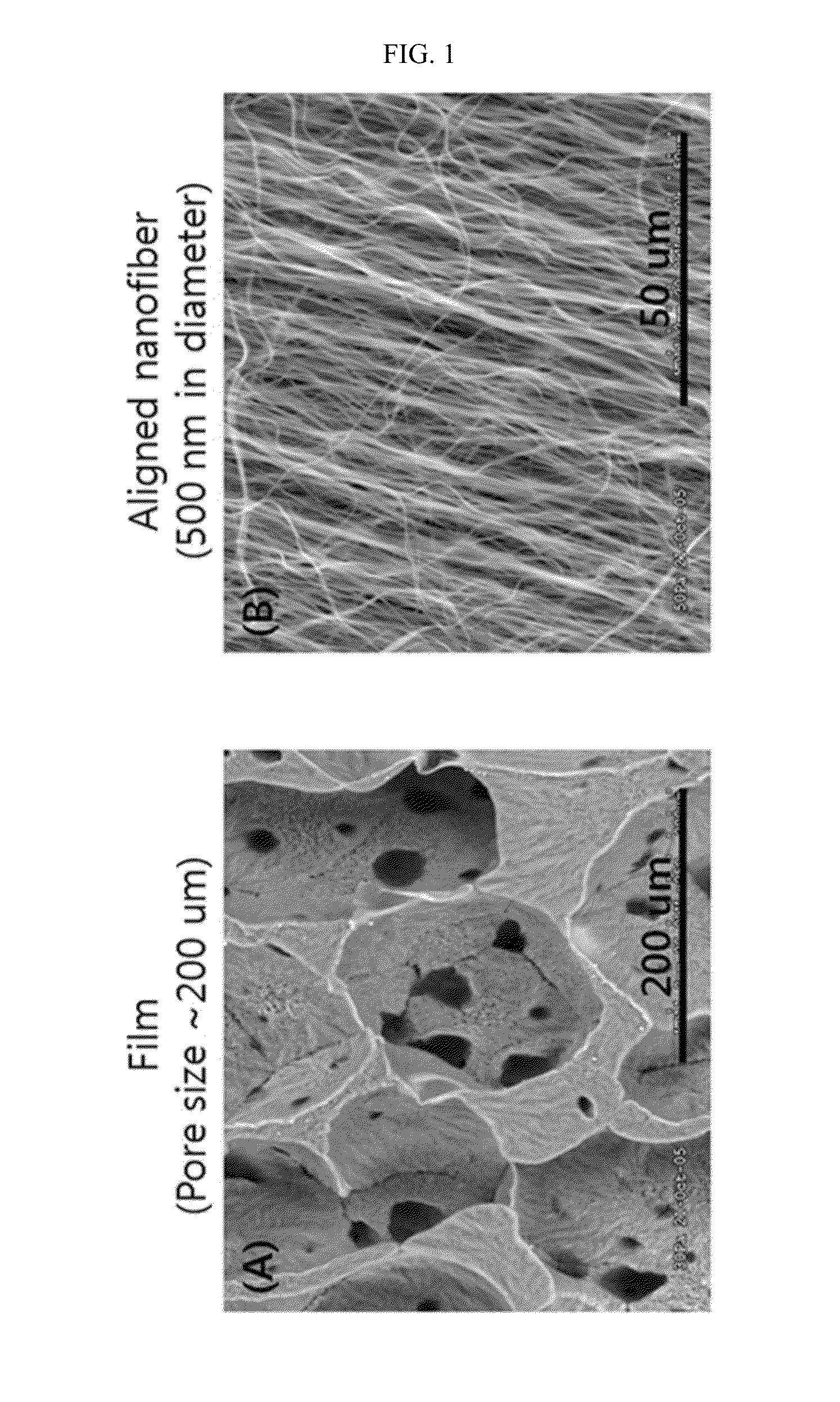
Preparation of Composite Scaffold Composed of Electrospun PCL Fiber Scaffold / Multiwalled Carbon Nanotube-Incorporated 3-D Collagen Type II-Based Hydrogel

[0074]The multiwalled carbon nanotube-incorporated 3-D collagen type II-based hydrogel prepared in Example 2 was pipetted in an amount of 400 μL onto the electrospun PCL fiber scaffold prepared in Example 1 and applied to the flat bottom of each well of 24-well plates to form 2 mm-thick constructs. Subsequently, they were completely set by incubation at 37° C. for 45 min to afford a bilayer composite scaffold of 2 mm in thickness in which the thin PLC fiber scaffold was firmly incorporated onto one side of the completely solidified 3-D collagen type II-based hydrogel. This composite scaffold was withdrawn from the 24-well plate, transferred into a petri dish filled with sterile water, prior to maintaining hydration with sterile water and AFM analysis. It was incubated at 37° C. for 30 min before AFM (atomic force microscope) analys...

experimental example 1

Physical Strength of 3-D Collagen Type II-Based Hydrogel

[0076]The physical strength of the 3-D collagen type II-based hydrogel was measured by AFM (atomic force microscopy). None of the hydrogel samples analyzed by AFM contained cells.

[0077]The ultrasonicated and multiwalled carbon nanotubes were added directly within 1 mL of the collagen hydrogel with pH of 7.5. For comparison, the control collagen hydrogel, EDC (1-ethyl-3-(3-dimethylaminopropyl)carbodiimide)-crosslinked collagen hydrogel, and multiwalled carbon nanotube-incorporated collagen hydrogel were employed. All samples were adjusted to have a final collagen concentration of 7 mg / mL. Samples were created by pipetting 50 μL of the multiwalled carbon nanotube-incorporated collagen hydrogel preparation onto a glass cover slip, and allowing the gel to set completely by incubating at 37° C. for 30 min. The collagen hydrogel samples were kept hydrated in sterile water prior to AFM analysis. AFM analysis was performed with an Atom...


PUM

PropertyMeasurementUnit
Temperatureaaaaaaaaaa
Fractionaaaaaaaaaa

Abstract

Disclosed are scaffolds for regeneration of articular cartilage which are applicable to both the superficial zone and the middle zone of articular cartilage, and a method for manufacturing the same. The scaffolds have sufficient mechanical properties to support the implantation and regeneration of chondrocytes, and allow cells to show high cell viability with a high content of sulfated glycosaminoglycans (GAGs). In addition, being applicable to both the superficial zone and the middle zone of articular cartilage, the scaffolds facilitate cell adhesion and provide biomimetic surface environments that are effective for growing and differentiating stem cells. Therefore, the scaffolds are helpful in regenerating damaged articular cartilage, thus finding applications in stem cell therapy for articular cartilage damage and disease. Also, the application of the scaffolds can be extended to prostheses of the ear and the nose in plastic surgery.

Description

TECHNICAL FIELD[0001]The present invention relates to a scaffold for the regeneration of articular cartilage which is applicable to the middle zone of articular cartilage or both the middle zone and the superficial zone of articular cartilage, and a method for manufacturing the same.BACKGROUND ART[0002]Once it is damaged, cartilage, a connective tissue found predominantly in the joints of vertebrates, is hardly apt to regenerate in the body. Persons with damaged articular cartilage can do only limited daily activities because of serious pain they endure. Chronically damaged articular cartilage may be further aggravated and develop into degenerative arthritis, which acts as a serious barrier to physical or vocational activities.[0003]Representative among the therapies for damaged articular cartilage are chondroplasty, osteochondral transplantation, and autologous chondrocyte transplantation.[0004]New tissue engineering-based therapies for damaged articular cartilage have recently gai...

Claims


Application Information

Patent Timeline
IPC IPC(8): C12N5/0775
CPCA61F2/30756C12N2533/54A61F2310/00365A61L27/24A61L27/3817A61L27/52A61L27/58A61L2430/06B82Y5/00C12N5/0675Y10S977/752Y10S977/923C12N5/0655C12N2533/10C12N2533/40A61F2002/30766A61L27/14B82B3/00C12N5/0662
Inventor CHO, MICHAEL
Owner TE BIOS
Why Eureka
Try Eureka