Novel VEGF mimetic peptide-based scaffolds for therapeutic angiogenesis and methods for their use

a mimetic peptide and scaffold technology, applied in the direction of peptide/protein ingredients, peptide sources, angiogenin, etc., can solve the problems of limited tissue-specific targeting, protein-based therapies, and the need for new therapeutic approaches, and achieve enhanced bioactivity, enhanced limb recovery, and improved blood flow measurement

Inactive Publication Date: 2013-04-25
NORTHWESTERN UNIV
View PDF7 Cites 25 Cited by
  • Summary
  • Abstract
  • Description
  • Claims
  • Application Information

AI Technical Summary

Benefits of technology

[0009]Accordingly, it is an object of the present invention to incorporate a VEGF-mimetic peptide sequence into the framework of a peptide amphiphile to yield a self-assembling structure having the requisite binding activity to activate VEGF receptors. As discussed in greater detail below, evaluation of the bioactivity in vitro and in vivo found the VEGF-PA structure of the present invention was observed to have enhanced bioactivity as compared to peptide alone. As a treatment for hind-limb ischemia, the VEGF-mimetic nanostructure of the present invention was found to enhance limb recovery following critical hind limb ischemia as assessed by tissue salvage and motor function of the ischemic hind-limb. These findings are associated with an improved blood flow measure by laser Doppler imaging. Immunohistological evaluation revealed a significantly enhanced microcirculation. Given that the major drawbacks of protein-based therapies lie in the excessive production costs and poor target tissue retention, the use of peptide amphiphiles to present bioactive VEGF-mimetic epitopes constitutes a significant improvement over the prior art, offering the benefits of large-scale synthesis, inexpensive production, enhanced tissue retention, increased bioavailability of the signal, and capacity to localize the signal within the nanofiber. The self-assembling VEGF-PA of the present invention is also able to efficiently signal cells in a manner consistent with VEGF signaling through a network of high aspect-ratio entangled fibers that can provide a more lasting signal in the host tissue due to prolonged residence time and high density of displayed epitopes. For example, whereas a single injection of VEGF has a short retention time within the tissue, the VEGF epitope-presenting PAs of the present invention remain in the tissue and bioavailable on the order of weeks. Further, this totally synthetic approach allows for these molecules to be produced through a highly controlled process on the scale of multiple grams for a cost much less than that associated with the preparation and purification of recombinant factors.

Problems solved by technology

However, revascularization is not consistently successful, leaving a tremendous medical need for new therapeutic approaches that regenerate ischemic tissue.
Protein-based therapies, while demonstrating success in several pre-clinical trials, are limited by a lack of tissue-specific targeting and inadequate temporal levels of the protein in the target zone [4, 7, 8].
Moreover, the costs associated with protein preparation and purification make these therapies, if feasible, extremely expensive at the clinical level [4].
While these vector-based approaches are not necessarily as expensive as recombinant protein, limitations range from inadequate transduction efficiency with some vectors to safety issues with others [9].
Moreover, clinical trials using gene therapy to deliver angiogenic factors are yet to demonstrate convincing efficacy for treatment of cardiovascular diseases [4].
While the synthetic oligopeptide addresses the major issue of protein production cost, it does not significantly improve on protein therapies in terms of tissue retention and sustained delivery.

Method used


Image

  • Novel VEGF mimetic peptide-based scaffolds for therapeutic angiogenesis and methods for their use
  • Novel VEGF mimetic peptide-based scaffolds for therapeutic angiogenesis and methods for their use
  • Novel VEGF mimetic peptide-based scaffolds for therapeutic angiogenesis and methods for their use

Examples


example 1

Design and Synthesis of a VEGF-Mimetic PA

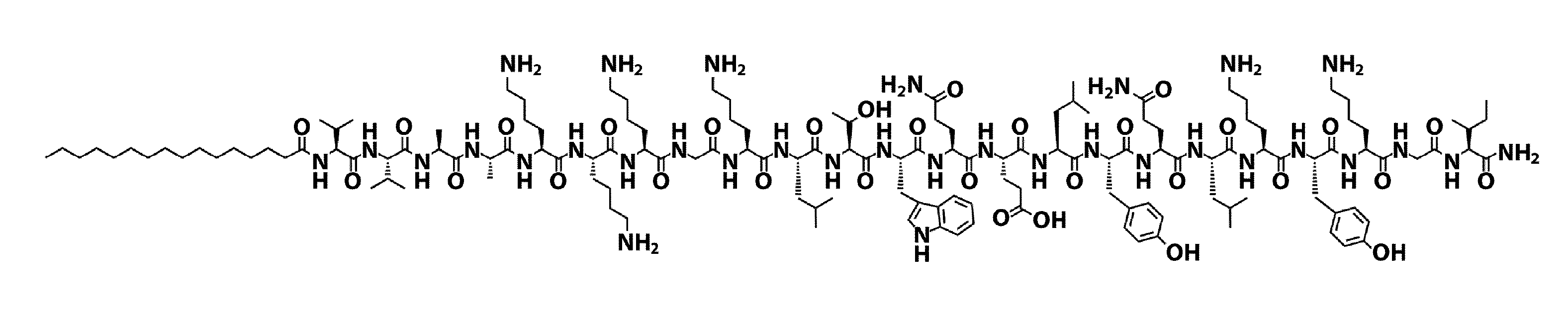
[0104]The VEGF-PA (FIG. 1A) utilized in the present examples was designed to display on surfaces of nanostructures a peptide sequence that mimics VEGF, namely KLTWQELYQLKYKGI-NH2 (SEQ ID NO: 2) [12]. A glycine (G) spacer was covalently attached to the N-terminus of this peptide, followed by a triple lysine (K3) sequence intended to improve solubility and trigger electrolyte-driven self-assembly. The K3G sequence was followed by a V2A2 β-domain and a C16 acyl chain to promote self-assembly into cylindrical nanostructures through intermolecular hydrogen bonding and hydrophobic collapse upon electrolyte screening of charged residues (FIG. 1G).

[0105]Stabilization of the bioactive conformation of the VEGF-mimetic peptide by conjugation to a PA could be advantageous to enhance the potency of the epitope. To demonstrate the improvement obtained a VEGF-PA and epitope-only peptide control were synthesized. In addition, as a nanostructure control for t...

example 2

Structural Characterization of a VEGF-Mimetic PA

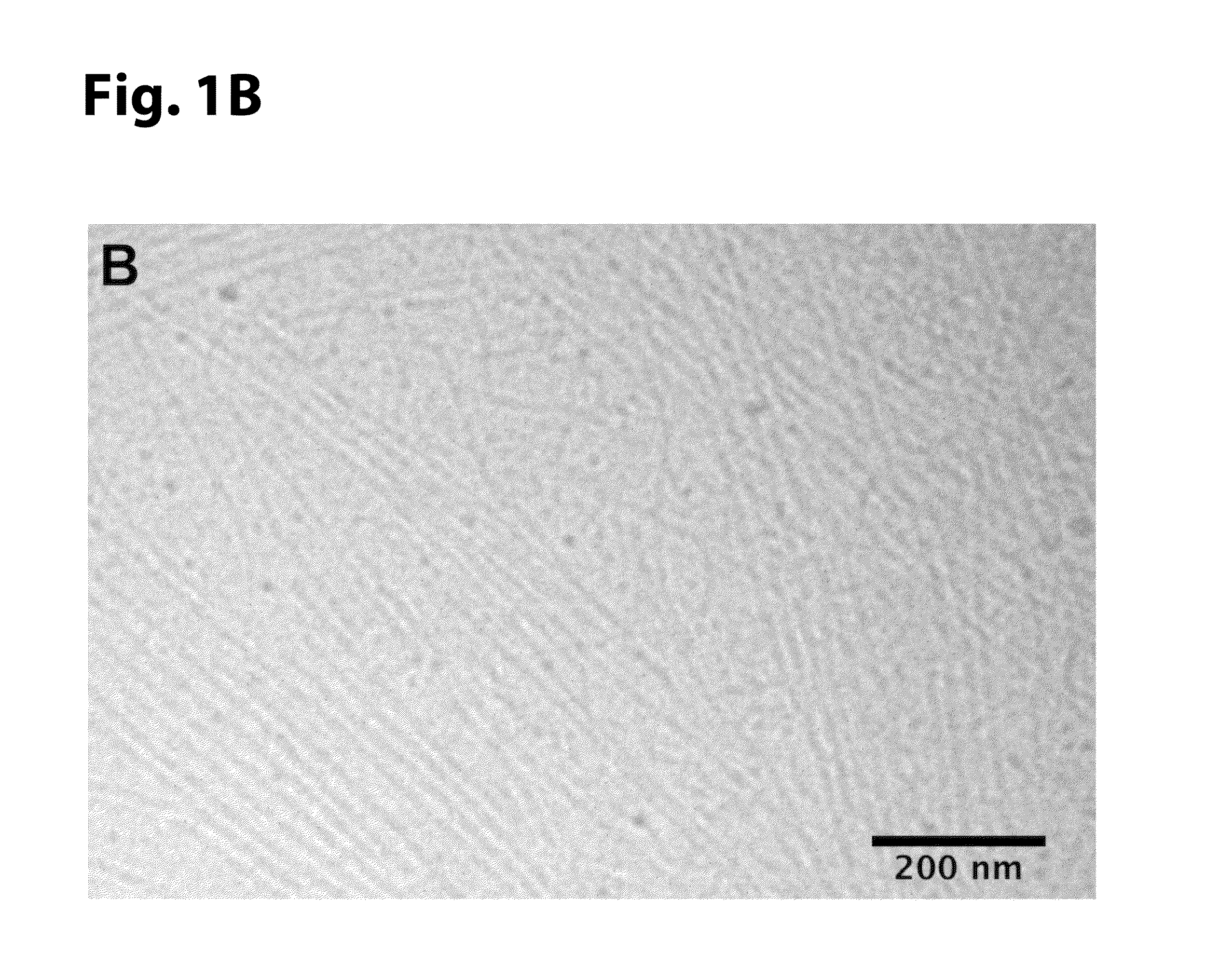
[0108]Cryogenic TEM was performed on a JEOL 1230 microscope with an accelerating voltage of 100 kV. A Vitrobot Mark IV equipped with controlled humidity and temperature was used for plunge-freezing samples. A small volume (7 μl) of 0.05 w / v % VEGF-PA dissolved in 0.5×PBS was deposited on a copper TEM grid with Quantifoil support film (Electron Microscopy Sciences) and held in place with tweezers mounted to the Vitrobot. The specimen was blotted in 90-95% humidity and plunged into a liquid ethane reservoir cooled by liquid nitrogen. The vitrified samples were transferred into liquid nitrogen and inserted into a Gatan 626 cryo-holder through a cryo-trarisfer stage. Samples were imaged using a Hamamatsu ORCA CCD camera.

[0109]SEM was performed using a Hitachi S4800 scanning electron microscope with a 5 kV accelerating voltage. To prepare samples for imaging, VEGF-PA was dissolved at 1.5 w / v % in water and mixed with 10 mM Na2HPO4 to induce...

example 3

VEGF-PA Specifically Activates VEGF Receptors

[0113]VEGF signal transduction is initiated by multiple tyrosine phosphorylation events on the intracellular domain of its receptors [29]. In order to determine if the VEGF-PA specifically signals in a manner consistent with VEGF, human umbilical vein endothelial cells (HUVEC) were stimulated with VEGF-PA and a sandwich ELISA was performed on cell lysates to quantify the amount of phosphorylated VEGF receptor 1 (VEGFR1 or Flt-1) or phosphorylated VEGF receptor 2 (VEGFR2 or KDR), the two primary VEGF receptors implicated in its angiogenic signaling.

[0114]Human umbilical vein endothelial cells (HUVECs) and complete endothelial cell growth media (EGM) were purchased (Genlantis, San Diego, Calif.), passaged two times following receipt, and cryo-preserved in media with 5% DMSO. Cells were thawed as needed and grown to confluence in a 75 mm2 flasks (VWR Falcon) prior to plating for experiments.

[0115]Phosphorylation of both VEGFRI and VEGFR2 was...


PUM

PropertyMeasurementUnit
diameteraaaaaaaaaa
diameteraaaaaaaaaa
accelerating voltageaaaaaaaaaa

Abstract

Disclosed herein is a completely synthetic cell-free therapy based on peptide amphiphile nanostructures designed to mimic the activity of vascular endothelial growth factor (VEGF), one of the most potent angiogenic signaling proteins. The VEGF-mimetic filaments disclosed herein were found to induce phosphorylation of VEGF receptors and induce pro-angiogenic behavior in endothelial cells, indicated by an enhancement in proliferation, survival and migration in vitro.

Description

RELATED APPLICATIONS[0001]This application is a non-provisional of U.S. Provisional Application No. 61 / 480,450, filed Apr. 29, 2011, which is incorporated herein by reference.STATEMENT OF GOVERNMENTAL SUPPORT[0002]This invention was made with U.S. Government support under NH-I Grant Nos. 1RO1-EB003806-04, HL-53354, HL-57516, HL-77428, HL-63414, HL-80137, PO1HL-66957. The U.S. Government has certain rights in this invention.REFERENCE TO SEQUENCE LISTING[0003]The official copy of the sequence listing is submitted herewith in both paper and electronic form. The latter contains an ASCII formatted sequence listing with a file named NAN—007P_SEQ_LIST.txt, created on Apr. 28, 2011, and having a size of 48 KB submitted concurrently with the specification. The sequence listing contained in this file is part of the specification and is herein incorporated by reference in its entirety.FIELD OF THE INVENTION[0004]The present invention relates generally to novel VEGF-mimetic peptide-based scaffo...

Claims


Application Information

Patent Timeline
Patent Type & Authority Applications(United States)
IPC IPC(8): C07K14/00
CPCC07K14/001C07K2319/00C07K14/52A61K38/18
Inventor WEBBER, MATTHEW J.TONGERS, JORNLOSORDO, DOUGLAS W.STUPP, SAMUEL I.
Owner NORTHWESTERN UNIV
Why Eureka
Try Eureka