Peripheral blood gene markers for early diagnosis of parkinson's disease

a gene marker and parkinson's disease technology, applied in the field of molecular risk marker profiles for the diagnosis of parkinson's disease, can solve the problems of insufficient pd specificity and sensitive diagnosis of tools, no laboratory blood test for pd is available, and the cost-effectiveness remains a problem, so as to achieve increased or decreased expression levels

Inactive Publication Date: 2013-08-22
MANDEL SILVA A +5
View PDF0 Cites 11 Cited by
  • Summary
  • Abstract
  • Description
  • Claims
  • Application Information

AI Technical Summary

Benefits of technology

[0010]As stated above, it has previously been found by the inventors of the present invention that certain genes show altered, i.e., increased or decreased, expression levels in brains of Parkinson's disease (PD) patients compared with control individuals. As later found, alterations in the expression levels of at least some of those genes relative to control individuals can also be detected in peripheral blood samples of PD patients, although not necessarily in the same direction shown in brains, and may therefore be used for diagnosis of PD in a tested individual.

Problems solved by technology

Imaging studies using positron emission tomography (PET) with [18F]-Dopa, single photon emission tomography (SPECT) with [123I]-β-CIT or diffusion-weighted MRI could improve differential diagnosis of Parkinsonism, but cost-effectiveness remains a problem.
Yet, these tools do not provide a specific and sensitive PD diagnosis (Jankovic et al., 2000).
Even more frustrating is the cognizance that PD remains undetected for years before early clinical diagnosis occurs and when this happens, the loss of dopamine neurons in the substantia nigra approaches already 68% in the lateral ventral tier and 48% in the caudal nigra (Fearnley and Lees, 1991).
No laboratory blood test for PD is available, let alone the detection of individuals at risk for developing PD, which is currently impossible.
Current treatment of PD is symptomatic and no truly neuroprotective drug having disease modifying activity has been developed.
Such biomarkers, if available, may further provide a measure of disease progression that can objectively be evaluated, while clinical measures are much less accurate.
However, since brain samples from live patients are usually not available, in order to use this approach tor diagnosing PD, it is still necessary to look for genes with altered expression patterns compared with controls in tissues such as blood, skin or saliva that can easily be obtained from living individuals.
A recent study has shown that after accounting for confounding variables, such as blood CSF contamination and age, alpha-synulelin and DJ-I protein levels were reduced in CSF from PD compared with healthy controls and AD individuals (Hong et al., 2010), although the test suffered from poor specificity and may have been affected by medication.

Method used


Image

  • Peripheral blood gene markers for early diagnosis of parkinson's disease
  • Peripheral blood gene markers for early diagnosis of parkinson's disease
  • Peripheral blood gene markers for early diagnosis of parkinson's disease

Examples


example 1

Evaluation of the Stability of Blood Reference Genes

[0126]The relative quantification of expression levels is based on the expression levels of target genes vs. one or more references i.e., reference or control, genes. The normalization procedure is mandatory in quantitative RT-PCR (qRT-FCR) studies and the reason for the choice of the most stably expressed reference genes is to avoid misinterpretation and low reproducibility of the final results.

[0127]We decided to determine the expression stability of live widely used reference genes, in particular, ACTB, GAPDH, ALAS1, PPIA and 60S RPL13A, in human leukocyte samples from PD and healthy age-matched controls, randomly divided between males and females.

[0128]RNA from blood samples of patients and controls was extracted and reverse transcribed, and expression was determined by quantitative Real-Time RT-PCR, as described in Materials and Methods.

[0129]The expression of the selected control genes in samples was analyzed with two widely ...

example 2

Identifying a PD Risk Marker Panel in Peripheral Blood

[0131]In order to identify a PD risk marker panel in peripheral blood with high probability to detect early PD, we have focused on non-medicated de novo PD patients to track for gene changes at very early stages of the disease and to ascertain no confounding bias that could arise from medication.

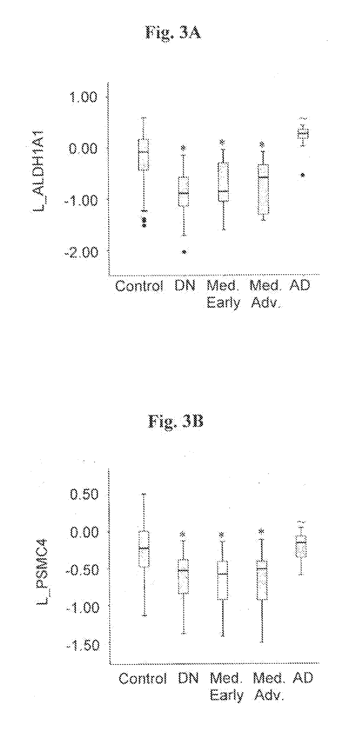
[0132]The transcriptional expression level of eight genes, in particular, ALDH1A1, PSMC4, SKP1A, HSPA8, CSK, HIP2 and EGLN1, which have previously been found to be altered in substantia nigra tissue from sporadic PD patients (Grünblatt et al., 2004); and CLTB, elected from the transeriptomic PD blood analysis of Scherzer et al., (2007), were assessed in blood samples from 38 individuals with de-navo PD, and 64 healthy age-matched controls without neurological dysfunction. (Table 1).

[0133]RNA from blood samples obtained from each one of those individuals was extracted and reverse transcribed, and expression level of each one of said genes ...

example 3

Characterizing Partial Risk Marker Panels

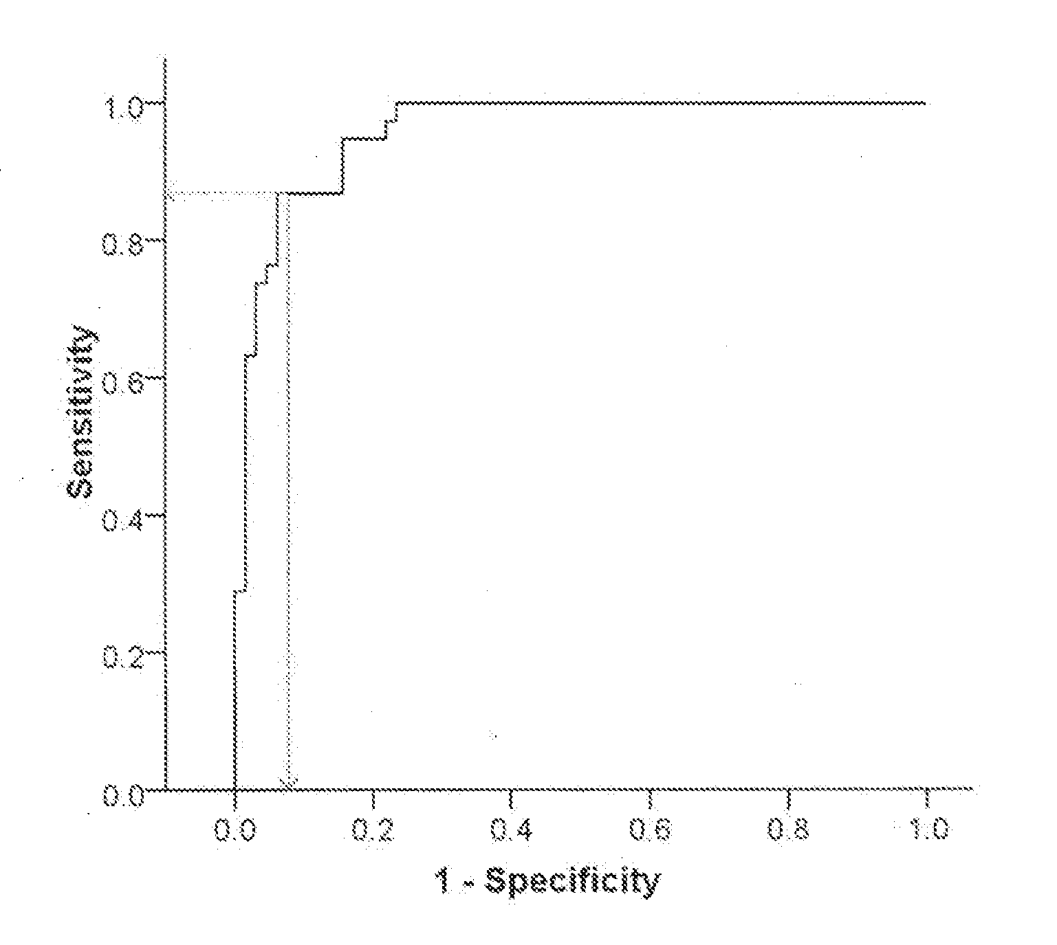
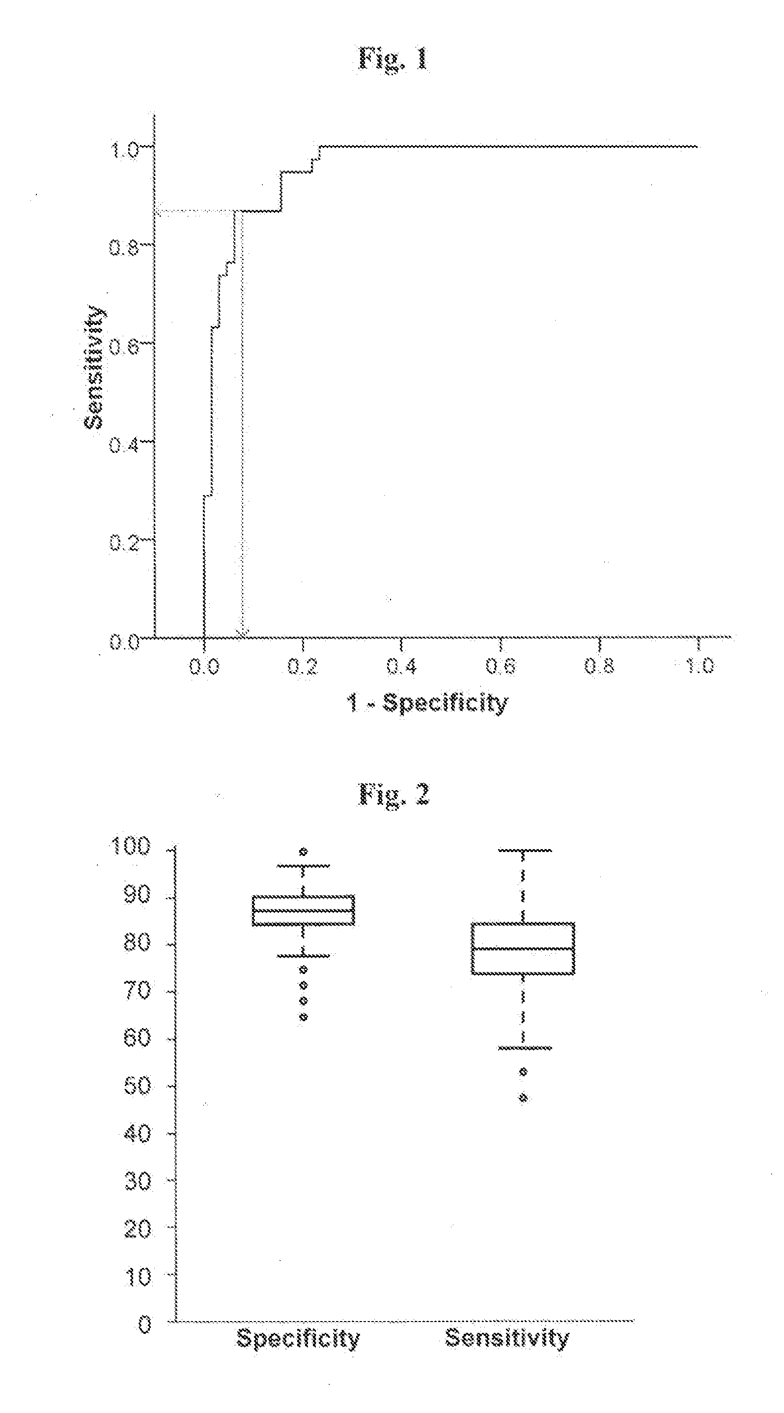
[0140]We next tested the ability of partial risk marker panels, including genes selected from the full six-genes risk marker panel, established and described in Example 2, to differentiate between PD de novo patients and healthy controls. A stepwise multivariate logistic regression analysis was conducted as described in Materials and Methods, and stopped after finding three, four, or five genes of the full six genes panel. A ROC curve was used to calculate the relationship between sensitivity and specificity for the de-novo PD group vs. healthy controls for each of the partial risk marker panels, and thus evaluate the diagnostic performance of the identified gene clusters. The results are presented in Table 5A and regression coefficient (B) values for the predicted probability equation for each partial panel are given in Table 5B.

[0141]The predicted probability for PD (p(PD)) for each one of these partial risk panels can be calculated using t...


PUM

PropertyMeasurementUnit
densityaaaaaaaaaa
phaseaaaaaaaaaa
rigidityaaaaaaaaaa

Abstract

The present invention relates to the use of molecular risk marker profiles for diagnosis of Parkinson's disease. More particularly, the invention provides methods for diagnosis of Parkinson's disease in an individual, utilizing certain profiles established based on the expression levels of certain genes, which together form a gene panel, in the peripheral blood of said individual, as well as kits for carrying out these methods. The profile encompass ALDH1A1.

Description

TECHNICAL FIELD[0001]The present invention relates to the use of molecular risk marker profiles for diagnosis of Parkinson's disease. More specifically, the invention provides methods and kits for diagnosis of Parkinson's disease utilizing expression profiles of particular gene panels in blood samples.Abbreviations[0002]ACTB, β-actin; AD, Alzheimer's disease; ALAS1, aminolevulinate delta synthase 1: ALDH1A1 aldehyde dehydrogenase 1 family, member A1: ARPP-21, 21-cyclic AMP-regulated phosphoprotein; CLTB, clathrin, light polypeptide; CNR2, Cannabinoid receptor 2; CSK, c-src tyrosine kinase; EGLN1 egl nine homolog 1; EIF4BP2, eukaryotic translation initiation factor 4E binding protein 2; GAPDH, glyceraldehyde-3-phosphate dehydrogenase; HIP2 / UBE2K, huntingtin interacting protein 2 / ubiquitin-conjugating enzyme E2K; HIST1H3E, histone cluster 1, H3e; HSPA8 / HSC70 / HSC54, chaperone heat shock 70 kDa protein 8; HS3ST2, heparan sulfate (glucosamine) 3-O-sulfotransferase 2; LAMB2, laminin, β2 (...

Claims


Application Information

Patent Timeline
Patent Type & Authority Applications(United States)
IPC IPC(8): C12Q1/68G06F19/20G16B25/10
CPCC12Q1/686G06F19/20C12Q2600/158C12Q1/6883G16B25/00G16B25/10
Inventor MANDEL, SILVA A.YOUDIM, MOUSSA B.H.RIEDERER, PETERGRUNBLATT, EDNARABEY, JOSE M.MOLOCHNIKOV, LEONID
Owner MANDEL SILVA A
Why Eureka
Try Eureka