Microfluidic Cell Trap and Assay Apparatus for High-Throughput Analysis

a microfluidic cell and assay technology, applied in the field of microfluidic devices, can solve the problems of high reagent cost, low throughput, and difficulty in accurately measuring low abundance transcripts, and achieve the effect of improving cell washing

Active Publication Date: 2015-01-15
THE UNIV OF BRITISH COLUMBIA
View PDF7 Cites 73 Cited by
  • Summary
  • Abstract
  • Description
  • Claims
  • Application Information

AI Technical Summary

Benefits of technology

[0039]The method may further include a cell sorting step prior to directing the cells to a cell capture chamber. The method may further include immobilizing the captured cell prior to washing. Immobilization may further enhance washing of the cells in the capture chamber.

Problems solved by technology

Of these, single cell RT-qPCR provides combined advantages of sensitivity, specificity, and dynamic range, but is limited by low throughput, high reagent cost, and difficulties in accurately measuring low abundance transcripts (6).
Unfortunately, such devices do not allow for high throughput analysis due to the manual effort involved in operating the valving mechanisms.
Moreover, the device does not allow isolated cells to be washed from the supernatant prior to treatment or analysis.
This in turn allows for contamination events, and further limits the downstream applications of the device.
Although these systems provide a high-throughput qPCR readout, they do not address the front end sample preparation and require single cell isolation by FACS or micropipette followed by off-chip processing and pre-amplification of starting template prior to analysis.
Despite the engineering complexity of this system, throughput was limited to four cells per run, cell capture required metabolic labeling of the cells, and the analysis was not quantitative.
Low trapping throughput and low trapping efficiency present a significant challenge to the goal of reliable and scalable analysis of single or small numbers of cells.
Low capture efficiencies necessitating tens of thousands of cells in order to make a few single cell measurements is not an issue when using cell lines, however it becomes a significant problem when using primary samples of rare cell types, such as stem cells.
Also, observations of both the trapped cells and those passing around the traps have indicated that the cell trapping efficiency was dependent on cell size, which could potentially introduce a bias into the single cell measurements.

Method used


Image

  • Microfluidic Cell Trap and Assay Apparatus for High-Throughput Analysis
  • Microfluidic Cell Trap and Assay Apparatus for High-Throughput Analysis
  • Microfluidic Cell Trap and Assay Apparatus for High-Throughput Analysis

Examples


example 1

Example 1.1

Materials and Methods

Nucleic Acid Detection and Quantification

[0131]DNA quantification through microarray analysis is a highly multiplexed, well established method to assay a sample for thousands of different targets. Specific DNA sequences that have been immobilized on a solid surface act as probes to target molecules in solution. The solution containing these target molecules is flowed over the surface of the microarray, and target molecules bind to the immobilized probes via standard DNA base pairing. Probe-target hybridization is detected and quantified by the detection of a fluorophore or a silver or chemiluminescent target. While microarray analysis has been used on single cells, it requires an amplification step (59) in order to generate the quantity of target molecules required to meet the assay detection limits.

[0132]Single molecule imaging techniques, including fluorescence in situ hybridization (FISH; 60, 61) and single fluorophore imaging (62) have been used t...

example 1.2

Cell Capture

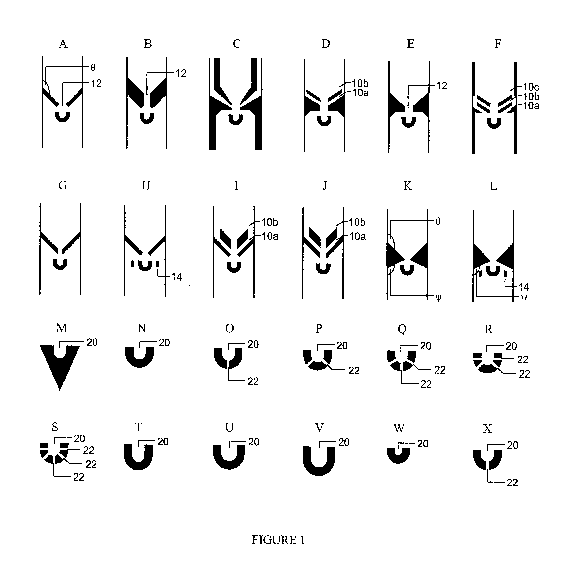
[0136]In order to increase the capture efficiency, decrease the size selectivity of the trap design, and attempt to characterize how trap dimensions affect capture efficiencies for different cell types, a variety of different trap geometries were designed and tested on two different cell lines: K562 cells, a human erythroleukemic cell line with an average diameter of 18 microns, and nBAF3, a murine pro-B-cell line with an average diameter of 12 microns.

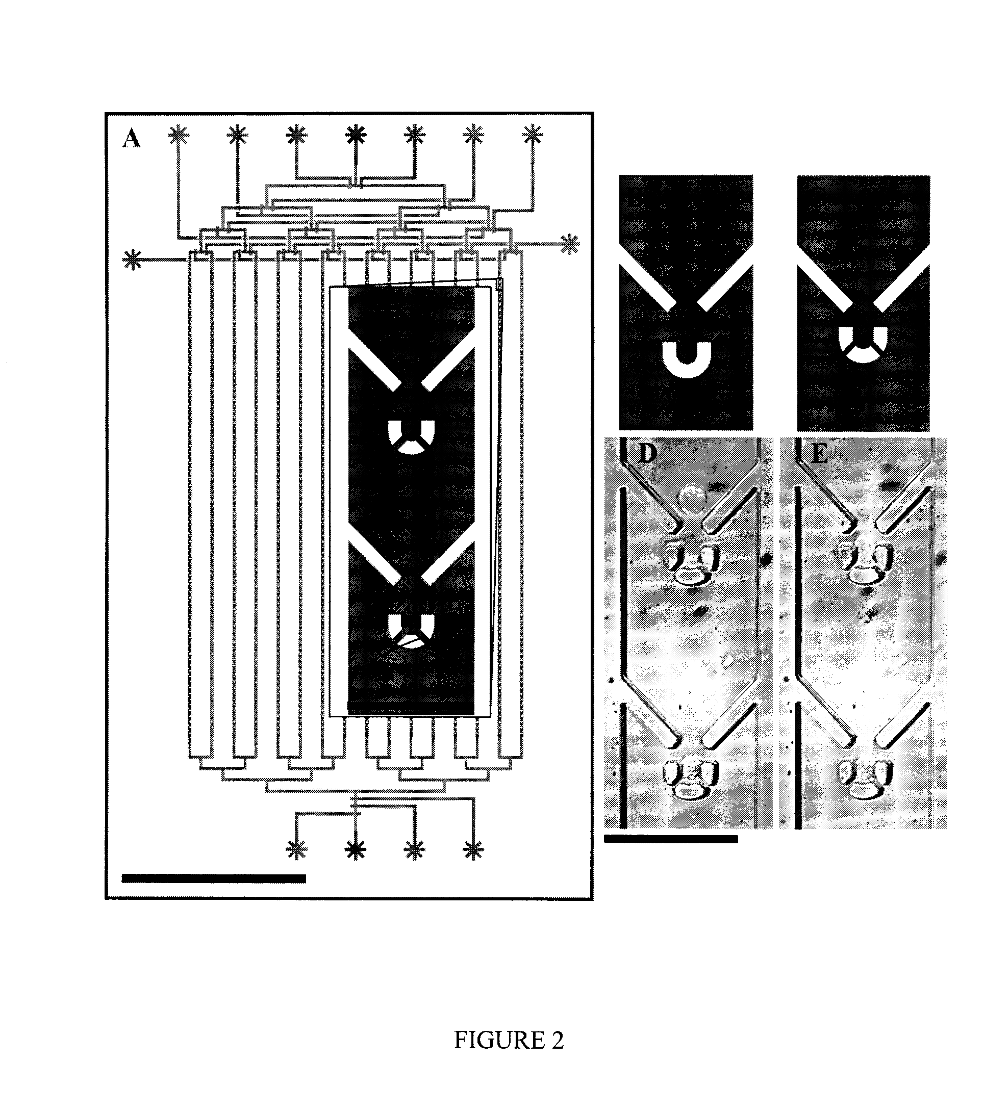
[0137]Ninety-six (96) different trap geometries were fabricated and tested in six independent devices. A schematic of cell capture testing device is shown in FIG. 2a. A single device is able to analyze 16 different trap geometries, each replicated 83 times in a single column, with 150 microns separating each trap. A binary demultiplexor (top of device) is used to select the desired trap geometry, and a microfluidic peristaltic pump (bottom of device) is used to pump the cells through the device. The scale bar is 500 micron...

example 1.3

Lysis

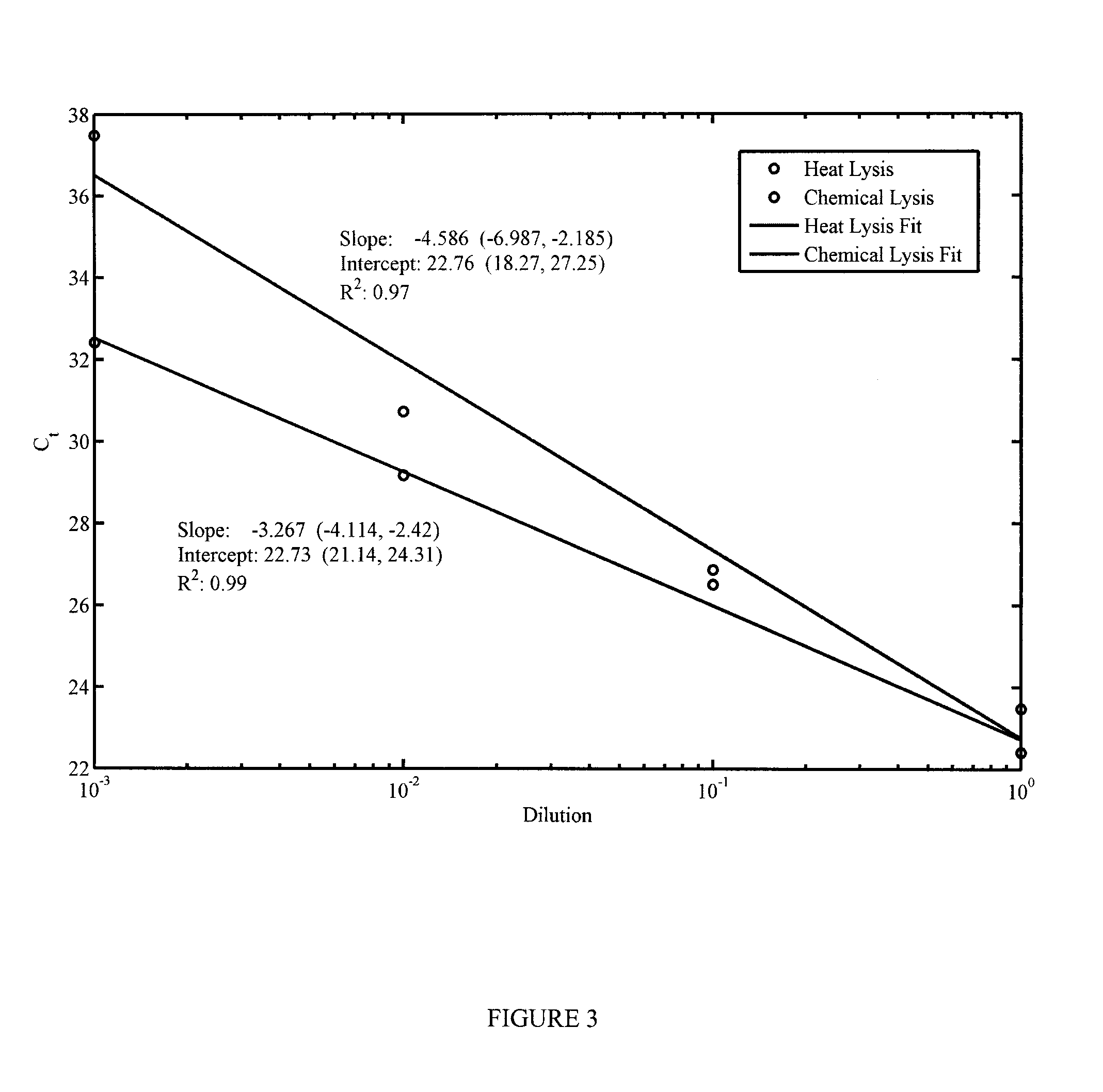
[0143]Two lysis methods were selected for their ease of integration into a microfluidic device: heat lysis, which involves heating the sample to 85° C. for 7 minutes, or a heat-inactivated chemical lysis buffer provided in the Invitrogen SuperScript® III CellsDirect cDNA Synthesis Kit. In order to assess the relative efficiency of each method on releasing miRNA from the cells and determine if further reactions would be inhibited by the chemical lysis buffer, a sample of K562 cells was serially diluted and lysed using each method. Released miRNA from these samples was subsequently reverse-transcribed into cDNA and the amount in each sample was quantified using qPCR.

[0144]The results from this test can be found in FIG. 3. The efficiency of the chemical lysis more closely resembles the expected value of approximately −3.3 cycles per 10-fold dilution. However, the increased benefit of the chemical lysis is reduced by the fact that using it requires the addition of an extra chamber ...


PUM

PropertyMeasurementUnit
volumeaaaaaaaaaa
volumesaaaaaaaaaa
volumeaaaaaaaaaa

Abstract

Microfluidic devices are provided for trapping, isolating, and processing single cells. The microfluidic devices include a cell capture chamber having a cell funnel positioned within the cell capture chamber to direct a cell passing through the cell capture chamber towards one or more a cell traps positioned downstream of the funnel to receive a cell flowing. The devices may further include auxiliary chambers integrated with the cell capture chamber for subsequent processing and assaying of the contents of a captured cell. Methods for cell capture and preparation are also provided that include flowing cells through a chamber, funneling the cells towards a cell trap, capturing a predefined number of the cells within the trap, interrupting the flow of cells, flowing a wash solution through the chamber to remove contaminants from the chamber, and sealing the predefined number of cells in the chamber.

Description

BACKGROUND OF THE INVENTION[0001]1. Field of Invention[0002]This invention relates to microfluidic devices. In particular, the invention relates to microfluidic devices and their uses and methods for assaying cells.[0003]2. Description of Related Art[0004]Single cells represent a fundamental biological unit. However, the vast majority of biological knowledge has emerged as a consequence of studying cell populations and not individual cells. Inevitably, there are fundamental and applied questions, such as those relating to transcriptional control of stem cell differentiation, intrinsic noise in gene expression, and the origins of disease, that may only be addressed at the single cell level. For example, single cell analysis allows for the direct measurement of gene expression kinetics, or for the unambiguous identification of co-regulated genes, even in the presence of de-synchronization and heterogeneity that could obscure population-averaged measurements. Similarly, single cell met...

Claims


Application Information

Patent Timeline
Patent Type & Authority Applications(United States)
IPC IPC(8): C12Q1/68G01N1/34
CPCG01N1/34C12Q1/6806C12M23/16C12Q1/6844G01N33/5005G01N33/54366G01N33/569B01L3/502753B01L7/52B01L2200/0668B01L2300/0816B01L2400/086B01L3/502761
Inventor HANSEN, CARL LARS GENOBLEVANINSBERGHE, MICHAELWHITE, ADAMPETRIV, OLEHLEAVER, TIMSINGHAL, ANUPAMBOWDEN, WILLIAMLECAULT, VERONIQUEDA COSTA, DANWU, LEORUSSELL, GEORGIASIKORSKI, DAREK
Owner THE UNIV OF BRITISH COLUMBIA
Why Eureka
Try Eureka