Skeletal muscle augmentation utilizing muscle-derived progenitor compositions, and treatments thereof

Active Publication Date: 2016-10-20
UNIVERSITY OF PITTSBURGH +1
View PDF3 Cites 0 Cited by
  • Summary
  • Abstract
  • Description
  • Claims
  • Application Information

AI Technical Summary

Problems solved by technology

These methods result in the enrichment of MDCs that have long-term survivability after transplantation or introduction into a site of soft tissue.

Method used


Image

  • Skeletal muscle augmentation utilizing muscle-derived progenitor compositions, and treatments thereof
  • Skeletal muscle augmentation utilizing muscle-derived progenitor compositions, and treatments thereof

Examples


example 1

MDC Enrichment, Isolation and Analysis According to the Pre-Plating Method

[0064]MDCs were prepared as described (U.S. Pat. No. 6,866,842 of Chancellor et al.). Muscle explants were obtained from the hind limbs of a number of sources, namely from 3-week-old mdx (dystrophic) mice (C57BL / 10ScSn mdx / mdx, Jackson Laboratories), 4-6 week-old normal female SD (Sprague Dawley) rats, or SCID (severe combined immunodeficiency) mice. The muscle tissue from each of the animal sources was dissected to remove any bones and minced into a slurry. The slurry was then digested by 1 hour serial incubations with 0.2% type XI collagenase, dispase (grade II, 240 unit), and 0.1% trypsin at 37° C. The resulting cell suspension was passed through 18, 20, and 22 gauge needles and centrifuged at 3000 rpm for 5 minutes. Subsequently, cells were suspended in growth medium (DMEM supplemented with 10% fetal bovine serum, 10% horse serum, 0.5% chick embryo extract, and 2% penicillin / streptomycin). Cells were then ...

example 2

MDC Enrichment, Isolation and Analysis According to the Single Plate Method

[0068]Populations of rapidly- and slowly-adhering MDCs were isolated from skeletal muscle of a mammalian subject. The subject may be a human, rat, dog or other mammal. Biopsy size ranged from 42 to 247 mg.

[0069]Skeletal muscle biopsy tissue is immediately placed in cold hypothermic medium (HYPOTHERMOSOL® (BioLife) supplemented with gentamicin sulfate (100 ng / ml, Roche)) and stored at 4° C. After 3 to 7 days, biopsy tissue is removed from storage and production is initiated. Any connective or non-muscle tissue is dissected from the biopsy sample. The remaining muscle tissue that is used for isolation is weighed. The tissue is minced in Hank's Balanced Salt Solution (HBSS), transferred to a conical tube, and centrifuged (2,500×g, 5 minutes). The pellet is then resuspended in a Digestion Enzyme solution (Liberase Blendzyme 4 (0.4-1.0 U / mL, Roche)). 2 mL of Digestion Enzyme solution is used per 100 mg of biopsy t...

example 3

Augmentation of Skeletal Muscle with MDCs

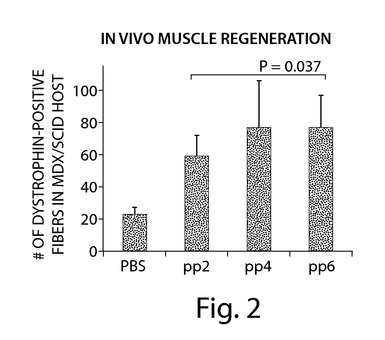
[0074]Populations of human muscle derived cells (hMDCs) isolated from human muscle biopsies by way of the preplate technique were tested to show that hMDCs had similar myogenic and regenerative characteristics to their murine counterparts.

[0075]Methods.

[0076]Pre-Plate Technique: This technique is disclosed throughout the application and specifically, above in Example 1.

[0077]Isolation and Cell Culture: Candidate populations were obtained using the pre-plate technique. These cells were grown in EGM™-2 media (Cambrex) at a density of 600 cells / cm2 and passaged every 72-96 hours before confluence under standard conditions (5.0% CO2, 37° C.). Flow Cytometry: hMDC were analyzed for the presence of the cell surface cluster of differentiation markers CD34, CD56, CD144, and CD146.

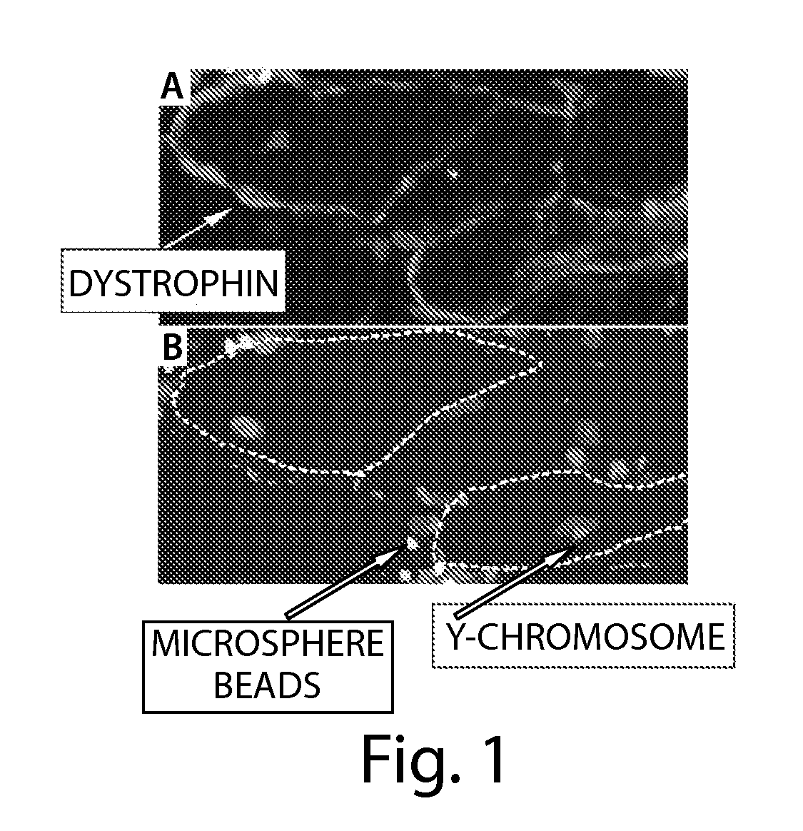
[0078]Immunochemistry: hMDC were stained for desmin, myosin heavy chain, and dystrophin.

[0079]Bioinformatic Live Cell Imaging: Cells were grown in a cell culture system with...


PUM

PropertyMeasurementUnit
temperatureaaaaaaaaaa

Abstract

The present invention provides muscle-derived progenitor cells that show long-term survival following transplantation into body tissues and which can augment soft tissue following introduction (e.g. via injection, transplantation, or implantation) into a site of soft tissue. Also provided are methods of isolating muscle-derived progenitor cells, and methods of genetically modifying the cells for gene transfer therapy. The invention further provides methods of using compositions comprising muscle-derived progenitor cells for the augmentation and bulking of mammalian, including human, soft tissues in the treatment of various cosmetic or functional conditions, including malformation, injury, weakness, disease, or dysfunction. The invention also relates to novel uses of muscle-derived progenitor cells for the treatment of cosmetic or functional conditions, including, but not limited to skeletal muscle weakness, muscular dystrophy, muscle atrophy, spasticity, myoclonus and myalgia. The invention also relates to the novel use of MDCs for the increase of skeletal muscle mass in athletes or other organisms in need of greater than average skeletal muscle mass.

Description

RELATED APPLICATIONS[0001]This application is a continuation-in-part of U.S. patent application Ser. No. 11 / 505,734, filed on Aug. 17, 2006, which is a divisional application of U.S. patent application Ser. No. 09 / 549,937, filed on Apr. 14, 2000; now U.S. Pat. No. 7,115,417; which is a continuation in part of U.S. patent application Ser. No. 09 / 302,896, filed on Apr. 30, 1999, now U.S. Pat. No. 6,866,842; which claims priority from U.S. Provisional Patent Application No. 60 / 083,917, filed on May 1, 1998. This application also claims benefit of priority from U.S. Provisional Application No. 60 / 977,450, filed on Oct. 4, 2007. All of the above applications are incorporated herein, by reference, in their entireties.FIELD OF THE INVENTION[0002]The present invention relates to muscle-derived progenitor cells (MDCs) and compositions of MDCs and their use in the augmentation of body tissues, particularly skeletal muscle. In particular, the present invention relates to muscle-derived progeni...

Claims


Application Information

Patent Timeline
Patent Type & Authority Applications(United States)
IPC IPC(8): A61K35/34A61P1/00A61P9/00A61P11/00A61P13/00A61P15/00A61P17/00A61P21/00A61P25/00A61P43/00C12N5/077
CPCA61K35/34C12N2510/00C12N2510/02C12N2501/70C12N2533/54C12N5/0659A61K35/12A61P1/00A61P1/02A61P1/16A61P1/18A61P11/00A61P13/00A61P13/02A61P13/10A61P13/12A61P15/00A61P17/00A61P17/02A61P21/00A61P25/00A61P43/00A61P9/00C12N5/0658C12N2500/84A61K9/0019
Inventor PAYNE, THOMASJANKOWSKI, RONALDPRUCHNIC, RYANCHANCELLOR, MICHAEL
Owner UNIVERSITY OF PITTSBURGH
Why Eureka
Try Eureka