

In vivo live 3D printing of regenerative bone healing scaffolds for rapid fracture healing
- Summary
- Abstract
- Description
- Claims
- Application Information
AI Technical Summary
Benefits of technology
Problems solved by technology
Method used
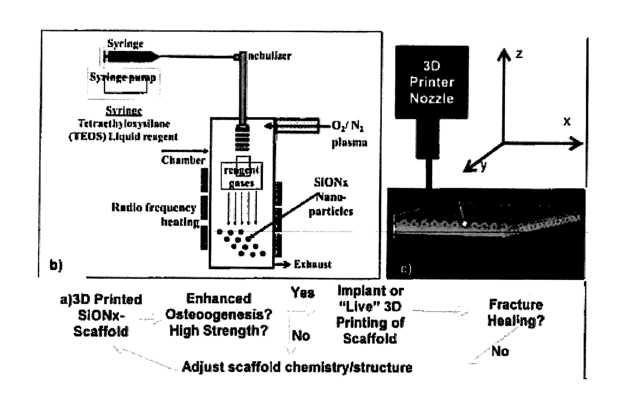
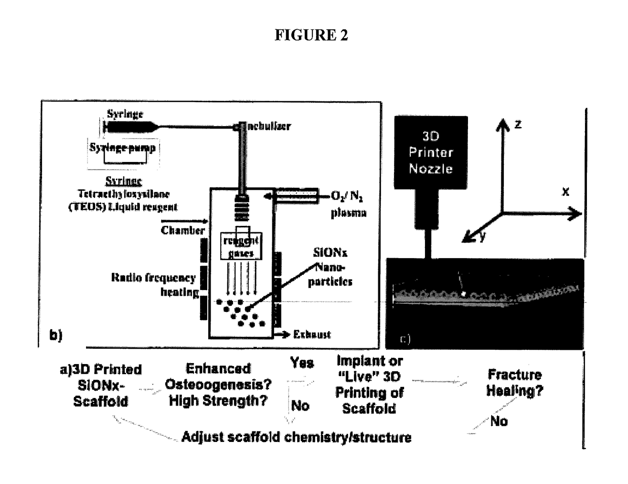
Image

Examples
example 1
3D Printed Biosilicate Scaffolds
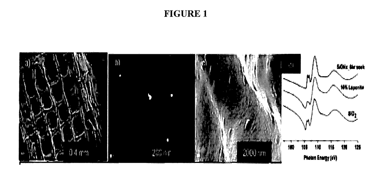
[0182]During initial attempts to synthesize porous scaffolds, materials were utilized that resembled the inorganic (synthetic HA) and organic (PLA) matrix for which they were later to be used. These scaffolds were able to promote the ingress of cells into their matrices, however, dense collagen and mineralized matrix formation was not observed within 28 days. Thus the central focus shifted to biosilicate nanoparticles (laponite nano-platelets) to fabricating structurally stable regenerative scaffolds to facilitate bone regeneration, ingress and complete replacement of lost bone.
[0183]Biosilicate nano-platelets offer unique advantages including the ability to modify the rheological properties of slurries to control scaffold fabrication during printing and also their afore-mentioned osteoinductive properties for the inorganic component of the scaffold. The inorganic component (biosilicate nanoparticles or laponite, 10-30 nm diameter) and organic compone...
example 2
In situ Printing into Calvarial Bone and Bone Defect Repair
[0188]The present example demonstrates the utility of the present invention for providing a method for repairing and / or reducing a bone defect in an animal by direct deposit of the repairative, biocompatible materials (“bio-Ink”) disclosed herein. The methods are demonstrated to provide high fabrication precision in formation of a tissue scaffold in vivo, the tissue scaffolds being characterized by pores having high resolution definition of between about 0.3 mm to 0.5 mm. The in situ bone tissue repair methods are demonstrated to provide an immediate point of care technique for providing bone repair and / or bone restoration at a defect, thus eliminating and / or reducing the several disadvantages and / or limitations associated with existing bone tissue defect repair methods.
[0189]Methodology
[0190]Micropatterning and microporosity in the scaffolds provided in the present techniques provide high resolution and precisely sized scaf...
example 3
The Effect of a Silicate-containing materials (Laponite) and MAG on Physical Properties of bio-Ink
[0285]In example 2, it is shown that by incorporating sucrose to an already established polymer nanosilicate, an ISP compatible material (bio-ink) was created. The in-situ printing concept is also demonstrated and validated as a useful bone-defect repair method.
[0286]The physical behavior of bio-ink and the role of its main components in this behavior are demonstrated. Printed scaffold is demonstrated to be resorbed faster than the rate that bone is healed. The degradation properties of the bio-ink is demonstrated in the present example.
[0287]It is hypothesized that degradation rate, swelling rate, and protein release of bio-ink can be adjusted by controlling LP and MAG concentration.
[0288]Implant integration has two steps; bony union between the surface of the implant and native bone segments, and then graft remodeling and resorption in coordination with new bone formation [46]. Graft ...
PUM
| Property | Measurement | Unit |
|---|---|---|
| Temperature | aaaaa | aaaaa |
| Length | aaaaa | aaaaa |
| Length | aaaaa | aaaaa |
Abstract
Description
Claims
Application Information