Disposable Single Cell Array for Personalized Diagnostics
- Summary
- Abstract
- Description
- Claims
- Application Information
AI Technical Summary
Benefits of technology
Problems solved by technology
Method used
Image
Examples
example 1
and Methods
[0079]PDAC-Modified PDMS Stamps.
[0080]Polydimethylsiloxane (PDMS Sylgard 184) stamps were prepared by casting PDMS pre-polymer and curing agent against solid masters generated using photolithography. The unmodified PDMS stamp had microposts with diameter of 10 μm. The unmodified PDMS stamp was immersed in polydiallyldimethyl ammonium chloride (PDAC) (100,000-200,000 Da) solution for 15 min at room temperature, rinsed by deionized water, and dried in a gentle nitrogen stream.
[0081]Cells.
[0082]Fibroblast cells, human glioblastoma cells (A172) and breast cancer cells (MCF7) were cultured in standard conditions (5% CO2 in air at 37° C.) in RPMI-1640 medium supplemented with 10% (v / v) fetal bovine serum and 1% (v / v) penicillin / streptomycin. For obtaining individual cells, cells were trypsinized.
example 2
n of Single-Cell Array Precursor
[0083]Bare paper was covered with ink by printing 1 to 5 dark layers on normal printing paper using a Cannon MF4890dw printer. Paper with different layers of ink was found to have different surface roughness. FIGS. 3A-D show scanning electron microscope (SEM) images of bare paper, and paper covered with one, three and five layers of ink. As layers of ink increase, the paper surface became smoother, and fibers in the paper could not be observed clearly. The PDAC-modified PDMS stamp containing 10 μm-microposts was brought into contact with the ink covered paper, and a slight pressure was applied on the stamp for 15 s to ensure conformal contact between the stamp and ink-covered paper, after which the stamp was peeled off from the paper. After removing the PDMS stamp, the PDAC layer was transferred on the ink-covered paper due to electrostatic attraction between the negatively-charged ink and the positively-charged PDAC.
example 3
of Single-Cell Array
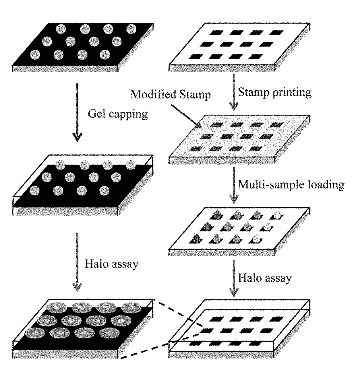
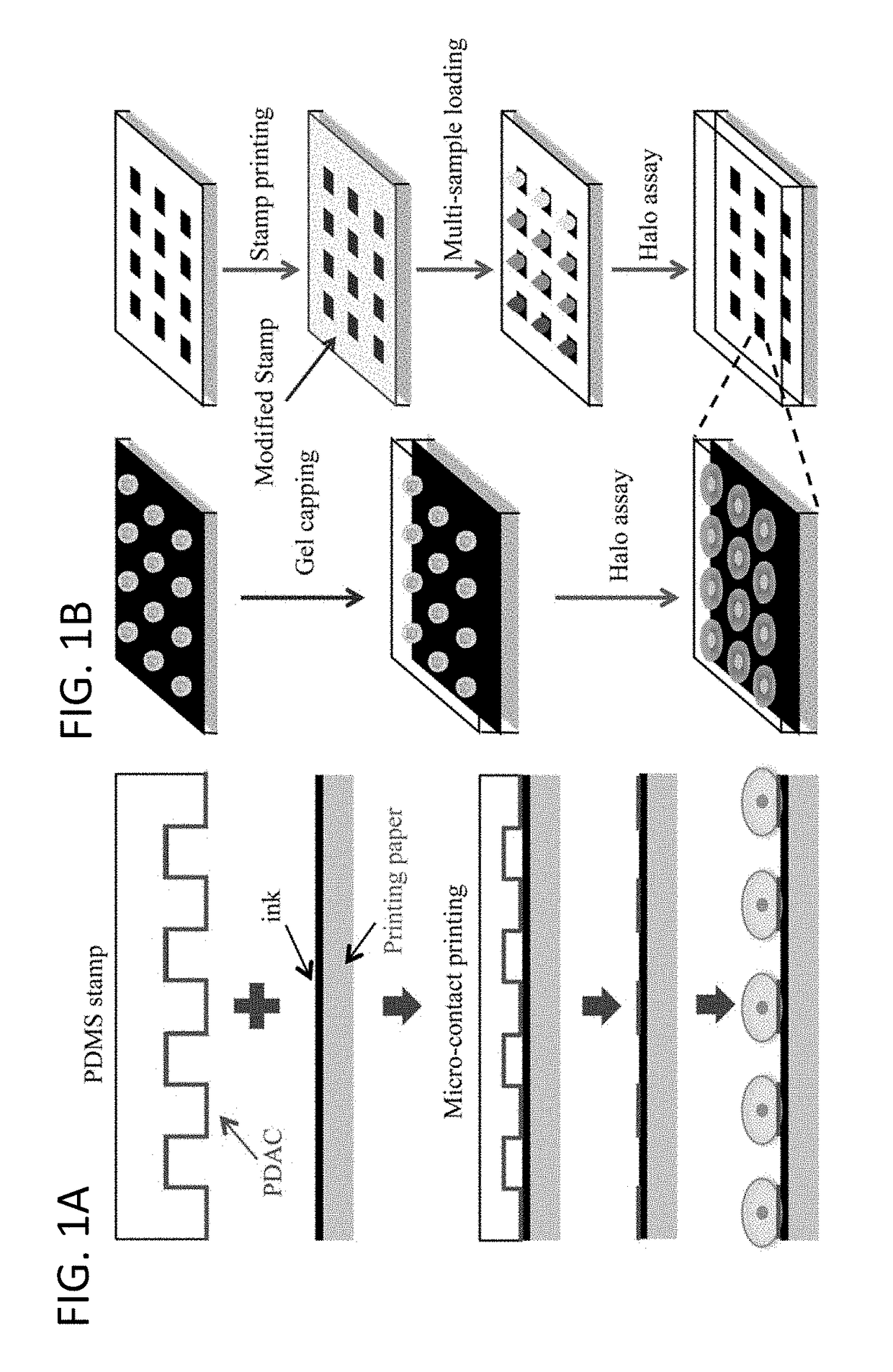
[0084]After exposure to anticancer treatment (radiation or chemotherapy drug), cells were seeded on the cell array precursor at a density of 1×10⁶ cells / ml, forming ordered single-cell arrays (FIG. 1A). The quality of single cell arrays was primarily determined by surface roughness of the substrate (FIGS. 3E-H). As the paper surface became smoother, more cells were attached, and the array became more ordered. The attachment probability was derived as the ratio between adsorbed cells and the number of micro-patches. The probability of single cell array increased as the number of layers increased from 25±3.1%, 70±3.5%, to 87±6.4% for 1, 3 and 5 layers of ink, respectively. Five layers of ink provided a high quality substrate for single cell array formation, making this method ideal for paper-based single-cell halo assay.
[0085]After incubation for 30 min, unattached cells were rinsed away by using PBS. In order to confirm that arrayed cells were still alive, cells w...
PUM
| Property | Measurement | Unit |
|---|---|---|
| Size | aaaaa | aaaaa |
| Size | aaaaa | aaaaa |
| Fluorescence | aaaaa | aaaaa |
Abstract
Description
Claims
Application Information
 - R&D
- Intellectual Property
- Life Sciences
- Materials
- Tech Scout
- Unparalleled Data Quality
- Higher Quality Content
- 60% Fewer Hallucinations
Browse by: Latest US Patents, China's latest patents, Technical Efficacy Thesaurus, Application Domain, Technology Topic, Popular Technical Reports.
© 2025 PatSnap. All rights reserved.Legal|Privacy policy|Modern Slavery Act Transparency Statement|Sitemap|About US| Contact US: help@patsnap.com


