Method and stationary phase for isolating extracellular vesicles from biological material

Pending Publication Date: 2020-10-08
ADVANCED EXTRACELLULAR VESICLE APPL SRL
View PDF0 Cites 1 Cited by
  • Summary
  • Abstract
  • Description
  • Claims
  • Application Information

AI Technical Summary

Benefits of technology

[0022]Several reports recently published show that in physiological solution of PBS the ZP of extracellular vesicles is between −17 and −35

Problems solved by technology

Therefore, many techniques for EV isolation from biological material have been proposed to date, although many of them are not very efficient or standardizable (Thery C, et al., Curr Protoc Cell Biol, 2006; Gardiner C, et al.
2016), due to the fact that alternative density gradient centrifugations and precipitation techniques use chemical agents interfering with EV yield, composition and integrity; immunoaffinity capture leads to differential isolation of EV subpopulations (hence low heterogeneity) and it is expensive because it involves antibodies; exclusion chromatography requi

Method used


Image

  • Method and stationary phase for isolating extracellular vesicles from biological material
  • Method and stationary phase for isolating extracellular vesicles from biological material
  • Method and stationary phase for isolating extracellular vesicles from biological material

Examples


example 1

on of the Nickel Ion Functionalized Beads

[0102]The procedure for beads functionalization is carried out as follows:[0103]commercially available NiNTA Sepharose High Performance beads (GE Healthcare product code 71-5027-67 or 17-5268-01) was taken as starting beads; the net charge values were measured and resulted in the range from 3 to 25 mV at 22-25° C. in PBS, as detected using the Malvern Zetasizer instrument.[0104]starting beads are subjected to stripping by one or more washing with an aqueous solution supplemented with 200-300 mM NaCl or KCl, 100-300 mM EDTA or EGTA, 300-500 mM Imidazole, or a solution containing cationic chelating agents with a wide pH range (generally between 5 and 8), and one or more washing with bi-distilled water (18.2 MO cm−1);[0105]A 40 mg / ml suspension of starting beads (NiNTA Sepharose High Performance, GE Healthcare, 17-5268-01, 34 μm known nominal size), previously subjected to stripping treatment aliquoted in 50 ml tubes is re-suspended in a double ...

example 2

ion Method from a Biological Sample

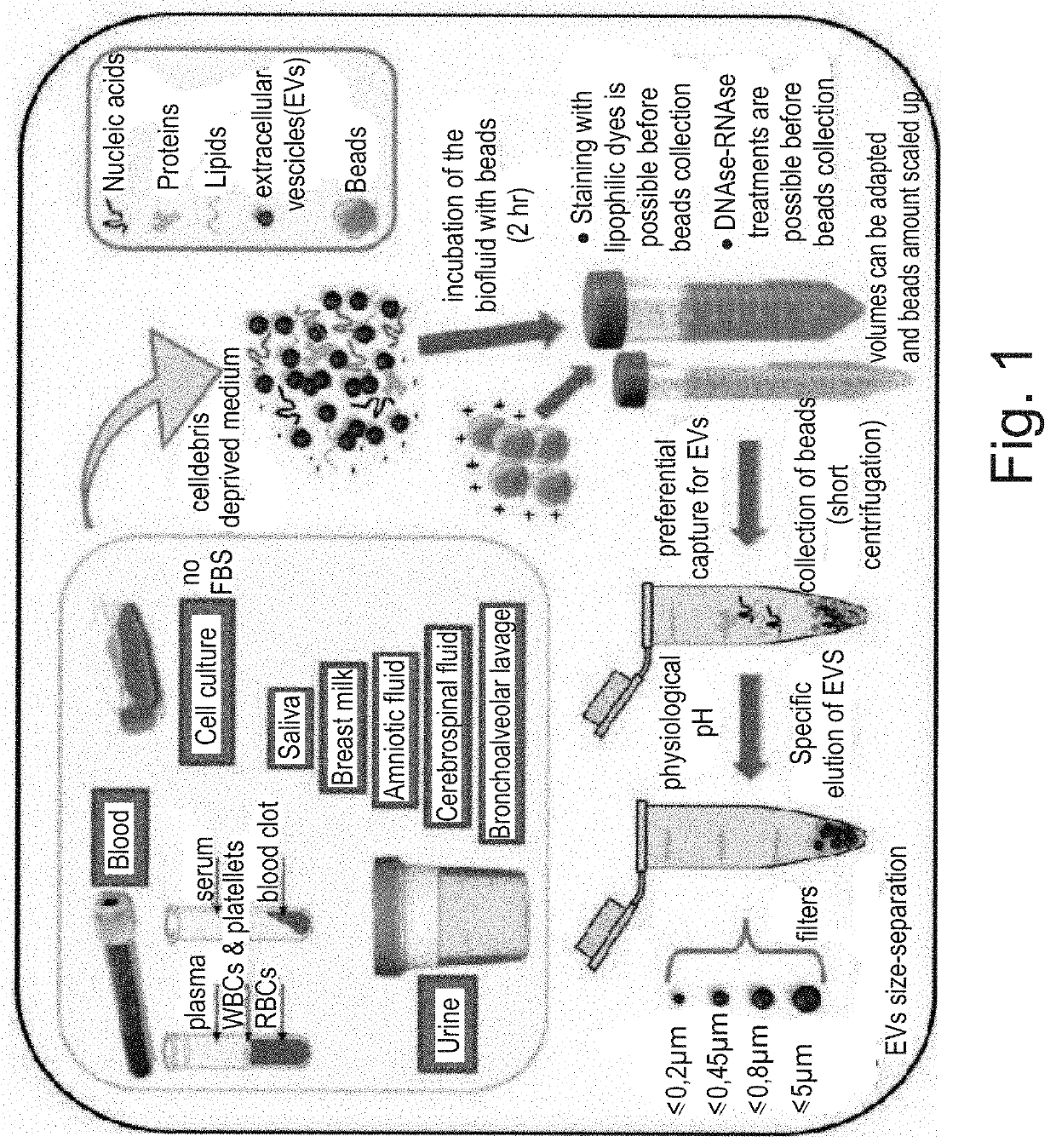
[0113]CBeads can be added dropwise to the surface of a biological liquid (clarified by cellular debris by 2800 rcf centrifugation) collected in tubes of any size and incubated at room temperature for 30 minutes in a volumetric ratio of 20 μl / ml.

[0114]Biological fluid may be cell culture medium mostly containing 1.5% fetal bovine serum (FBS)-PBS at pH 7.4 dilution is allowed if FBS percentage is higher; liquid biopsy sample (whole blood or serum or plasma, urine, cerebrospinal fluid, milk, saliva).

[0115]EV isolation from the biological sample is carried out as follows:

[0116]CBeads are incubated with the biological sample with gentle orbital rotation (300-600 rpm) for 30 minutes at room temperature, at the end of which the tube is stabilized in a vertical position to allow gravity settling or weak centrifugation (100-400 rcf) of the CBeads (7-15 minutes) at the bottom of the tube.[0117]The supernatant is completely sucked up and discarded.

[0118]EV pu...

example 3

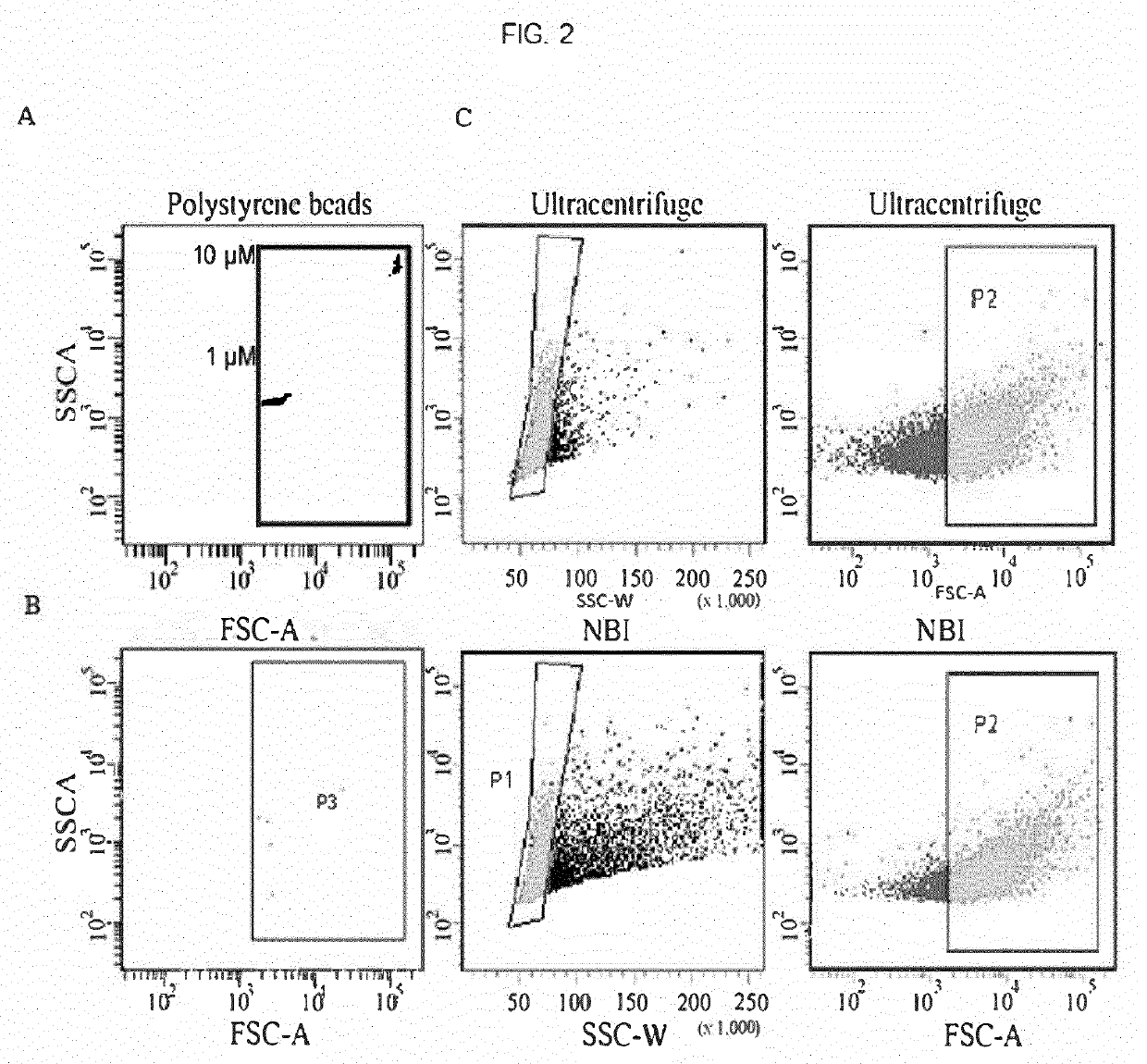
n of Particle Number (>0.5 μm) Isolated by UC or by NBI

[0128]NBI was applied to isolate vesicles released from U87 gliomas cells and the particle number with ≥0.5 μm in diameter (as estimated by flow cytometry) is comparable to that obtained using differential UC, unlike few events captured by non-functionalized beads (FIG. 2A).


PUM

PropertyMeasurementUnit
Temperatureaaaaaaaaaa
Molar densityaaaaaaaaaa

Abstract

The present invention describes a method for isolating extracellular vesicles (EVs) from different biological fluids, said nickel-based isolation method (NBI) is fast, scalable and allows for the purification of dimensionally heterogeneous EVs at physiological pH, preserving their integrity and stability in solution.

Description

FIELD OF THE INVENTION[0001]The present invention relates to the field of extracellular vesicles isolation from cell culture media or biological fluids.BACKGROUND[0002]Extracellular vesicles (EVs) are membranous particles that include exosomes (80-200 nm), microvesicles (100-600 μm) and apoptotic bodies (800-5000 nm). This classification is mainly based on vesicle size, although different mechanisms have been proposed for their biogenesis. In oncology, EVs hold potential to study the modulation of tumor microenvironment and immune surveillance, to capture information and / or biomarkers released from the tumor, or to be exploited as carriers of therapeutics.[0003]Biological and biomedical research is increasingly focused on the role of EVs in different physiological and pathological processes. Therefore, many techniques for EV isolation from biological material have been proposed to date, although many of them are not very efficient or standardizable (Thery C, et al., Curr Protoc Cell...

Claims


Application Information

Patent Timeline
IPC IPC(8): B01J47/014C12N15/10C12Q1/686G01N1/40B01J39/19B01J39/07B01D15/36B01D15/38
CPCB01D15/3828C12N15/1003G01N1/4077C12N15/1096B01J47/014B01D15/362C12Q1/686B01J39/19B01J39/07B01J20/286B01J20/3265C12Q1/6806C12Q1/6886B01J20/3212C12Q2527/125C12Q2563/149
Inventor D'AGOSTINO, VITO GIUSEPPEPROVENZANI, ALESSANDROQUATTRONE, ALESSANDROZUCAL, CHIARANOTARANGELO, MICHELAMODELSKA, ANGELIKAPESCE, ISABELLA
Owner ADVANCED EXTRACELLULAR VESICLE APPL SRL
Why Eureka
Try Eureka