In vitro diagnosis device comprising beads and uses thereof

a technology of in vitro diagnosis and beads, which is applied in the field of immunodeficiency diagnosis to achieve the effect of improving the sensitivity and specificity of detection and reducing the quantity of used analytes

Pending Publication Date: 2020-12-31
DIAGAST
View PDF1 Cites 1 Cited by
  • Summary
  • Abstract
  • Description
  • Claims
  • Application Information

AI Technical Summary

Benefits of technology

[0063]As indicated above, in order to improve the sensitivity and the specificity of the detection and / or the identification of an antigen and / or an antibody and to reduce the quantity of used analytes, the inventors implemented a device similar to the one disclosed in the patent application WO2013 / 186482 which further comprises beads disposed on the reaction area, wherein antibodies and / or antigens are fixed or adsorbed on the bead surface.

Problems solved by technology

The principal risk is the possibility of bringing together an antibody and its erythrocyte antigen in the recipient's body (person receiving the transfusion).
The consequences of such an immunological reaction may range from inefficient transfusion with no clinical sign, to a slight clinical reaction (anxiety, shivers), serious clinical reaction (shock, hemoglobinurea, renal failure) or dramatic clinical reaction (shock, disseminated intravascular hemolysis) resulting in death.
Blood groups become even more complex and numerous when several antigen systems are used.
In practice, it is not possible to take into consideration all these antigens when carrying out a transfusion, as obtaining the right blood group at the right moment would not be possible, especially as some antigenic combinations are extremely rare.
However, it is an insensitive method.
The test cannot be conducted for weakly or rarely reactive antigens from which the results are difficult to interpret, and additionally, a low titer of anti-A (anti-ABO1) or anti-B (anti-ABO2) could lead to false positive or false negative results.
However, it is not reliable enough to completely ensure safe transfusion.
The purpose of centrifugation is to ensure enhanced chemical interactions, particularly for weaker antibodies to react, thus leading to agglutination.
However, in infants, reverse grouping is somewhat difficult to perform, since they produce insufficient amounts of antibodies to be determined.
Since this test is a manual method, its use for high throughput analysis is very limited.
However, since microplating techniques need a centrifuging phase followed by an agitation step, there is therefore the risk of undoing weak agglutination without succeeding in resuspending strong agglutination.
However, the main risk consists of not detecting some agglutinations especially during the plasmatic test of the ABO group due to dissociation by shearing forces of small agglutinates as they pass into the gel.
Also, there is a major disadvantage to all these techniques because they need centrifuging step to decant the red blood cells or have them pass through the gel, a restricting step which adds considerable time and analysis costs and which needs the use of bulky centrifuges which are difficult to handle.
This method has significant sensitivity and specificity problems, because when the sample is deposited it spreads and imbibes the porous membrane, and many analytes are lost in the dead volume of the porous membrane or pass outside the capture area.
However, with these devices there is still the problem of centrifugal diffusion from a spot, and the revelation elements which pass through the capture spot and which are not fixed to it will be able to diffuse centrifugally in the hydrophilic porous membrane and be stored at the periphery of the spot.
Another major problem of existing immunofiltration devices today is control of the motion speed of the sample through the membrane and in some cases pre-incubation time.
Such mechanical approaches are difficult to execute using a robot since they need development and use of dedicated systems.
Another method, described in EP0334015, consists in using an extra membrane subjacent to the first to control flow, but the proposed device fails to resolve the problems linked to diffusion of the sample and the revelation element in the hydrophilic porous membrane.
The value of serological HPA phenotyping is limited as often too few platelets can be obtained from thrombocytopenic patients, and reliable serotyping reagents for HPA antigens, other than HPA-1a and HPA-5b, are rare.
However, unmatched platelets, however, are often used due to the unavailability of matched platelets.
It is a common complication in patients receiving multiple platelet transfusions.
However, proficiency in anti-platelet antibody detection depends on a variety of factors, including techniques, cell panels and operator experience which may render some of the above-mentioned techniques costly, difficult to implement and less reliable.
Nevertheless, these tests do not suit all the situations and every test has limits, which must be known and taken into account during its selection.
The major disadvantages are the health and safety risks posed by the use of radiation and the time and expense associated with maintaining a licensed radiation safety and disposal program.
However, these tests are not suitable for the screening of large numbers of samples.
It remedies several disadvantages arising from the above-mentioned tests, particularly arising from immunofiltration tests by:preventing returns of revelation agent responsible for false positivesheightened sensitivity from using lower volumesminiaturisation and automation of the systemcontrol of the kinetics of reactions without need for mechanical handling of the device.
To compensate this “loss” of capturing agent, it is necessary to add a big quantity of capturing agent which may increase the cost of this diagnosis test.
However, the described method does not use a device comprising a hydrophilized porous membrane and does not deal with the problem relating to the absorption of the capturing agent through the membrane.

Method used


Image

  • In vitro diagnosis device comprising beads and uses thereof
  • In vitro diagnosis device comprising beads and uses thereof
  • In vitro diagnosis device comprising beads and uses thereof

Examples


example 1

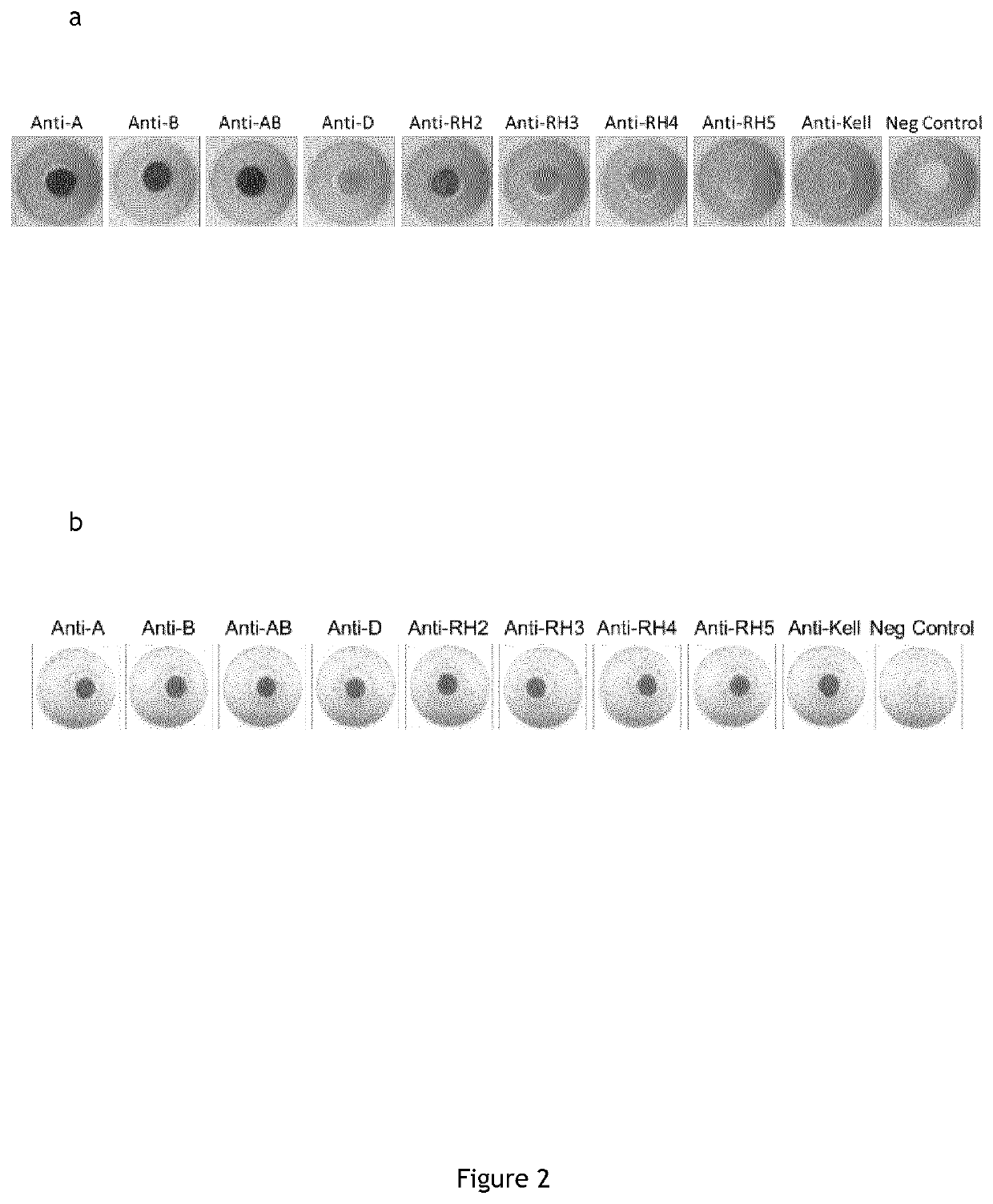
Cell Typing and Phenotyping in the In Vitro Device With Direct and Indirect Coupling of Anti-Red Blood Cells Antibodies on Beads Via Anti-Immunoglobulins IgM and IgG

1. Protocol for Preparing the In Vitro Device That Comprises on the Surface of the Reaction Area the Anti-Red Blood Cells Antibodies Directly Spotted on the Membrane

1.1.Materials for the Preparation of the In Vitro Device

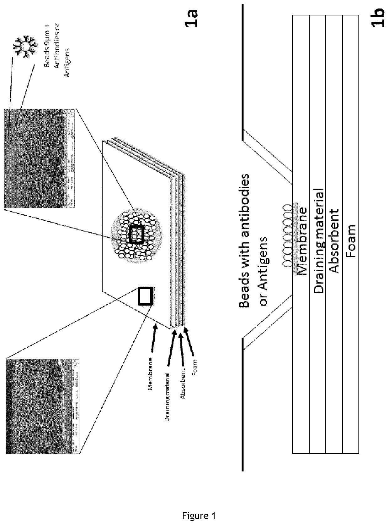
[0317]Membrane “POREX” 0.6 mm hydrophobic with 9 to 12 μm porosity; Cotton;[0318]Cleanis;[0319]Hydrophilization buffer (1% triton, 1% Green dye, phosphate buffered saline).[0320]Wash Buffer (TLA composition);[0321]Dilution buffer (Chromasolcoombs);[0322]Antibodies (Anti-ABO01, Anti-ABO02, Anti-ABO03, Anti-RH1, Anti-RH2, Anti-RH3, Anti-RH4, Anti-RHS, Anti-KEL1, Negative Control).

1.2.Preparation Mode of the Reaction Medium

[0323]Deposit 1 μl of the hydrophilization solution in each well;[0324]Drying 16 hours at 37° C. and 10% humidity;[0325]Deposit 2 μl of antibody per well;[0326]Drying 72 hours at 37° C. a...

example 2

Coupling of Red Blood Cells Antibodies Via Affinity Proteins for Antibodies

1. Indirect Antibodies Coupling

[0349]On 1 ml of homogenized 4% latex beads (Thermo Fisher latex Aldehyde / Sulfate 9 μm 4% w / v), 5 or 2.5 μg of Protein AGL are dissolved in 3 ml PBS1× and are added. The solution is end over end mixed overnight to ensure coupling.

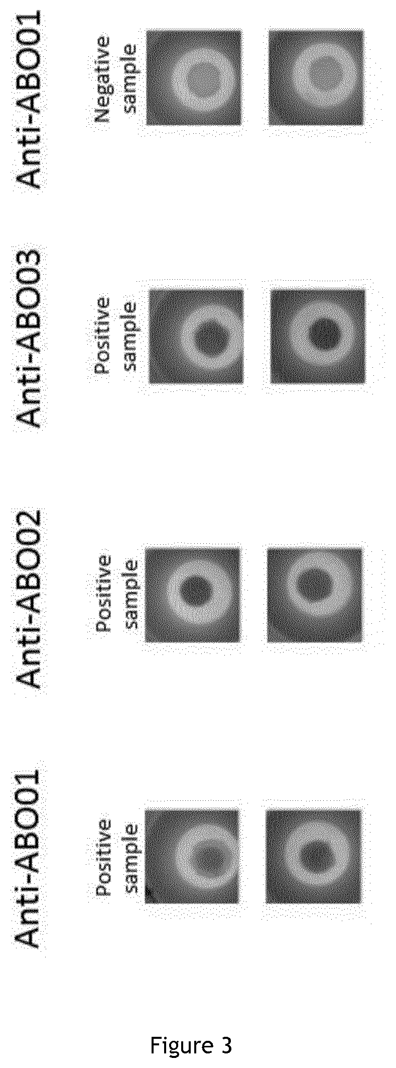
[0350]On 1 ml of centrifugated 4% beads (Thermo Fisher latex Aldehyde / Sulfate 9μm 4% w / v) coupled with protein AGL, 3 ml of 1× phosphate buffered saline are added and 1 ml of Anti-ABO01 antibody (91 13 D10 Diagast clone) or Anti-ABO02 antibody (96 21 A8 Diagast clone) or Anti-ABO03 (152D12 Diagast clone) are dissolved. The solution is end over end mixed overnight to ensure coupling.

[0351]The coupled beads are then centrifuged 5 min at 2500 g. Finally the bead pellet is resuspended in the conservation buffer (phosphate buffered saline supplemented with 0.5% bovine serum albumin and 0.25M saccharose) to get back the introduced bead solution weight.

2. Reac...

example 3

of Antibodies of Red Blood Cells on Beads Having Different Chemical Functional Groups on Their Surface and Different Size in Different Reactional Conditions

1. Chloromethyl Group

1.2. Antibodies Coupling

[0357]On 500 μl of homogenized chloromethyl beads (Thermo Fisher chloromethyl latex beads 4% w / v, 2.0 μm) 1.5 ml of phosphate buffer is added and the obtained solution is centrifuged at 3000 g for 20 min. The supernatant is discarded and the bead pellet is then resuspended with a mix of 950 μl of phosphate buffered saline and 50 μl of concentrated CPC51 or CP6D4 (Diagast AHG).The antibody bead solution is incubated overnight at room temperature on a tube rocker.

[0358]After AHG chemical coupling, the bead solution is centrifuged at 3000 g for 15 min and the supernatant is discarded. The beads are washed with 1.5 ml of phosphate buffered saline and the solution is centrifuged at 3000 g for 10 min. The supernatant is discarded and the excess active groups are blocked by adding 1 ml of 1 M...


PUM

PropertyMeasurementUnit
sizeaaaaaaaaaa
sizeaaaaaaaaaa
sizeaaaaaaaaaa

Abstract

The present invention relates to an in-vitro diagnosis device for detecting and/or identifying antigen and/or antibody from a sample of biological fluid, particularly from a sample of blood or sample of blood components. The invention also relates to different uses of this device such as the detection and/or the identification of red blood cells antigens, platelet antigens, viralantigens, bacterial antigens, parasite antigens, the detection and/or the identification of anti-red blood cells antibodies, antiplatelet antibodies, antiviral antibodies, antibacterial antibodies and antiparasitic antibodies.

Description

FIELD OF THE INVENTION[0001]The present invention is in the field of immune diagnosis. Particularly, the present invention relates to an in vitro diagnosis device for detecting and / or identifying antigen and / or antibody from a sample of biological fluid, particularly from a sample of blood or sample of blood components. The invention also relates to various uses of this device such as the detection and / or the identification of red blood cells antigens or anti-red blood cells antibodies, platelet antigens or antiplatelet antibodies, viral antigens or antiviral antibodies, bacterial antigens or antibacterial antibodies, parasite antigens or antiparasitic antibodies. In vitro methods using the device of the invention are also a part of the present invention.BACKGROUND OF THE INVENTION[0002]Immunodiagnostic assays are methods that are available in several formats of test and usable to detect antibodies, antigens or combination of both. Commonly, these detection tests are based on the us...

Claims


Application Information

Patent Timeline
Patent Type & Authority Applications(United States)
IPC IPC(8): G01N33/543G01N33/538G01N33/68
CPCG01N33/5436G01N33/6854G01N33/538G01N2333/02G01N2333/20G01N2333/445
Inventor BETREMIEUX - MERLIER, CHRISTINEAMNIAI, LAZIZASCHELPE, JULIEN
Owner DIAGAST
Why Eureka
Try Eureka