

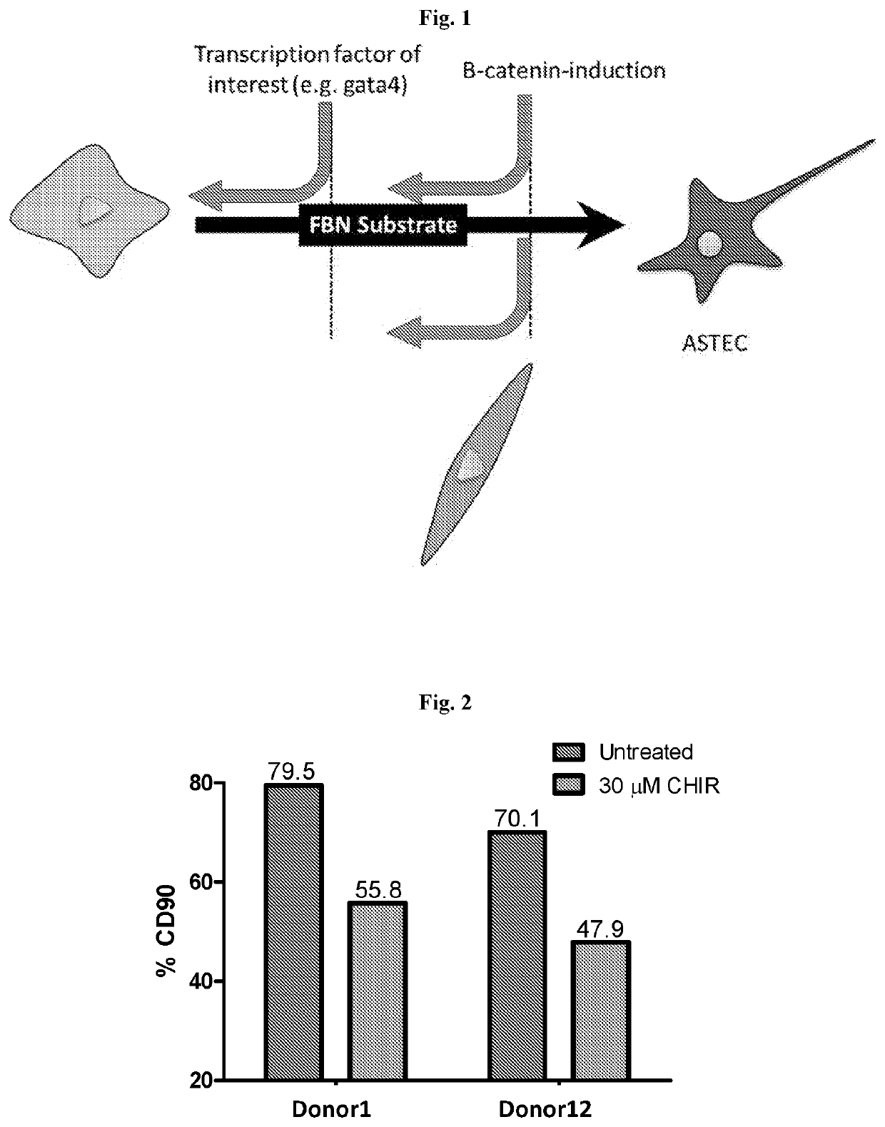
Activation-induced tissue-effector cells suitable for cell therapy and extracelluar vesicles derived therefrom
a technology of extracelluar vesicles and activation-induced tissue, which is applied in the direction of genetically modified cells, skeletal/connective tissue cells, drug compositions, etc., to achieve the effects of increasing the level of a transcription factor, improving a particular disease state, and increasing the left ventricular ejection fraction
- Summary
- Abstract
- Description
- Claims
- Application Information
AI Technical Summary
Benefits of technology
Problems solved by technology
Method used
Image

Examples
example 1
re
[0060]CDCs were prepared as described in U.S. Patent Application Publication No. 2012 / 0315252, the disclosures of which are herein incorporated by reference in their entirety.
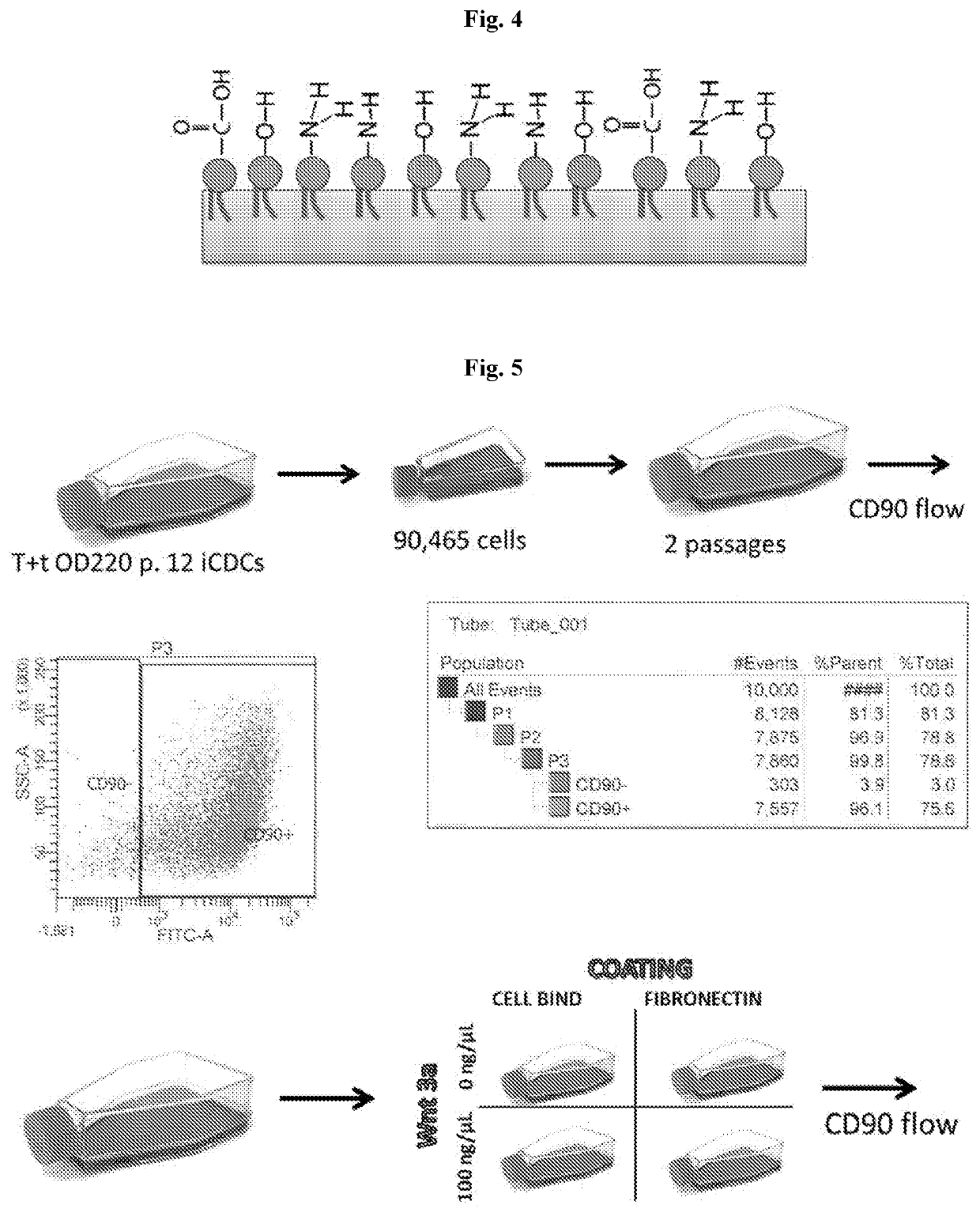
[0061]In brief, heart biopsies were minced into small fragments and briefly digested with collagenase. Explants were then cultured on 20 mg / mL fibronectin-coated dishes. Stromal-like flat cells and phase-bright round cells grew out spontaneously from tissue fragments and reached confluency by 2-3 weeks. These cells were harvested using 0.25% trypsin and were cultured in suspension on 20 mg / mL poly-d-lysine to form self-aggregating cardiospheres. CDCs were obtained by plating and expanding the cardiospheres on a fibronectin-coated flask as an adherent monolayer culture. All cultures were maintained at 5% O2, 5% CO2 at 37° C., using IMDM basic medium supplemented with 10% FBS, 1% penicillin / streptomycin, and 0.1 mL 2-mercaptoethanol. CDCs were grown to 100% confluency on a fibronectin-coated flask to passage 5....
example 2
talization
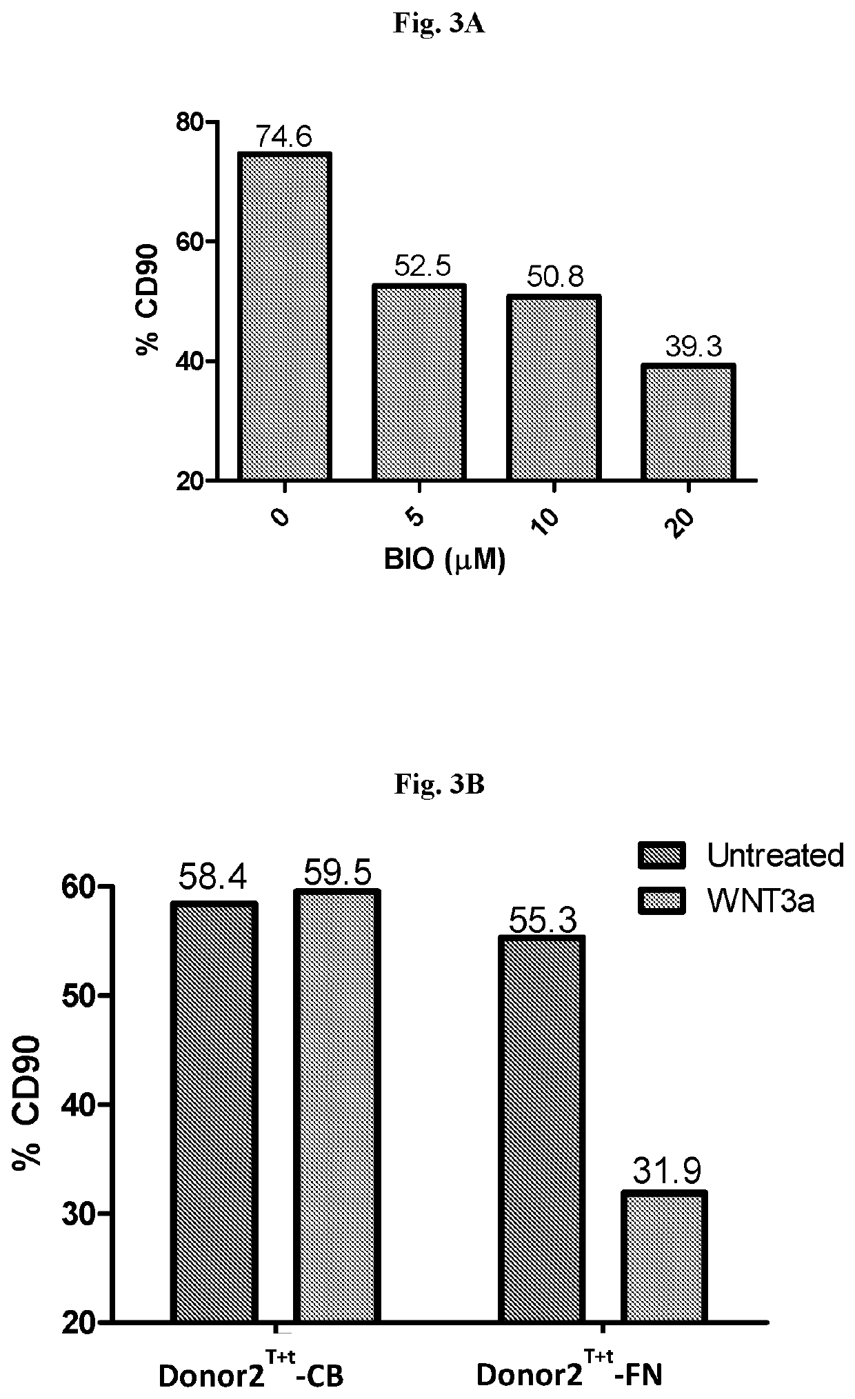
[0062]CDCs were transduced with lentivirus containing genes for telomerase (hTert), simian virus serotype 40 large and small T antigens (SV40 T+t) or the cellular myelocytomatosis (c-Myc) gene. Briefly, 5×10⁴ CDCs (passage 2) were plated on a fibronectin-coated plate in a 24-well plate format. The cells were then treated with the aforementioned viruses at an MOI of 20 in complete media (10% FBS, pen / strep, 1-glut, and β-mercaptoethanol). The cells were transduced in the absence of a transduction reagent (namely polybrene) as previous observations have shown that it interferes with cell growth. The cells were passed as they became confluent (using complete media). At passage 5, the cells were passed from a T75 flask to a T25 flask, this time in the presence of the selection factor puromycin (5 μg / ml). When the cells were recovered and colonies began to form in the flask, the cells were passed and growth behavior was characterized well past the senescent stage of CDCs (passa...
example 3
Exosomes from Immortalized CDCs
[0064]Exosomes were derived from immortalized iCDCs in the same manner as described herein. Briefly, the cells were grown in T175 flasks. At confluence, the cells were washed twice with 30 ml of Iscove's Modified Dulbecco's Medium (IMDM). The cells were then conditioned in 32 ml of IMDM for a period of 15 days. At 15 days of conditioning, media was harvested and cleaned by spinning at 3000 g for 15 minutes. Conditioned media (CM) was aliquoted and stored at −80° C.
[0065]Exosomes size and concentration was measured using diffusion light scattering using a Malvern Nanosight instrument. Briefly, CM was diluted 1:10 in phosphatebuffered saline. To ensure accurate measurements, five (5)-60 sec videos were taken for each sample and batched together, and the data was pooled from all five videos of the same samples.
[0066]RNA from exosomes was isolated with a starting volume of 10 ml of CM using a Norgen Biotek Urine Exosome Isolation Kit. RNA was eluted in 50 ...
PUM
| Property | Measurement | Unit |
|---|---|---|
| diameter | aaaaa | aaaaa |
| diameters | aaaaa | aaaaa |
| sizes | aaaaa | aaaaa |
Abstract
Description
Claims
Application Information