Methods for tissue generation

a tissue and tissue technology, applied in the field of tissue generation, can solve the problems of sudden cardiac death, limited success of current treatment approaches, and inadequate platform for atrial drugs in the human ventricular myocardium, and achieve the effect of improving electrical function

Pending Publication Date: 2021-12-09
RADISIC MILICA
View PDF0 Cites 0 Cited by
  • Summary
  • Abstract
  • Description
  • Claims
  • Application Information

AI Technical Summary

Benefits of technology

The present patent relates to a chamber-specific cardiac tissue that includes a hydrogel and cells, such as cardiomyocytes and cardiac fibroblasts, that are aligned in a specific direction. The tissue can be used for the treatment of cardiac disease and can be derived from atrial or ventricular tissue. The hydrogel can be made from collagen or a collagen derivative, intestinal submucosa or a derivative thereof, cellulose or a cellulose derivative, a proteoglycan, heparin sulfate, chondroitin sulfate, keratin sulfate, hyaluronic acid, elastin, fibronectin, thrombin, laminin, fibrin, chitosan, alginate, Matrigel®, Geltrex®, agarose, decellularized extracellular matrix, polyethylene glycol or a derivative thereof, silicone or a derivative thereof, or a combination thereof. The bioreactor can support the tissue and apply electrical stimulation. The invention provides a new method for creating chamber-specific cardiac tissue with improved function and alignment of cells.

Problems solved by technology

Moreover, the ventricles and atria have unique chamber-specific defects and drug-induced myopathies, making human ventricular myocardium an inadequate platform for discovery of atrial drugs.
The atrial-specific platforms for drug screening are especially important, given that atrial fibrillation is the most common cardiac arrhythmia which is reaching epidemic levels in our aging population, and for which current treatment approaches have limited success.
Indeed, currently used pharmacological agents for treating atrial fibrillation have deadly unwanted side-effects on ventricular CMs which predispose to sudden cardiac death.
Yet, some of the most common cardiac diseases are complex, polygenic conditions that are strongly influenced by environmental factors.

Method used


Image

  • Methods for tissue generation
  • Methods for tissue generation
  • Methods for tissue generation

Examples


example 1

and Methods

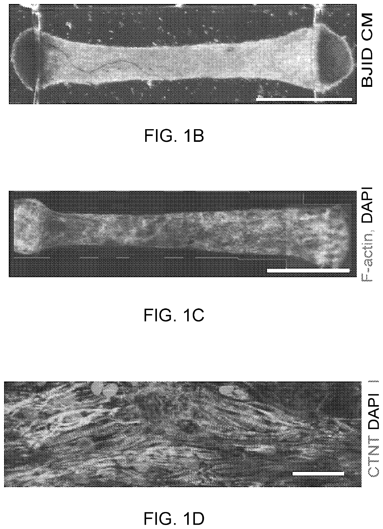
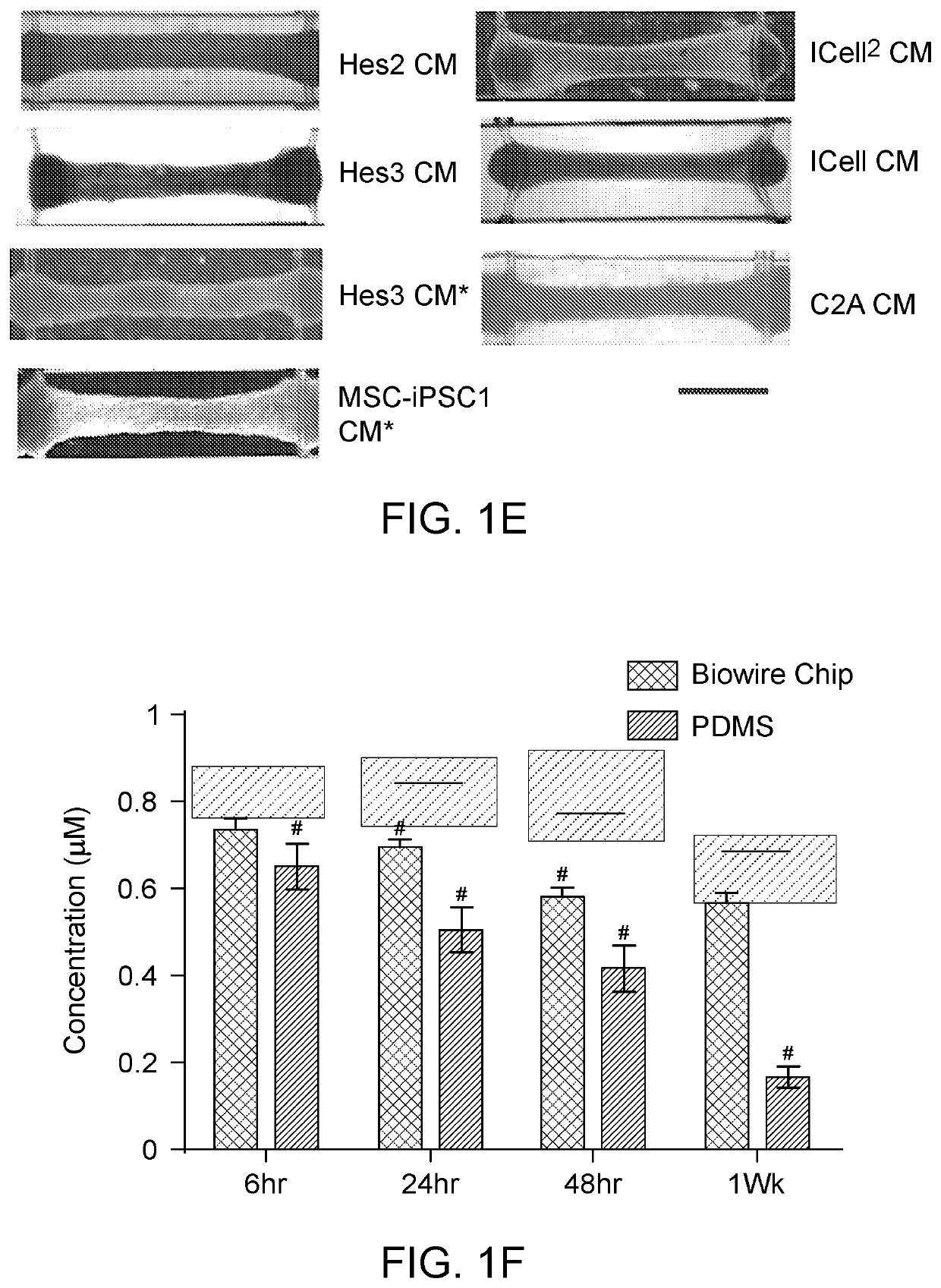
[0267]Predominantly ventricular cardiomyocytes (CMs) were derived from the human embryonic stem cell (hESC) lines Hes2 and HES3-NKX2-5gfp / w using the using embryoid body (EB) differentiation protocol and the human induced pluripotent stem cell (hiPSC) line BJ1D using the monolayer differentiation protocols as previously described. Ventricular cell populations contained 74.7±6.3% (n=9) of CMs on average, based on cardiac troponin T expression analysis using flow cytometry, at the end of directed differentiation protocols.

[0268]C2A hiPSC derived cardiomyocytes were differentiated with EB protocol as previously described.

[0269]iCell and iCell2 cardiomyocytes were purchased from CDI and used according to the manufacturer's instructions.

[0270]Cardiomyocytes of Non-affected A, Non-affected B, Non-Affected C, affected D, affected E, and affected F were provided by Cellular Dynamics Inc, Madison, Wis.

[0271]Predominantly atrial cardiomyocytes were derived from Hes3-NKX2-5gfp / w hES...

example 2

[0323]Generating Heart Tissues From Multiple Cell Sources

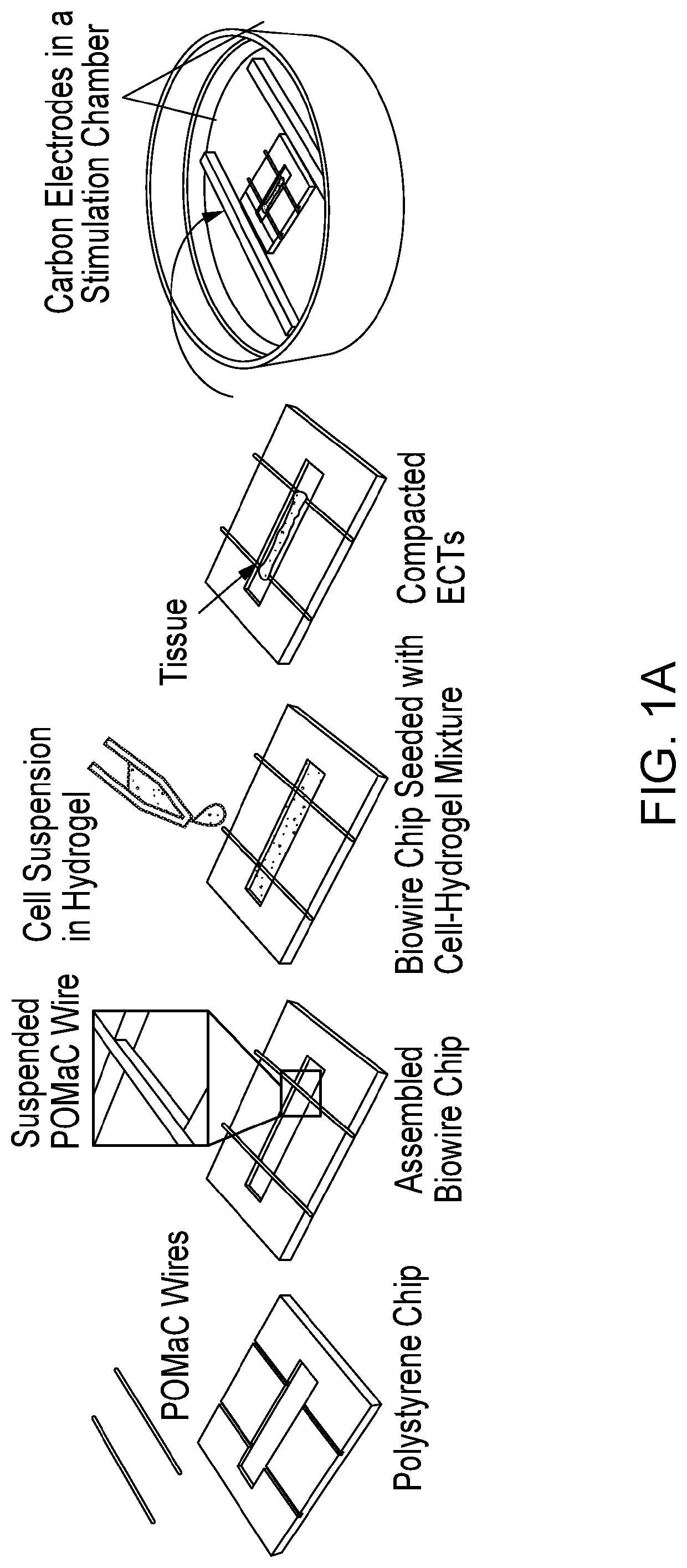
[0324]FIG. 1A and FIG. 8A illustrate the general features of our “Biowire” platform which consists of an array of microwells (5 mm×1 mm×0.3 mm) patterned onto polystyrene sheets. Two flexible wires manufactured from a POMaC polymer are secured with adhesive glue along either end of the microwells. Myocardial tissues are created by combining 100,000 CMs (either ventricular or atrial or both) and cardiac fibroblasts (usually at a 10:1 ratio) with hydrogel within the microwells. During the next ˜7 days, cells undergo “compaction” thereby forming cylindrical trabecular strips (called Biowires II) that are suspended in the microwell but physically attached to the POMaC wires (FIGS. 8A, 8C, and 8D). Beginning at 1 week, the suspended Biowires are electrically conditioned for weeks with electrical field stimulation via a pair of carbon electrodes connected to a stimulator with platinum wires (FIG. 8A).

[0325]Details of the conditionin...

example 3

[0459]We investigated the impact of four microenvironmental cues: cell seeding density, types and percentages of non-myocyte populations, the types of hydrogels used for tissue inoculation and the electrical conditioning regimes on the structural and functional assembly of human pluripotent stem cell-derived cardiac tissues. Utilizing a heart-on-a-chip system that is capable of continuous non-invasive monitoring of tissue contractions, we were able to study how different micro-environmental cues affect the assembly of the cardiomyocytes into a functional cardiac tissue. We have defined conditions that resulted in tissues exhibiting hallmarks of the mature human myocardium, such as positive force-frequency relationship and post-rest potentiation.

[0460]Our goal here was to characterize and optimize a user-friendly organ-on-a-chip platform for the cultivation of human pluripotent stem cell-derived cardiac tissues. Unlike common organ-on-a-chip platforms, the described chip is construct...


PUM

PropertyMeasurementUnit
lengthaaaaaaaaaa
frequencyaaaaaaaaaa
frequencyaaaaaaaaaa

Abstract

The present disclosure provides ex vivo chamber-specific cardiac tissues, methods for generating the cardiac tissues in a bioreactor, and methods of using the cardiac tissues. Examples of cardiac tissues that can be generated include, but are not limited to, atrial tissues, ventricular tissues, and composite tissues having an atrial tissue connected to a ventricular tissue.

Description

RELATED APPLICATIONS[0001]This application claims priority to and the benefit of U.S. Provisional Application No. 62 / 772,515, filed Nov. 28, 2018, the contents of which are incorporated herein by reference in their entireties.FIELD OF THE INVENTION[0002]The present invention relates to ex vivo tissues, methods of generating the tissues, and methods of using the tissues.BACKGROUND OF THE INVENTION[0003]Profound physiological differences among species, in action potentials, ion current profiles, and contractile rates, motivate the development of human cardiac tissues for evaluation of the structural and functional changes induced by pharmacological compounds and disease modelling.[0004]Although ideal platforms for human cardiac studies should yield distinct atrial and ventricular tissues of high biological fidelity, most cardiac tissue engineering approaches using human pluripotent stem cells (hPSC) have focused on generating ventricular myocardium and assessing adverse ventricular ev...

Claims


Application Information

Patent Timeline
Patent Type & Authority Applications(United States)
IPC IPC(8): C12N5/071C12N5/077C12N13/00G01N33/50
CPCC12N5/0697C12N5/0657C12N5/0656C12N13/00G01N33/5061C12N2513/00C12N2502/1323C12N2502/1329C12N2533/54C12N2533/56G01N33/5088C12N2502/1388C12N2506/45C12N2529/00C12N2531/00C12N2506/02G01N33/5082C12M21/08C12M25/16C12M41/00C12M35/02
Inventor RADISIC, MILICAZHAO, YIMUBACKX, PETERRAFATIAN, NAIMEH
Owner RADISIC MILICA
Why Eureka
Try Eureka