Agents for promoting tissue regeneration by recruiting bone marrow mesenchymal stem cells and/or pluripotent stem cells into blood

a technology of stem cells and blood, applied in the direction of drug compositions, peptide/protein ingredients, peptide sources, etc., can solve the problems of high treatment cost, poor cosmetic outcome, and qol in remarkably poor condition, so as to promote cell growth, promote functional regeneration/repair of damaged tissues, and promote cell growth

Pending Publication Date: 2022-02-17
STEMRIM INC +1
View PDF0 Cites 0 Cited by
  • Summary
  • Abstract
  • Description
  • Claims
  • Application Information

AI Technical Summary

Benefits of technology

The present invention provides methods for promoting repair of damaged tissues by recruiting bone marrow pluripotent stem cells to the peripheral blood. By administering certain recruitment factors, such as HMGB1, HMGB2, HMGB3, S100A8, and S100A9, these stem cells can differentiate into various types of cells and induce tissue repair. This method is safe and simple, as it does not require removal of stem cells from the body for artificial manipulation. The invention also involves the use of HGF, EGF, VEGF, and FGF, which are known pharmaceutical agents for promoting cell growth, as well as bone marrow-derived pluripotent stem cells, which have the potential to differentiate into various types of cells.

Problems solved by technology

This is expected to lead to the problem of high treatment costs.
However, it is known that if damaged areas are large, they become filled with nonfunctional scar tissues.
Damage healing with such scar tissues becomes an inhibitory factor for nerve regeneration in cerebral infarction or spinal cord damage, becomes a causative factor for cardiac rupture in myocardial infarction, or results in keloid formation in surgical wounds or extensive burns, thereby causing remarkably poor prognosis and QOL in the cosmetic aspect.

Method used


Image

  • Agents for promoting tissue regeneration by recruiting bone marrow mesenchymal stem cells and/or pluripotent stem cells into blood
  • Agents for promoting tissue regeneration by recruiting bone marrow mesenchymal stem cells and/or pluripotent stem cells into blood
  • Agents for promoting tissue regeneration by recruiting bone marrow mesenchymal stem cells and/or pluripotent stem cells into blood

Examples


example 1

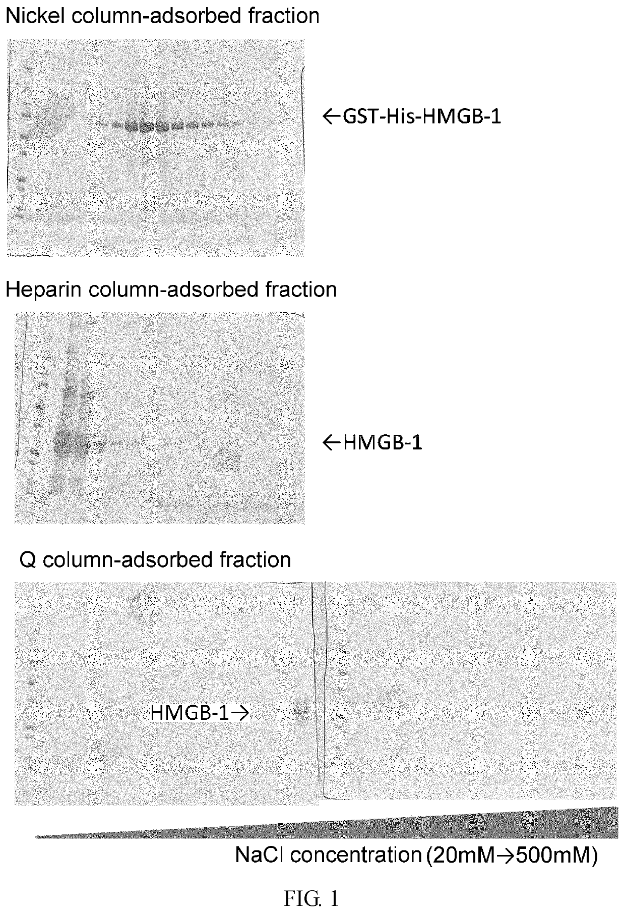
Purification of HMGB-1 and S100A8

[0350]RNA was extracted from newborn mouse skin using Trizol (Invitrogen), and then cDNA was synthesized using SuperScript III cDNA synthesis kit (Invitrogen). Using this cDNA as a template, HMGB1 cDNA was amplified by polymerase chain reaction (PCR). The resulting cDNA was inserted into pCAGGS, a plasmid vector for protein expression in mammalian cells, such that the vector would express the protein attached with GST tag and 6×His tag sequences at the N terminus of its amino acid sequence for the convenience of purification.

[0351]pCAGGS-Flag-His-S100A8 was transfected into a human fetal kidney cell-derived cultured cell line HEK 293 using polyethyleneimine (PEI). After 48 hours, the cells and culture supernatant were separately collected by centrifugation at 4,400 G at 4° C. for five minutes. Then, the collected supernatant was filtered through a cellulose acetate filter having pores with a diameter of 0.8 μm and then through a nitrocellulose filter...

example 2

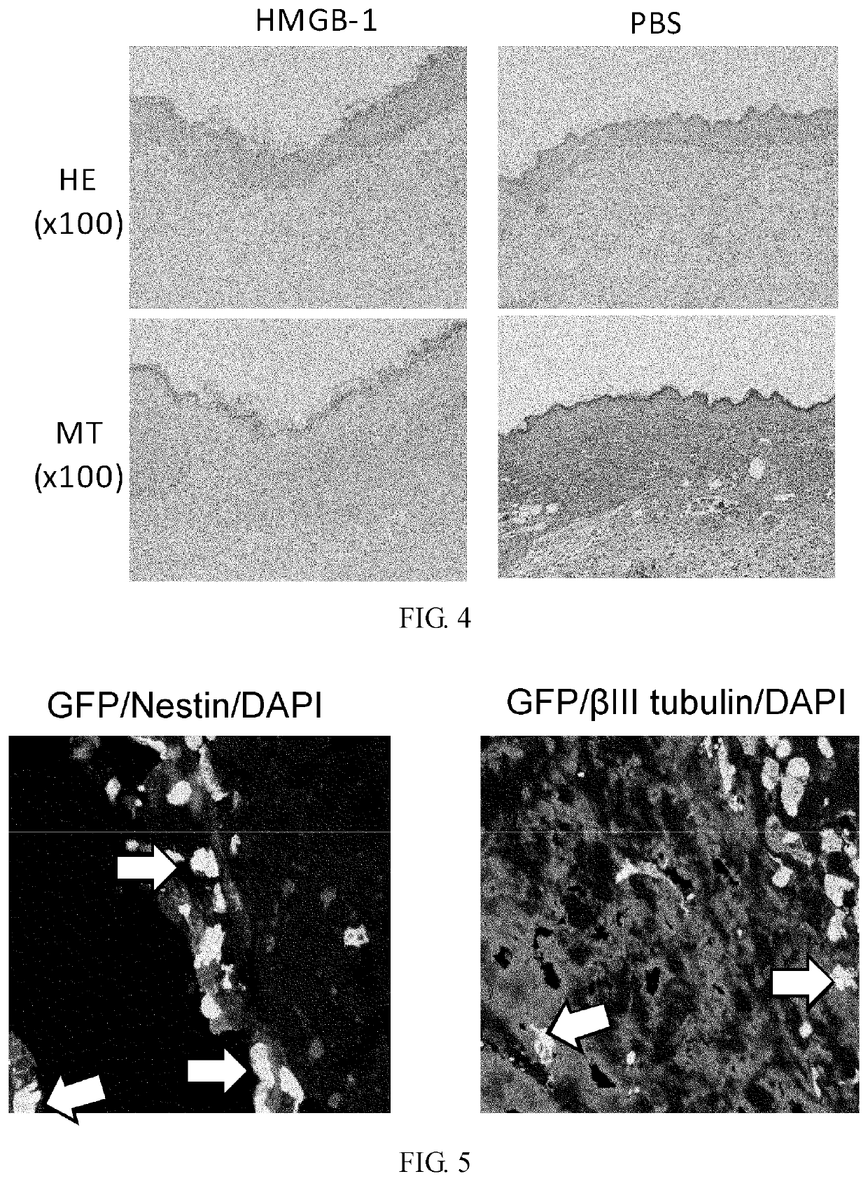
Effect of Intravenous Administration of HMGB-1 and S100A8 in Recruiting Bone Marrow-Derived Cells to Skin Ulceration Site During Skin Ulcer Healing Process

[0355]Male C57BL / 6 mice (6 weeks old) were irradiated at a lethal dose (10 Gy). Immediately, bone marrow cells (5×10⁶ cells / 0.1 ml physiological phosphate buffer (pH 7.4)) derived from a green fluorescent protein (GFP) transgenic mouse (Okabe M. et al., FEBS Lett. 407, 313-319, 1997) were transplanted via the caudal vein. After 8 weeks, a round-shaped skin ulcer with a diameter of 6 mm was created on the back. To prevent shrinkage of the skin of the mice, a silicone ring with an outer diameter of 10 mm, inner diameter of 6 mm, and thickness of 1 mm was attached to the ulcer site using two-sided adhesive tape and medical adhesive Aron alpha A (Sankyo). The ulcer was covered with a silicone disc with a diameter of 10 mm and a thickness of 1 mm to prevent desiccation and bacterial infection at the ulcer. In addition, the ulcer was ma...

example 3

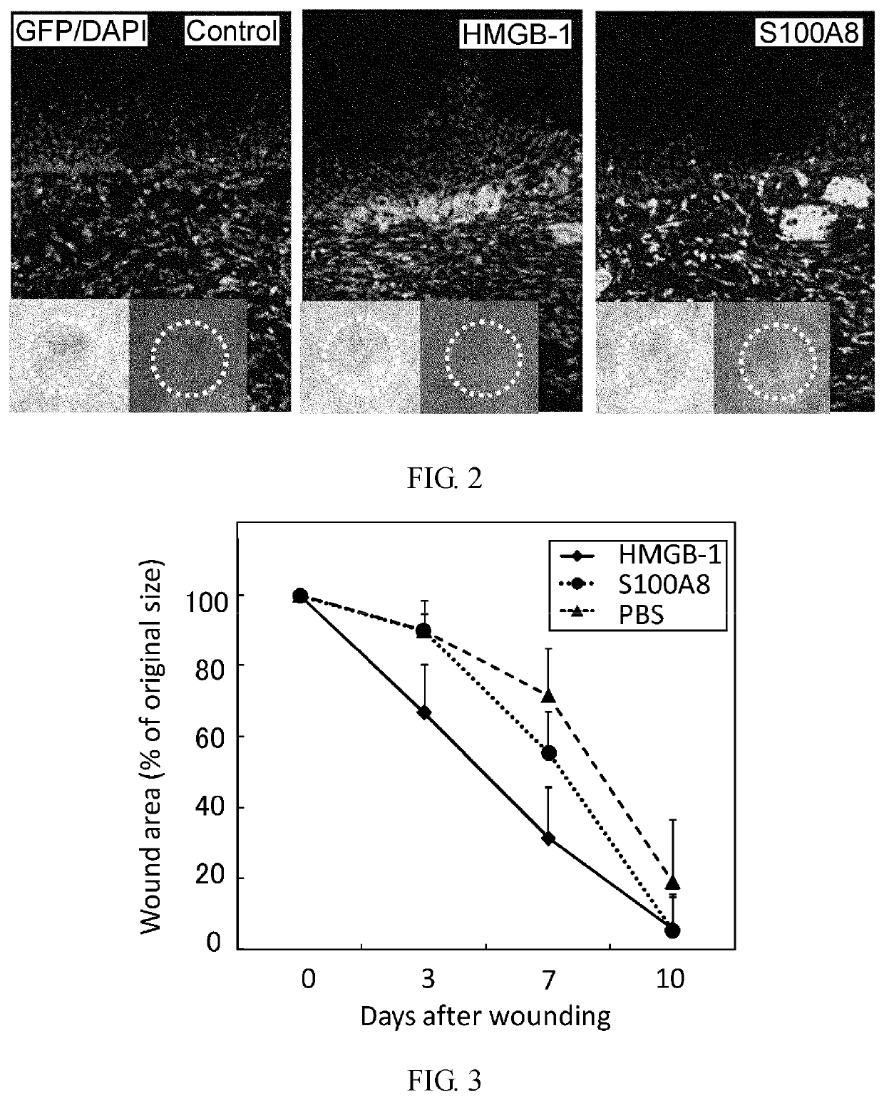
Effect of Intravenous Administration of HMGB-1 and S100A8 in Promoting Skin Ulcer Healing

[0359]In male C57BL / 6 mice (8 weeks old), a round-shaped skin ulcer with a diameter of 6 mm was created on the back. To prevent shrinkage of the skin of the mice, a silicone ring with an outer diameter of 10 mm, inner diameter of 6 mm, and thickness of 1 mm was attached to the ulcer site using two-sided adhesive tape and medical adhesive Aron alpha A (Sankyo). The ulcer was covered with a silicone disc with a diameter of 10 mm and a thickness of 1 mm to prevent desiccation and bacterial infection at the ulcer site. In addition, the ulcer was masked with Tegaderm (3M) for protection.

[0360]HMGB-1 (40 μg) or S100A8 (250 ng) was administered via the caudal vein five times at 24-hour intervals from the day of skin ulcer creation. The ulcer size was measured on days 3, 5, and 10 after creation of ulcer.

[0361]The result is shown in FIG. 3. HMGB-1 reduced the ulcer size on day 3 after creation of ulcer ...


PUM


Abstract

It was revealed that the intravenous administration of HMGB-1 and S100A8 promoted the healing of skin ulcer by recruiting bone marrow-derived cells to the site of skin ulcer. Furthermore, when HMGB-1 was intravenously administered to cerebral infarction model mice after creation of cerebral infarction, bone marrow-derived cells expressing nerve cell markers were detected in their brain. A marked cerebral infarct-reducing effect was observed in mice intravenously administered with HMGB-1 as compared to the control. The post-cerebral infarction survival rate was increased in the intravenous HMGB-1 administration group. The involvement of bone marrow pluripotent stem cells in the process of bone fracture healing was assessed using mice, and the result demonstrated that bone marrow-derived cells distant from the damaged site migrated to the bone fracture site to repair the damaged tissue.

Description

CROSS REFERENCE TO A RELATED APPLICATION[0001]This application is a continuation of U.S. patent application Ser. No. 13 / 503,329, filed Jun. 20, 2021; which is a National Stage Application of International Application Number PCT / JP2010 / 069133, filed Oct. 28, 2010; which claims priority to Japanese Application No. 2009-247143, filed Oct. 28, 2009; all of which are incorporated herein by reference in their entirety.[0002]The Sequence Listing for this application is labeled “As-filed_ST25.txt”, which was created on Apr. 19, 2012, and is 34 KB. The entire content is incorporated herein by reference in its entirety.TECHNICAL FIELD[0003]The present invention relates to tissue regeneration-promoting agents that are administered to a tissue other than a tissue in need of regeneration.BACKGROUND ART[0004]Regenerative medicine aims at functional and structural regeneration of damaged organs, utilizing cells or tissues cultured and processed ex vivo. For example, a cultured skin sheet is produc...

Claims


Application Information

Patent Timeline
Patent Type & Authority Applications(United States)
IPC IPC(8): A61K35/28C07K14/47
CPCA61K35/28A61K48/00C07K14/47A61P17/00A61P19/00A61P19/08A61P25/00A61P25/28A61P43/00A61P9/10A61K9/0019A61K38/17
Inventor TAMAI, KATSUTOKANEDA, YASUFUMIYAMAZAKI, TAKEHIKOCHINO, TAKENAOSAGA, KOTAROENDO, MAYUMI
Owner STEMRIM INC
Why Eureka
Try Eureka