Electrophoretic biological sample staining method and staining apparatus using

a biological sample and staining method technology, applied in the field of electrotrophoretic biological sample staining method and staining apparatus, can solve the problems of difficult image of the inner tissue of difficult to image the thick biological tissue sample, and inability to accurately measure the thickness of the cleared tissue, etc., to achieve accurate measurement, low electrical resistance, and high electrical efficiency

Pending Publication Date: 2022-02-17
CRAYON TECH INC
View PDF0 Cites 0 Cited by
  • Summary
  • Abstract
  • Description
  • Claims
  • Application Information

AI Technical Summary

Benefits of technology

[0037]According to the staining method of the biological sample provided in the present specification, while moving a staining reagent (for example, an antibody) for a biological sample to a thick tissue sample by using electric force, it is possible to use a sample chamber with an ion conductive film to prevent leakage of all organic molecules having extremely low electrical resistance and including macromolecules.
[0038]In addition, it is possible to provide a sample chamber of which a shape may be freely implemented according to a type and size of a biological sample, in which no osmotic pressure phenomenon occurs, and in which unnecessary heat is not generated during electrophoresis to have high electrical efficiency.
[0039]Further, since an amount of electricity applied from the electrode is almost the same as an amount of electricity applied to a sample by using an ion conductive film with almost no electrical resistance, the electric force applied to the sample may be accurately measured, thereby realizing high reproducible and efficient staining.
[0040]Further, it is possible to accelerate movement of an antibody by using electric force to speed up staining of a sample, thereby enabling efficient staining.
[0041]Furthermore, it is possible to minimize degeneration of an antibody responsible for staining and damage to a stained biological sample tissue, by cooling around an ion conductive film.
[0042]That is, according to the biological sample staining technique provided in the present specification, it is possible to stain the inside of a thick biological sample, to remarkably shorten a time required for staining a biological sample, and to effectively stain a biological tissue even when a small amount of staining reagent for a biological sample is used.

Problems solved by technology

When a thick biological tissue sample is observed with an optical device such as a microscope, there is a problem that light scattering severely occurs and a resolution is geometrically deteriorated, so it is difficult to image an inner tissue of the thick biological tissue sample.
In the cleared tissue, since large and small pores are formed inside a structure thereof, a fluorescent antibody may pass through it, so immunostaining is theoretically possible, but when a typical immunostaining method is used through traditionally used passive diffusion, an immunostaining time exponentially increases in proportion to a thickness of the cleared tissue, so that practical immunostaining is impossible.
Recently, research related to this has been continued, but satisfactory results have not been achieved in terms of efficiency and effectiveness.
First, as described above, the most common tissue staining technique is the diffusion method. The diffusion method is a method in which an antibody diffuses into tissue by immersing the tissue in a solution containing an immune antibody, and is passive staining. The diffusion method is the simplest and easiest method, but when a thickness of a biological tissue sample is 50 to 100 μm or more, it takes a long time to stain the inside of the tissue only by diffusion, and it is impossible to commercialize it because it requires a high concentration of a large amount of antibodies.
Second, as a technique designed to stain thick biological samples, there is a method of moving an immune antibody into a sample by using centrifugal force. As such, the method using the centrifugal force may move an antibody into a relatively thick biological sample, but damage is applied to the tissue by the centrifugal force, so there is a limitation in observing a shape of intact tissue.
Third, as a technique that performs immunostaining by forming an electric field, there are limitations in efficiency and practicality in that the highest resistance is applied to a center of a sphere due to a straightened electric field, and because of this, a large difference occurs in antibody staining between an edge and a center, making uniform staining impossible; in that a nano-pore membrane is used to limit movement of a sample, but as staining proceeds, an antibody concentration is diluted due to inflow of a solution due to osmotic pressure; in that because a frame is fixed, there are physical limitations in staining tissues of various shapes and sizes; in that since a pore size of a commercially available nano-pore membrane is not varied, a stain or buffer component with very small molecular weights flow out of a chamber; in that because a pore size of a nano-pore membrane is not constant, the reproducibility of staining is degraded; and in that a large amount of immune antibodies must be used to perform one staining.

Method used


Image

  • Electrophoretic biological sample staining method and staining apparatus using
  • Electrophoretic biological sample staining method and staining apparatus using
  • Electrophoretic biological sample staining method and staining apparatus using

Examples


example 1

on of Sample

[0139]A cleared brain tissue sample was prepared by a typical CLARITY method using the brain of an untransformed rat (SD Rat, 4-6 weeks old).

[0140]Through heart perfusion, blood was drained from the brain microvessels. The brain was excised from the rat, was immersed in a hydrogel monomer solution in which 4% (w / v) acrylamide, 0.25% (w / v) VA-044, and 4% (w / v) PFA (paraformaldehyde) was dissolved in a phosphate buffer saline (PBS) solution, and was incubated for 2 days at 4° C.

[0141]Then, the brain was placed in a vacuum state for 2 to 4 hours in the dark while raising a temperature to 37° C. by using a specially manufactured machine (CLARITY Easy-Imbedding, LCI).

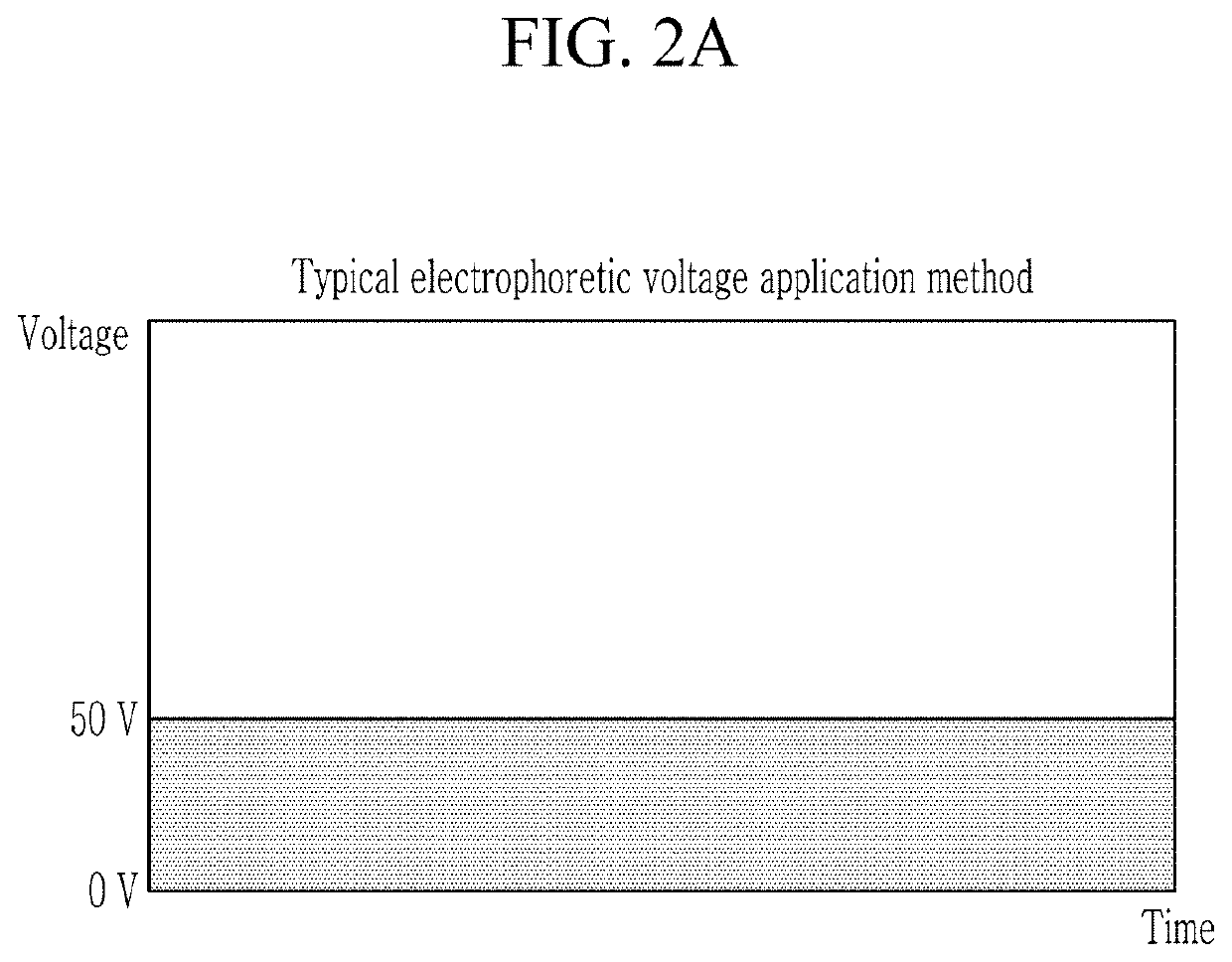
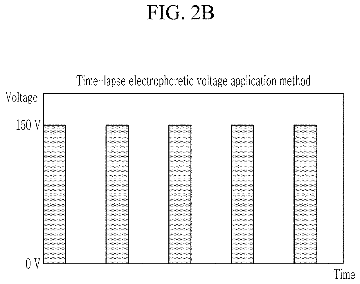
[0142]Thereafter, after slicing it at a desired sample size (thickness: 500 μm, 1 mm, 1.5 mm. 2 mm, 5 mm; diameter: 5 mm, 10 mm), Electro-Tissue Clearing (ETC) was performed by using a CLARITY machine (CLAIRT Easy-Clearing, LCI). In this case, a buffer solution containing 4% of SDS, 50 mM of LiOH, and 25 mM of bo...

example 2

on of Biological Sample Staining Apparatus Using Electrophoretic Method of Staining Biological Sample with Ion Conductive Film Applied

[0144]The manufactured sample chamber was installed between respective electrode parts of a device including a power supply, both electrode parts (each electrode part includes a buffer inlet at a lower portion of one side and a buffer outlet at an upper portion of the opposite side), a buffer supply part (borate buffer; 50 mM of LiOH, 25 mM of boric acid) connected to a buffer inlet and a buffer outlet of the both electrode parts, and a cooler connected to the buffer supply part. The device includes a cooling water circulation channel that is disposed in full contact with two sides of the sample chamber except for two sides thereof in contact with the electrodes, a lower surface, and an upper surface, and that has an inner space through which cooling water may circulate, and a cooling water supplier connected to the cooling water circulation channel.

example 3

ining Test Through Electrophoresis Technique Using Ion Conductive Film (Primary Antibody and Secondary Antibody Test)—Glial Cell Staining

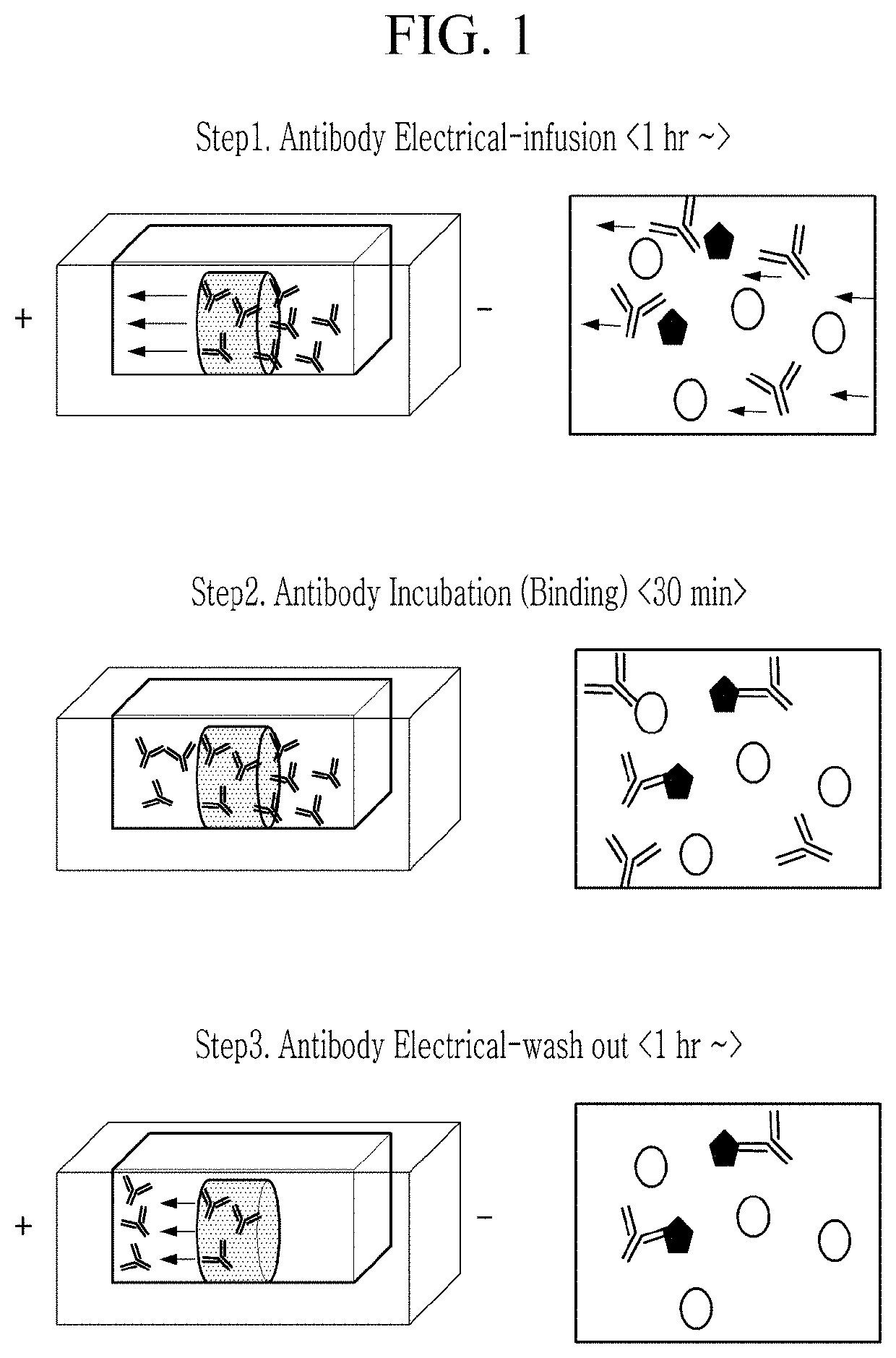
[0145]Immunostaining was performed on the CLARITY sample of the brain tissue of the untransformed rat (SD Rat, 4-6 weeks old) obtained in Example 1 by using a glial fibrillary acidic protein (GFAP) antibody through an electrophoresis technique using the ion conductive film, and then was imaged.

[0146]The brain tissue sample with the diameter of 10 mm and the thickness of 1 mm prepared in Example 1 was inserted into the holder body of the sample chamber in the device prepared in Example 2, and was inserted into the biological sample fixing part to be fixed, and then each space of the electrode part and the sample chamber was filled with a borate buffer (50 mM of LiOH, 25 mM of boric acid). An antibody (1st Antibody, Abcam, UK) targeting GFAP, which is a glia cell marker of the brain, in the immunostaining reagent part (antibody supply part), which is...


PUM

PropertyMeasurementUnit
currentaaaaaaaaaa
thicknessaaaaaaaaaa
thicknessaaaaaaaaaa

Abstract

A biological sample staining method according to an embodiment of the present invention may include: positioning a biological sample to be adjacent to a staining reagent for a biological sample, and separating the biological sample and the staining reagent for the biological sample from an outer buffer by using an ion conductive film; and forming an electric field so that a current flows through the ion conductive film to the staining reagent for the biological sample and the biological sample. In this case, the biological sample is separated from a living body.

Description

TECHNICAL FIELDBackground Art(a) Field of the Invention[0001]The present disclosure relates to a biological sample staining method and staining apparatus, and more particularly, to a biological sample staining method and staining apparatus using an electrophoresis technique.(b) Description of the Related Art[0002]When a thick biological tissue sample is observed with an optical device such as a microscope, there is a problem that light scattering severely occurs and a resolution is geometrically deteriorated, so it is difficult to image an inner tissue of the thick biological tissue sample. In order to overcome these limitations, tissue clearing techniques have been continuously researched.[0003]In the cleared tissue, since large and small pores are formed inside a structure thereof, a fluorescent antibody may pass through it, so immunostaining is theoretically possible, but when a typical immunostaining method is used through traditionally used passive diffusion, an immunostaining ...

Claims


Application Information

Patent Timeline
Patent Type & Authority Applications(United States)
IPC IPC(8): G01N1/31
CPCG01N1/31G01N2001/302G01N1/30G01N33/582
Inventor PARK, HWEONLEE, HOSEUNGLIM, SEUNGHYUNNA, MYUNGSU
Owner CRAYON TECH INC
Why Eureka
Try Eureka