

Method and kit for extracting prion protein
a technology of prion protein and kit, which is applied in the field of methods and kits for extracting prion protein, can solve the problems of reducing the sensitivity of any assay, requiring unfavorable veterinary diagnostic laboratories to have instruments readily available, and aggregates are difficult to dissolve and detect in subsequent steps, so as to achieve rapid isolating
- Summary
- Abstract
- Description
- Claims
- Application Information
AI Technical Summary
Benefits of technology
Problems solved by technology
Method used
Image

Examples
example 2
Analysis of Abnormal Prion Protein Extracted and Purified From Infected Sheep Brain and Lymph Nodes.
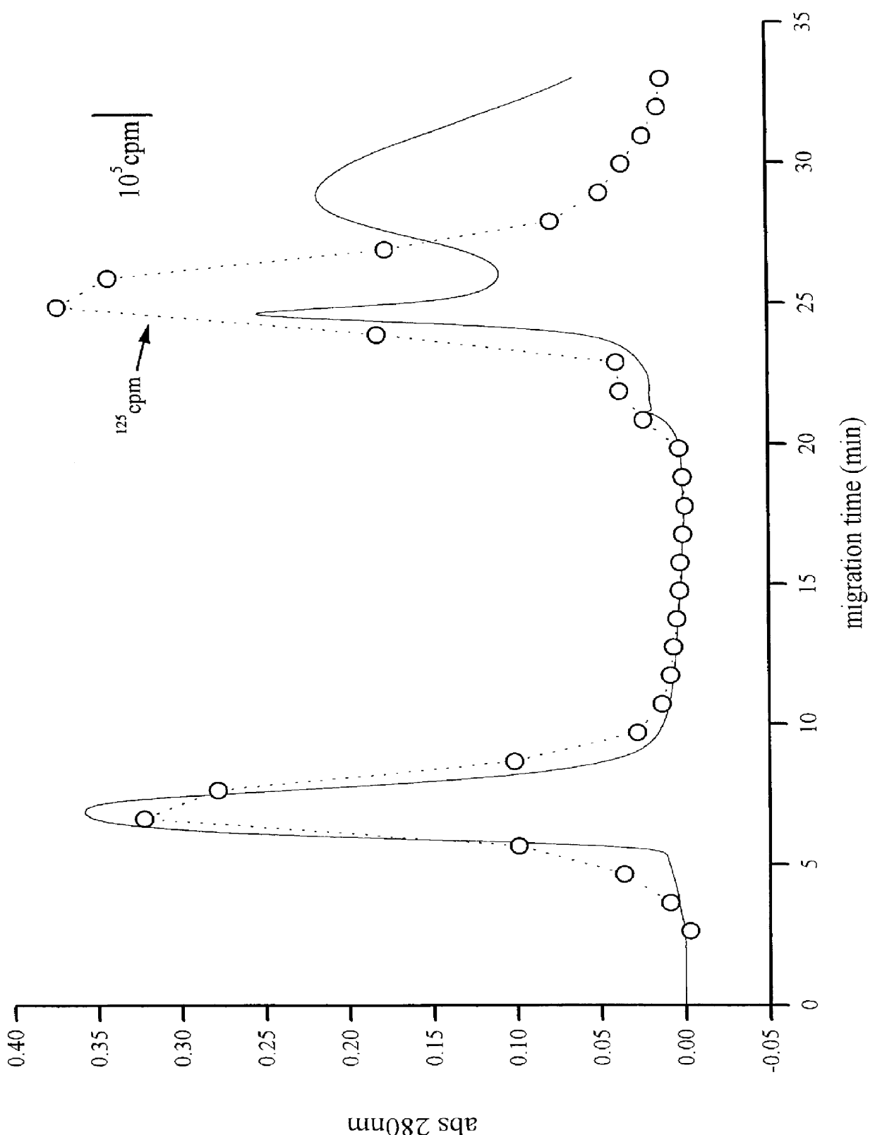
This example shows further purification and analysis of abnormal (scrapie) prion protein (PrPsc) using hydrophilic interaction chromatography (HILIC). Tissue samples including sheep brain and lymph nodes were processed with detergent and proteinase K as previously described. The resulting extracts were applied to a HILIC column and eluted with a decreasing gradient of acetonitrile in 0.1% trifluoroacetic acid and 50 mM hexafluoro-2-propanol. Recovery from the column was approximately 75% as determined with a radioiodinated prion protein. After drying, the collected peak fractions were resuspended in water and assayed with antibodies specific for the prion protein. The method permitted efficient purification of the prion protein as well as testing by immunoassay, since interfering detergents were removed.
example 3
Analysis of Abnormal Prion Protein Extracted From Infected Sheep Brain.
Preparation of Sheep Brain Material.
Scrapie infected sheep brains were obtained from field cases that were positive for the abnormal prion by Western blot (Race et al., Am. J. Vet. Res. 53:883, 1992). A pool was made of 3 positive brains. The same pool was used for all the experiments presented here. Normal brains came from sheep from a scrapie-free flock and were negative for abnormal prion protein by Western blot. The brain material was prepared for chromatography by a modification of the method of Bolton et al. (J. Virol. 53:596, 1985). Briefly, the brain stems were dissected out, weighed and placed in 0.32 M sucrose (10% w / v). The material was then homogenized for 60 s with a Brinkman Polytron (Kinematica AG, Lucerne Switzerland) using a 0.7 cm stainless steel generator at the highest speed. The homogenate was centrifuged at 10,000 g for 20 min to remove particulates, and the resultant supernatant fluid was c...
example 4
Analysis of Abnormal Prion Protein Extracted From Infected Sheep Blood.
Buffy coat centrifuge fractions from blood samples from TSE-infected sheep were diluted with Tris buffered saline (10% tissue:90% buffer). The samples were then treated with proteinase K to digest the normal host prion protein but not the altered abnormal form of prion protein. After digestion, the treated sample was mixed with an equal volume of hexafluoro-2-propanol (HFIP) and incubated at 56.degree. C. for five minutes. An equal volume of 0.5 M sodium sulfate was added and the phases were allowed to separate. The layer containing HFIP was removed and the sample dried in a vacuum centrifuge.
The pellet was resuspended in water and the suspension was put in an organic chromatography mobile phase containing 95% acetonitrile, 5% water, 0.1% trifluoroacetic acid and 50 mM HFIP. The mobile phase was applied to a solid phase extraction cartridge of PolyHYDROXYETHYL Aspartamide.TM. (PolyLC, Inc.). Abnormal prion protei...
PUM
| Property | Measurement | Unit |
|---|---|---|
| Angle | aaaaa | aaaaa |
| Angle | aaaaa | aaaaa |
| Molar density | aaaaa | aaaaa |
Abstract
Description
Claims
Application Information