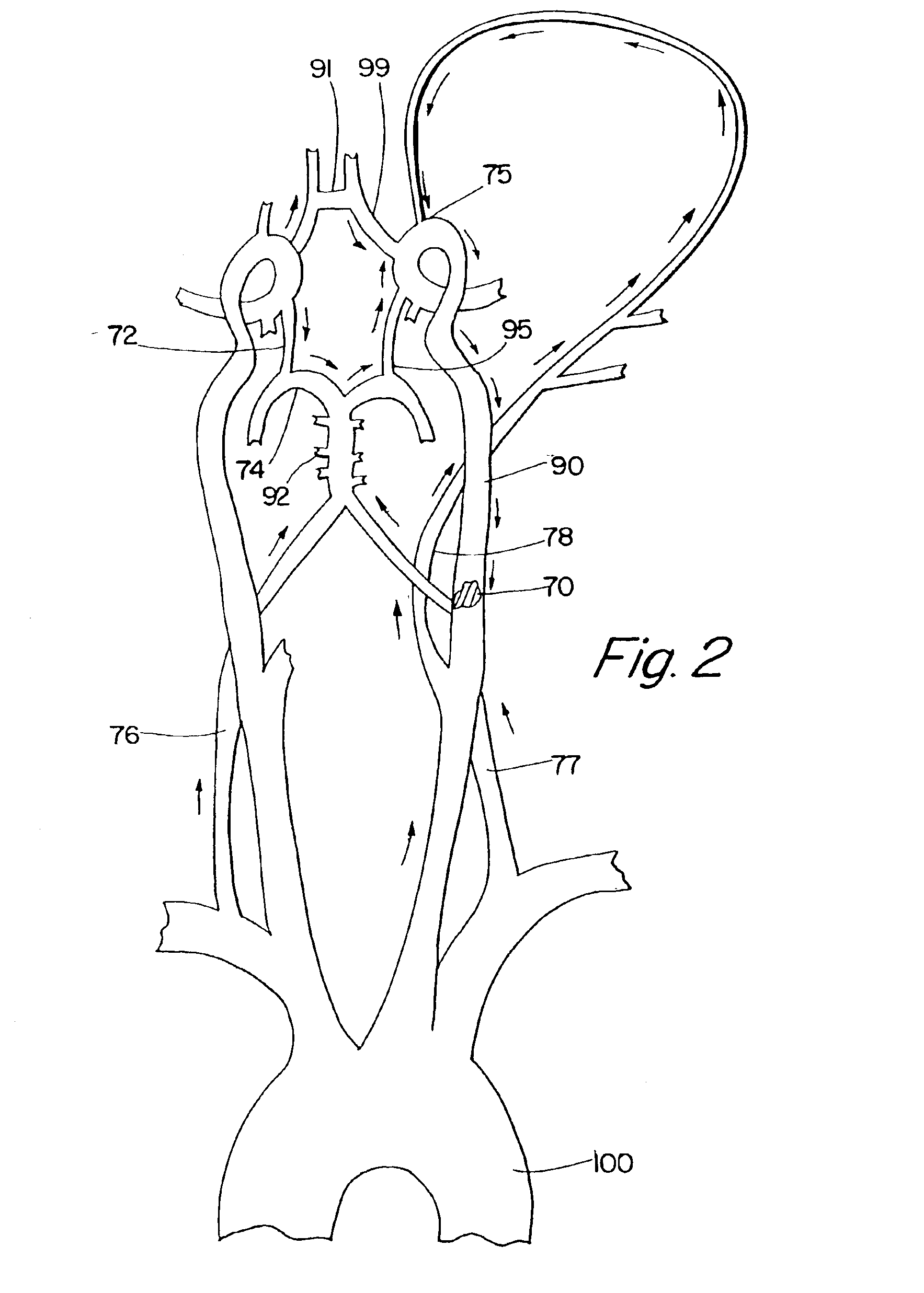
In a first method, the distal end of the first tubular member is inserted through an incision into a
peripheral artery, such as a
femoral artery. The occluding lesion in the symptomatic artery is localized with an angiogram or
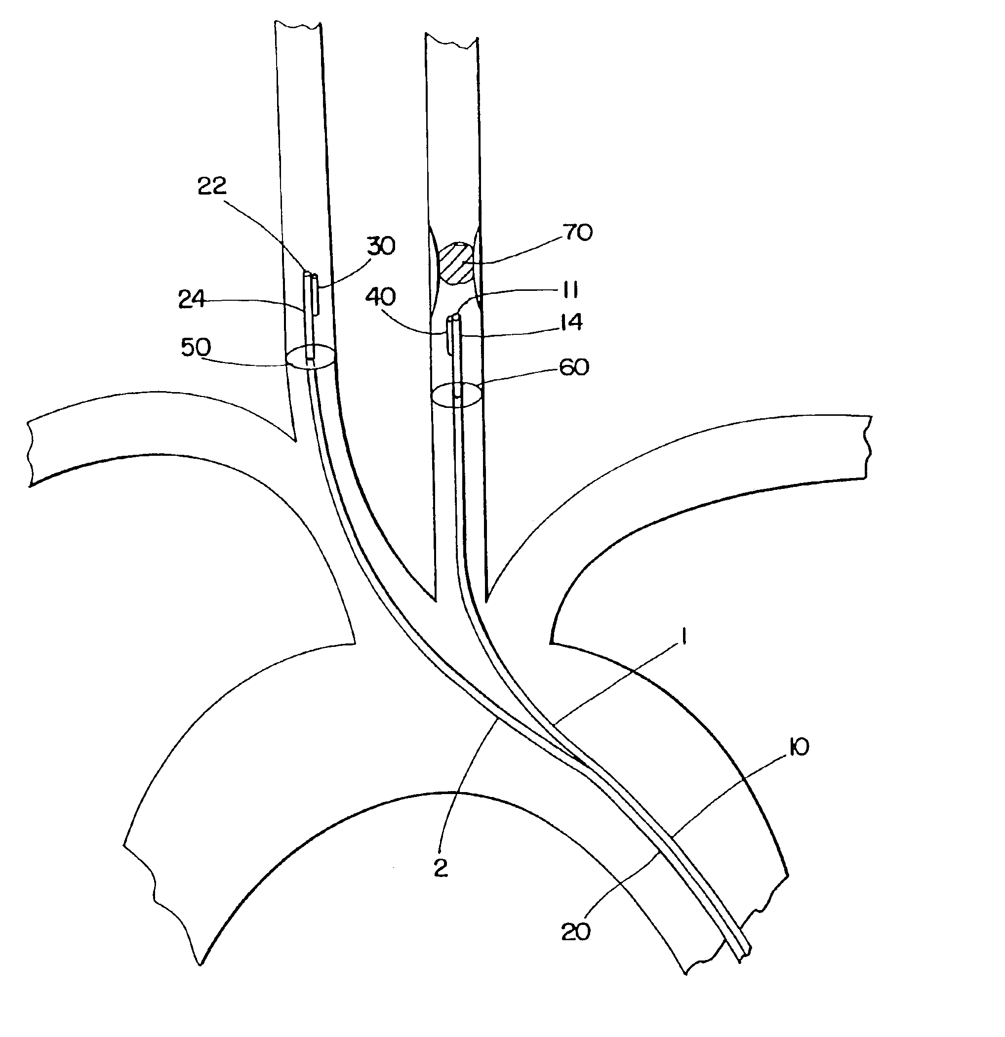
intravascular ultrasound (IVUS). With assistance of a guide wire, the distal end of the second tubular member is inserted through the same incision or a different incision into the contralateral carotid artery. Oxygenated blood is aspirated from the artery through the lumen and port of the first tubular member and perfused into the contralateral carotid artery through the lumen and port of the second tubular member. An expandable occluder, e.g., a
balloon occluder, may be expanded on the second tubular member proximal to the distal port to control the flow rate more effectively. In this manner, augmented contralateral perfusion provides enhanced reversal of
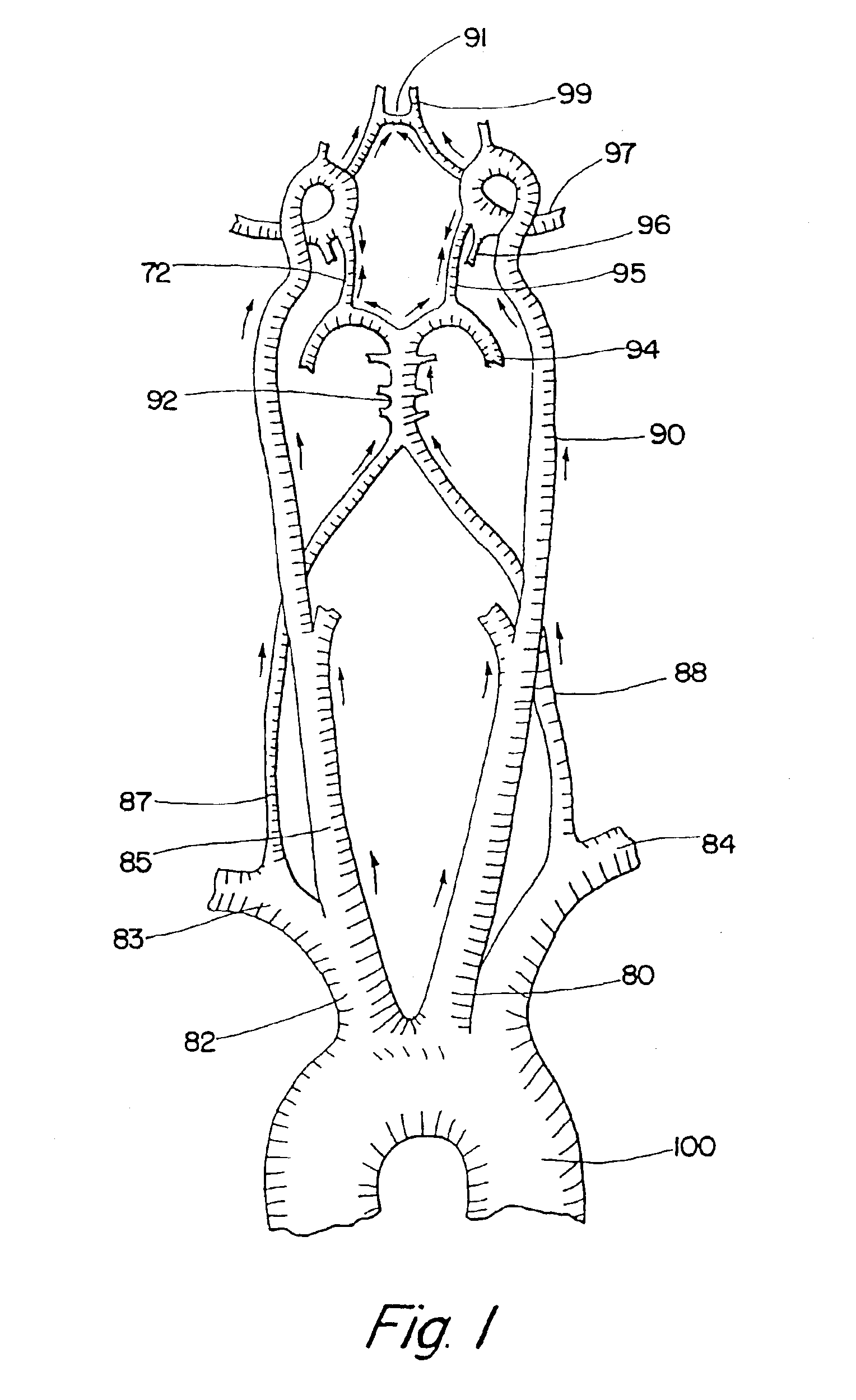
blood flow across the Circle of Willis to compensate for the sudden decrease of flow in the occluded artery.
In another method, the distal end of the first tubular member is inserted through an incision on a peripheral artery, such as a
femoral artery, and advanced into the symptomatic carotid or cerebral artery proximal to the occluding lesion. In an emergency, the device can also be inserted into a patient's carotid artery as a direct stick after localizing the
occlusion with the assistance of IVUS or standard carotid doppler and / or
transcranial doppler (TCD). The distal end of the tubular member can be advanced as far as the occluding site which could be in the
common carotid artery,
internal carotid artery,
middle cerebral artery, anterior cerebral artery, carotid
siphon, terminal
internal carotid artery, or any other part of the cerebral vasculature. The distal end of the second tubular member is then inserted through the same incision or a different incision, and advanced into the contralateral carotid artery. When present, the
balloon occluder mounted on the first tubular member proximal to the distal port is inflated to partially occlude the
arterial lumen. The proximal end of the first tubular member is attached to a
vacuum pump and blood is aspirated from the symptomatic carotid artery through the lumen and port of the first tubular member, and delivered to the contralateral carotid artery through the lumen and port of the second tubular member. The flow rate can be controlled by deflating or inflating the
balloon, e.g., the flow rate increases as the balloon is deflated. The augmented contralateral hemispheric
blood flow, which helps to reverse flow across the Circle of Willis, provides (1) retrograde arterial collateral enhancement to the ischemic area distal to the
occlusion and (2) enhances the pressure differential across the occluding lesion, which may be sufficient to dislodge any thromboembolic material. Blood aspirated from the symptomatic artery is, in certain embodiments, passed through a blood filter optionally included in the proximal end of the first or second tubular member or in the pump to entrap any embolic debris before the blood is returned to the contralateral carotid artery.
It will be understood that there are several advantages in using the devices and methods disclosed herein for management of
acute stroke. For example, the devices can be used (1) in a majority of
stroke patients, including those with
contraindication to using systemic t-PA, (2) to administer neuroprotective agents locally into an occluded vessel, thereby providing greater local benefit and fewer systemic side effects, (3) to infuse hypothermic fluid or blood to the ischemic area, thereby providing protective focal
hypothermia, (4) with standard
atherectomy to remove arterial
atheroma, (5) as an
angioplasty device by inflating the balloon over the stenotic
arterial lumen to enlarge the luminal
diameter, (6) by any invasive radiologist or cardiologist, (7) in the angiogram or
fluoroscopy suite available in most hospitals, (8) in treating
acute stroke patients with few systemic side effects, (9) to treat symptomatic vertebral artery
occlusion, (10) to maintain cerebral perfusion in patients with
asymptomatic flow limiting carotid
stenosis undergoing major cardiothoracic surgeries or in patients with hemodynamic
instability, e.g., cardiogenic or
septic shock, and (11) to maintain perfusion to the distal ischemic area, even without removal of the occlusion, to minimize neurologic damage while alternative intervention is being considered.

