
Methods of treating undesired angiogenesis with 2-methyl-EM-138

a technology of 2-methyl-em-138 and angiogenesis, which is applied in the direction of biocide, heterocyclic compound active ingredients, organic chemistry, etc., can solve the problems of slow and linear growth rate of tumor implants, rapid and nearly exponential growth rate, and slow and linear growth ra

Inactive Publication Date: 2006-09-26
CHILDRENS MEDICAL CENT CORP
View PDF41 Cites 32 Cited by
  • Summary
  • Abstract
  • Description
  • Claims
  • Application Information

AI Technical Summary

Benefits of technology

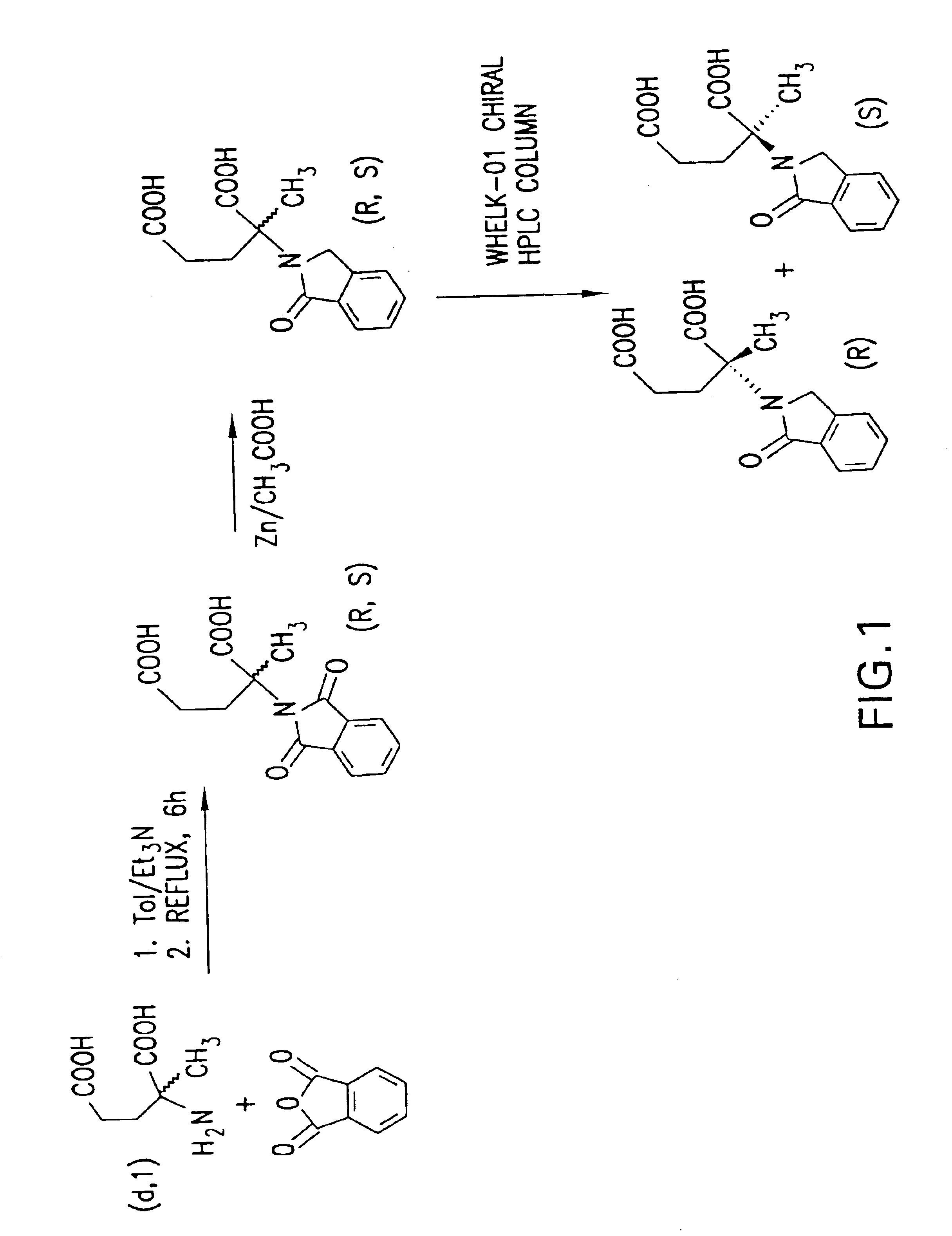
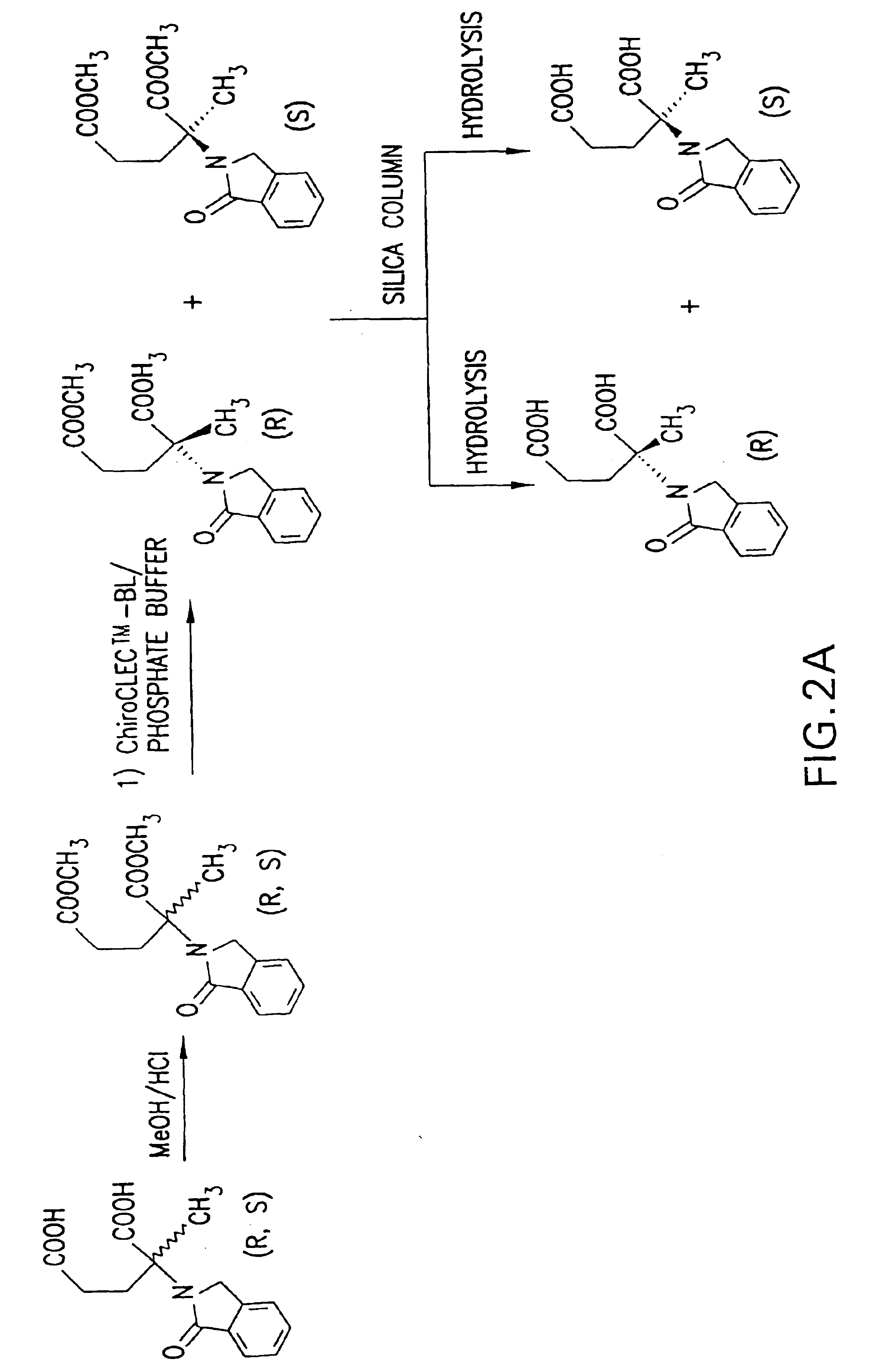
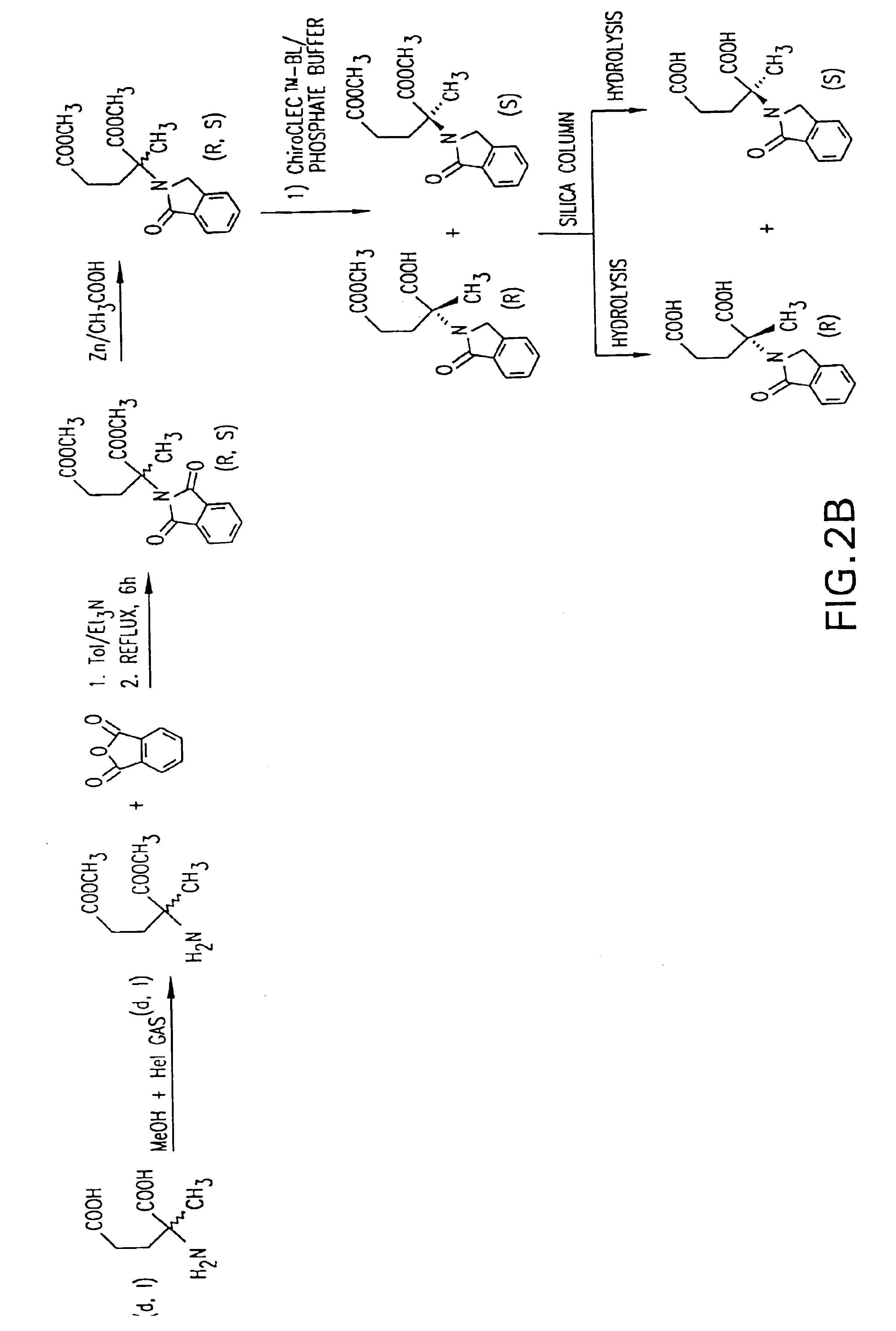
[0045]It is another object of the present invention to provide a facile and economic method for producing the compound DL-2-methyl-2-phthalimidinoglutaric acid.
[0052]It is an object of the present invention to provide a safe and effective method for inhibiting angiogenesis in a human or animal.
[0053]It is a further object of the invention to provide a safe and effective method for inhibiting angiogenesis in a human or animal by administering analogs of 2-phthalimidinoglutaric acid.
[0054]It is a further object of the invention to provide a safe and effective method for inhibiting angiogenesis in a human or animal by administering DL-2-methyl-2-phthalimidinoglutaric acid, (R)-(+)-2-methyl-2-phthalimidinoglutaric acid, or (S)-(−)-2-methyl-2-phthalimidinoglutaric acid.
[0055]It is a further object of the invention to provide a safe and effective method for inhibiting angiogenesis in a human or animal by administering hydroxylated analogs of 2-phthalimidinoglutaric acid.

Problems solved by technology

(1) The growth rate of tumors implanted in subcutaneous transparent chambers in mice is slow and linear before neovascularization, and rapid and nearly exponential after neovascularization. (Algire, et al., J. Nat. Cancer Inst., 6:73-85 (1945)).
(2) Tumors grown in isolated perfused organs where blood vessels do not proliferate are limited to 1-2 mm3 but expand rapidly to >1000 times this volume when they are transplanted to mice and become neovascularized. (Folkman, et al., Annals of Surgery, 164:491-502 (1966)).
(3) Tumor growth in the avascular cornea proceeds slowly and at a linear rate, but switches to exponential growth after neovascularization. (Gimbrone, Jr., et al., J. Nat. Cancer Inst., 52:421-27 (1974)).
(4) Tumors suspended in the aqueous fluid of the anterior chamber of the rabbit eye remain viable, avascular, and limited in size to 3. Once they are implanted on the iris vascular bed, they become neovascularized and grow rapidly, reaching 16,000 times their original volume within 2 weeks. (Gimbrone, Jr., et al., J. Exp. Med., 136:261-76).
(5) When tumors are implanted on the chick embryo chorioallantoic membrane, they grow slowly during an avascular phase of >72 hours, but do not exceed a mean diameter of 0.93+0.29 mm. Rapid tumor expansion occurs within 24 hours after the onset of neovascularization, and by day 7 these vascularized tumors reach a mean diameter of 8.0+2.5 mm. (Knighton, British J. Cancer, 35:347-56 (1977)).
(6) Vascular casts of metastases in the rabbit liver reveal heterogeneity in size of the metastases, but show a relatively uniform cut-off point for the size at which vascularization is present. Tumors are generally avascular up to 1 mm in diameter, but are neovascularized beyond that diameter. (Lien, et al., Surgery, 68:334-40 (1970)).
(7) In transgenic mice which develop carcinomas in the beta cells of the pancreatic islets, pre-vascular hyperplastic islets are limited in size to Nature, 339:58-61 (1989)).
(8) A specific antibody against VEGF (vascular endothelial growth factor) reduces microvessel density and causes “significant or dramatic” inhibition of growth of three human tumors which rely on VEGF as their sole mediator of angiogenesis (in nude mice). The antibody does not inhibit growth of the tumor cells in vitro. (Kim, et al., Nature, 362:841-44 (1993)).
(9) Anti-bFGF monoclonal antibody causes 70% inhibition of growth of a mouse tumor which is dependent upon secretion of bFGF as its only mediator of angiogenesis. The antibody does not inhibit growth of the tumor cells in vitro. (Hori, et al., Cancer Res., 51:6180-84 (1991)).
(10) Intraperitoneal injection of bFGF enhances growth of a primary tumor and its metastases by stimulating growth of capillary endothelial cells in the tumor. The tumor cells themselves lack receptors for bFGF, and bFGF is not a mitogen for the tumors cells in vitro. (Gross, et al., Proc. Am. Assoc. Cancer Res., 31:79 (1990)).
(11) A specific angiogenesis inhibitor (AGM-1470) inhibits tumor growth and metastases in vivo, but is much less active in inhibiting tumor cell proliferation in vitro. It inhibits vascular endothelial cell proliferation half-maximally at 4 logs lower concentration than it inhibits tumor cell proliferation. (Ingber, et al., Nature, 48:555-57 (1990)). There is also indirect clinical evidence that tumor growth is angiogenesis dependent.
(12) Human retinoblastomas that are metastatic to the vitreous develop into avascular spheroids which are restricted to less than 1 mm3 despite the fact that they are viable and incorporate 3H-thymidine (when removed from an enucleated eye and analyzed in vitro).
(13) Carcinoma of the ovary metastasizes to the peritoneal membrane as tiny avascular white seeds (1-3 mm3). These implants rarely grow larger until one or more of them becomes neovascularized.
(14) Intensity of neovascularization in breast cancer (Weidner, et al., New Eng. J. Med., 324:1-8 (1991); Weidner, et al., J Nat. Cancer Inst., 84:1875-87 (1992)) and in prostate cancer (Weidner, et al., Am. J. Pathol., 143(2):401-09 (1993)) correlates highly with risk of future metastasis.
(15) Metastasis from human cutaneous melanoma is rare prior to neovascularization. The onset of neovascularization leads to increased thickness of the lesion and an increased risk of metastasis. (Srivastava, et al., Am. J. Pathol., 133:419-23 (1988)).
(16) In bladder cancer, the urinary level of an angiogenic protein, bFGF, is a more sensitive indicator of status and extent of disease than is cytology. (Nguyen, et al., J. Nat. Cancer Inst., 85:241-42 (1993)).
In more severe cases, the tumors progress to large cavernous and infiltrative forms and create clinical complications.
Systemic forms of hemangiomas, hemangiomatoses, have a high mortality rate.
Therapy-resistant hemangiomas exist that cannot be treated with therapeutics currently in use.
In wound healing, excessive repair or fibroplasia can be a detrimental side effect of surgical procedures and may be caused or exacerbated by angiogenesis.
Adhesions are a frequent complication of surgery and lead to problems such as small bowel obstruction.
The toxicity of protamine limits its practical use as a therapeutic.
Thus, there are concerns regarding its use in women of child-bearing age.
Although minimal, there are a number of side effects which limit the desirability of thalidomide as a treatment.
One such side effect is drowsiness.
In a number of therapeutic studies, the initial dosage of thalidomide had to be reduced because patients became lethargic and had difficulty functioning normally.
Another side effect limiting the use of thalidomide is peripheral neuropathy, in which individuals suffer from numbness and disfunction in their extremities.

Method used


Image

  • Methods of treating undesired angiogenesis with 2-methyl-EM-138
  • Methods of treating undesired angiogenesis with 2-methyl-EM-138
  • Methods of treating undesired angiogenesis with 2-methyl-EM-138

Examples


example 1

Inhibition of Metastasis through Intraperitoneal Administration of EM-138

[0105]B16-BL6 melanoma cells (5×10⁴) were injected intravenously into the tail veins of C57B1 / 6 mice. Three days later, the mice were treated intraperitoneally with increasing doses of thalidomide or 2-phthalimidinoglutaric acid (EM-138) on alternate days. Fourteen days after tumor cell inoculation, the lungs were removed from the mice and the surface pulmonary metastases were counted. The results of this experiment are shown in FIG. 4. The values shown are the mean of 5 mice per group. The bars on the graph represent the standard deviation.

example 2

Inhibition of Metastasis through Oral Administration of EM-138

[0106]B16-BL6 melanoma cells (5×10⁴) were injected intravenously into the tail veins of C57B1 / 6 mice. Three days later, the mice were treated orally with increasing doses of thalidomide or 2-phthalimidinoglutaric acid (EM-138) on alternate days. Fourteen days after tumor cell inoculation, the lungs were removed from the mice and the surface pulmonary metastases were counted. The results of this experiment are shown in FIG. 5. The values shown are the mean of 5 mice per group. The bars on the graph represent the standard deviation.

example 3

Effect of the Number of Treatments on EM-138 Activity

[0107]B16-BL6 melanoma cells (5×10⁴) were injected intravenously into the tail veins of C57B1 / 6 mice. Three days later, the mice received a gavage treatment with 0.8 mmol / kg of 2-phthalimidinoglutaric acid (EM-138). The mice received either a single treatment, five treatments on alternate days, or one treatment every day for eleven days. Fourteen days after tumor cell inoculation, the lungs were removed from the mice and the surface pulmonary metastases were counted. The results of this experiment are shown in FIG. 6. The values shown are the mean of 5 mice per group. The bars on the graph represent the standard deviation.


PUM

PropertyMeasurementUnit
volumeaaaaaaaaaa
diameteraaaaaaaaaa
sizeaaaaaaaaaa

Abstract

The invention provides new and useful analogs of 2-phthalimidinoglutaric acid. These analogs include DL-2-methyl-2-phthalimidinoglutaric acid and hydroxylated analogs of 2-phthalimidinoglutaric. The invention also provides processes for making these analogs. The invention also provides the two individual enantiomers of DL-2-methyl-2-phthalimidinoglutaric acid, (R)-(+)-2-methyl-2-phthalimidinoglutaric acid and (S)-(−)-2-methyl-2-phthalimidinoglutaric acid, and processes for separating these individual enantiomers from the racemate. Further, the invention provides methods for inhibiting angiogenesis and treating angiogenesis-associated diseases, including cancer and macular degeneration, by administering one or more of these compounds.

Description

CROSS REFERENCE TO RELATED APPLICATIONS[0001]This application is a divisional of application Ser. No. 09 / 309,464, filed May 11, 1999, now U.S. Pat. No. 6,673,828, which claims priority to U.S. Provisional Patent Application No. 60 / 085,037, filed on May 11, 1998, U.S. Provisional Patent Application No. 60 / 097,384, filed on Aug. 21, 1998 and U.S. Provisional Patent Application No. 60 / 108,037, filed on Nov. 12, 1998 the disclosure of each of which is incorporated by reference herein in its entirety.FIELD OF THE INVENTION[0002]This invention is related to derivatives of 2-methyl glutamic acid, namely 2-methyl-2-phthalimidinoglutaric acid and hydroxylated derivatives of 2-phthalimidinoglutaric. More particularly, the invention relates to the preparation of analogs of 2-phthalimidinoglutaric acid and the separation of the enantiomers of 2-methyl-2-phthalimidinoglutaric acid. Further, the invention relates to the use of such compounds for the treatment of cancer and for the treatment of an...

Claims


Application Information

Patent Timeline
Patent Type & Authority Patents(United States)
IPC IPC(8): A61K31/40C07D209/46
CPCC07D209/46
Inventor D'AMATO, ROBERT J.GREEN, SHAWN J.MADSEN, JOHN W.FOGLER, WILLIAMSWARTZ, JR., GLENN M.PAPATHANASSIU, ADONIA E.SHAH, JAMSHED H.
Owner CHILDRENS MEDICAL CENT CORP
Learn More