

Tumor marker for urothelial carcinoma
a tumor marker and urothelial carcinoma technology, applied in the protein field, can solve the problems of unsuitable mass screening, likely to indicate false positive for gross hematuria or cystitis, etc., and achieve the effect of effective detection of urothelial carcinoma
- Summary
- Abstract
- Description
- Claims
- Application Information
AI Technical Summary
Benefits of technology
Problems solved by technology
Method used
Image

Examples
example 1
Screening of Proteins Changed in Cancer by the Proteome Analysis
(1) Preparation of Samples
[0061]Three specimens of bladder transitional cell carcinoma obtained through surgical operations (surgical operations: two examples of transurethral resection of bladder tumor and one example of total cystectomy; grade of atypism: one example of grade 2 and two examples of grade 3) were used as samples. Normal mucous membranes of urinary bladders obtained through retropubic prostatectomy for prostatic hyperplasia (two examples) and a normal ureter obtained through nephrectomy for renal cell carcinoma (one example) were used as controls. A tissue lysate comprising 8M urea, 2% CHAPS, 1% dithiothreitol (DTT), 0.5% Pharmalyte 3-10 (Amersham bioscience), 10% glycerol, and 1% protease inhibitor cocktail (Nacalai Tesque Inc.) was added, the mixture was homogenized, and the supernatant obtained by centrifugation at 15,000 rpm for 10 minutes was determined to be a solution of solubilized tissues. In or...
example 2
Examination of Usefulness as a Tumor Marker
(1) Confirmation of a Calreticulin Protein Spot by Two-Dimensional Immunoblotting
[0072]The gel subjected to two-dimensional electrophoresis was transferred to the Immobilon PVDF membrane (Millipore), and the membrane was subjected to immunoblotting using the monoclonal antibody SPA-601 (StressGen) against recombinant human calreticulin proteins and the polyclonal antibody SPA-600 (StressGen) having the synthetic peptide of the human calreticulin protein-C-terminus as an immunogen. Blocking was carried out using the Super Block Blocking Solution in TBS (Pierce) at 4° C. overnight. The dilution rate for the SPA-601 antibody was 10,000-fold, that for the SPA-600 antibody was 20,000-fold, that for the HRP labeled anti-mouse IgG antibody (MBL) was 10,000-fold, and that for the HRP labeled anti-rabbit IgG antibody (Vector) was 50,000-fold. 10 mM Tris-HCl (pH 7.4), 100 mM NaCl, and 0.1% Tween 20 were used for washing and dilution of antibody. Band...
example 3
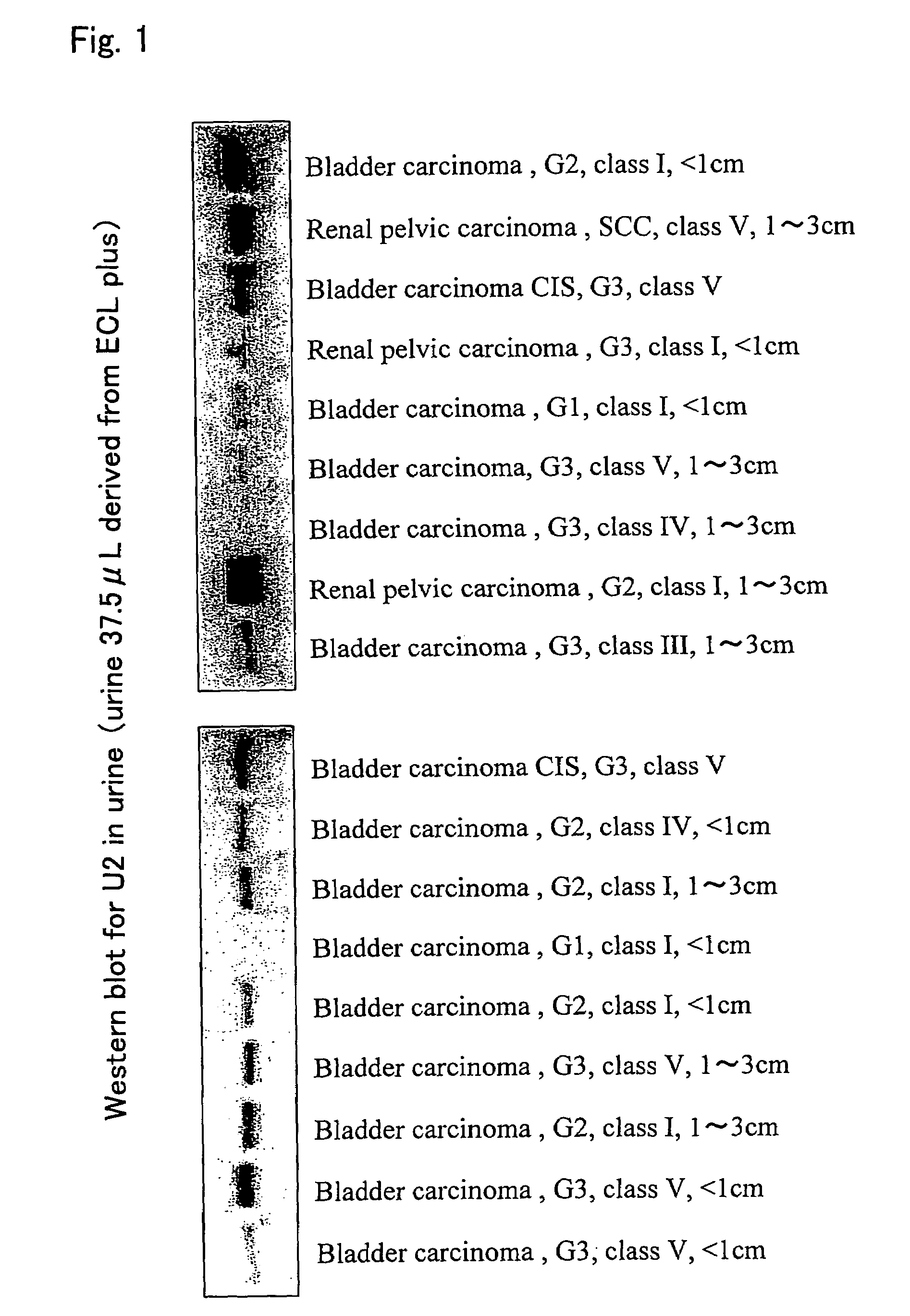
Detection of Calreticulin Protein in Urine
[0081]In order to detect calreticulin proteins in urine, one-dimensional electrophoresis was carried out in the same manner as described above, the resultant was transferred to the PVDF membrane, and immunoblotting was then carried out using the SPA-600 antibody.
[0082]Twenty seven urine specimens of patients with urothelial carcinomas and 123 urine specimens of patients with non-urothelial carcinomas were used to compare the expression levels of calreticulin proteins in urine. These specimens were cryopreserved until the assay after they were collected. As urine of patients with non-urothelial carcinomas, urine of patients who had, for example, prostatic hyperplasia, prostate carcinoma, urinary tract infection, renal cell carcinoma, urolithiasis, or idiopathic hematuria but did not have urothelial carcinoma were used.
[0083]As a result, weak calreticulin protein bands were detected by immunoblotting of only 22 examples (17.9%) among the 123 u...
PUM
| Property | Measurement | Unit |
|---|---|---|
| molecular weight | aaaaa | aaaaa |
| temperature | aaaaa | aaaaa |
| temperature | aaaaa | aaaaa |
Abstract
Description
Claims
Application Information