
Nanoparticles for targeting hepatoma cells

a technology of nanoparticles and hepatoma cells, applied in the field of nanoparticles for targeting hepatoma cells, can solve the problems of limited chemotherapy for cancer, and restricted therapeutic efficacy

Inactive Publication Date: 2008-03-25
GP MEDICAL
View PDF1 Cites 20 Cited by
  • Summary
  • Abstract
  • Description
  • Claims
  • Application Information

AI Technical Summary

Benefits of technology

[0013]The results obtained by the TEM and AFM examinations show that the morphology of the prepared nanoparticles is spherical in shape with a smooth surface. In the stability study, no aggregation or precipitation of nanoparticles was observed during storage for up to 1 month, as a result of the electrostatic repulsion between the negatively charged nanoparticles. With increasing the galactosamine content conjugated on the rhodamine-123-containing nanoparticles, the intensity of fluorescence observed in HepG2 cells increased significantly. Additionally, the intensity of fluorescence observed in HepG2 cells incubated with the nanoparticles with or without galactosamine conjugated increased approximately linearly with increasing the duration of incubation. In contrast, there was no fluorescence observed in Hs68 cells (without ASGP receptors) incubated with the nanoparticles with galactosamine conjugated. The aforementioned results indicated that the galactosylated nanoparticles prepared in the study had a specific interaction with HepG2 cells via ligand-receptor recognition.
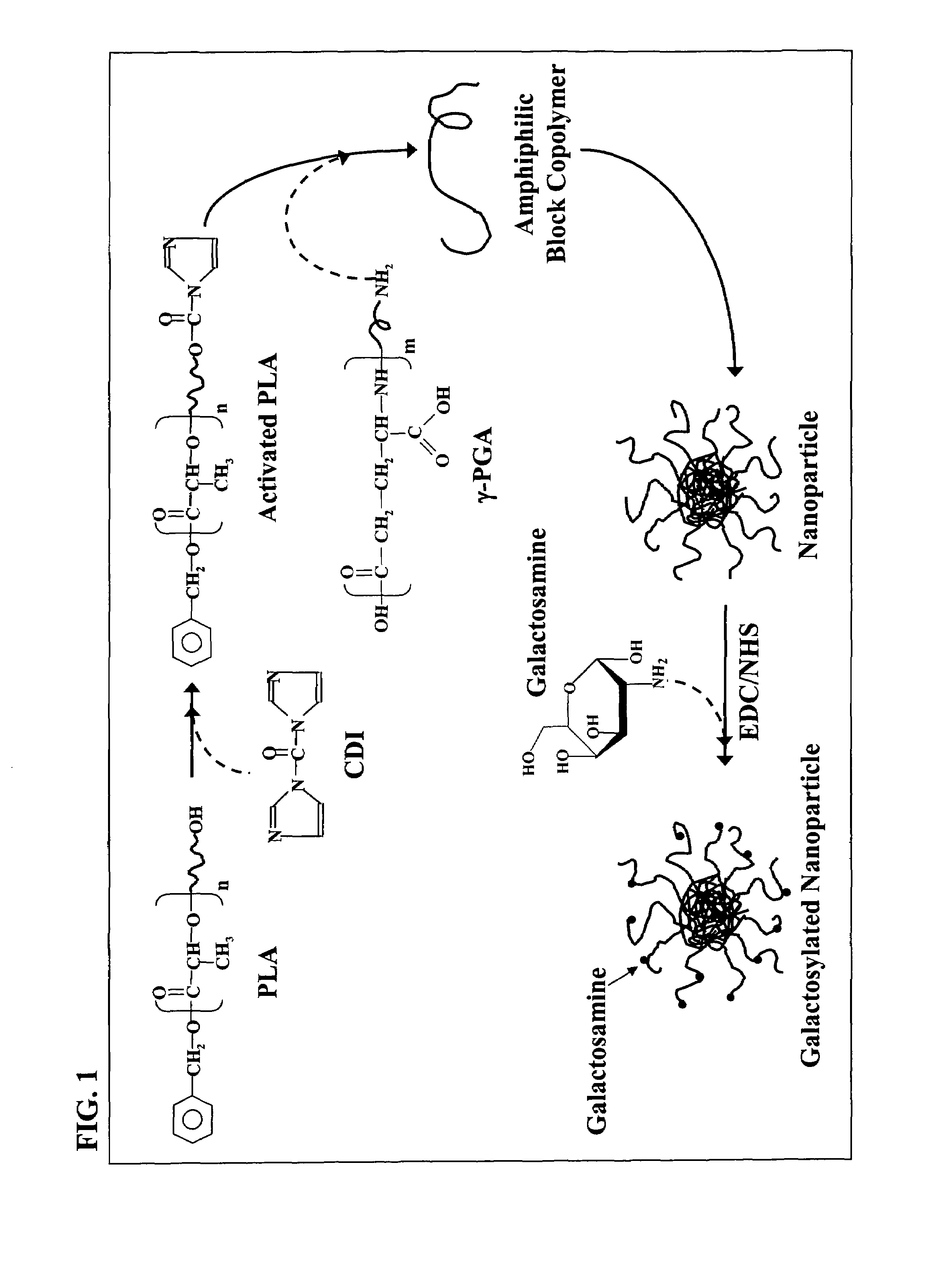
[0019]It is one object of the present invention to provide paclitaxel-loaded formulations using a novel type of self-assembled nanoparticles (NPs). In one embodiment, the NPs are composed of block copolymers synthesized by poly(γ-glutamic acid) and poly(lactide). In a further embodiment for the potential of targeting liver cancer cells, galactosamine is conjugated on the prepared nanoparticles (Gal-NPs). In the in vitro studies, it was found that both the NPs and the Gal-NPs have a similar release profile of paclitaxel. The activity in inhibiting the growth of HepG2 cells by the Gal-NPs was comparable to that of a clinically available paclitaxel formulation (Phyxol®), while the NPs of the present invention display a significantly less toxicity. The biodistribution and anti-tumor efficacy of the NPs and the Gal-NPs were studied in hepatoma-tumor-bearing nude mice. It was found that the groups injected with Phyxol®, the NPs or the Gal-NPs significantly delayed the tumor growth as compared to the control group injected with PBS.

Problems solved by technology

Chemotherapy for cancers is usually limited by the toxicity of drugs to normal tissues.
Additionally, short circulation half-life in plasma, limited aqueous solubility, and non-selectivity are usually encountered by most of the currently available anticancer drugs and thus restrict their therapeutic efficacy (Adv.
It is known that chemotherapy for cancers is usually limited by the toxicity of drugs to normal tissues (Adv.
However, the hydrophobicity of paclitaxel causes great difficulties in preparing formulations.
This binding stabilizes microtubules and greatly interferes with microtubular reorganization necessary, among other factors, for spindle formation and cell division (Cancer Lett.

Method used


Image

  • Nanoparticles for targeting hepatoma cells
  • Nanoparticles for targeting hepatoma cells
  • Nanoparticles for targeting hepatoma cells

Examples


example no.1

Example No. 1

Materials and Methods of Nanoparticles Preparation

[0052]PLA is herein an abbreviated term representing poly(L-lactide), Mn at 10 kDa, with a polydispersity of 1.1 determined by the GPC analysis. Dimethyl sulfoxide (DMSO99%), N-(3-dimethylaminopropyl)-N′-ethylcarbodiimide (EDC), N-hydroxysuccinimide (NHS), galactosamine, hydrophobic dye rhodamine-123, and sodium cholate were purchased from Sigma, USA. Pyrene as a fluorescence probe was acquired from Aldrich, USA. 4-Dimethylaminopyridine (DMAP) and 1,4-dioxane was purchased from ACROS (Janssen Pharmaceuticalaan, Belgium). All other chemicals used were reagent grade.

example no.2

Example No. 2

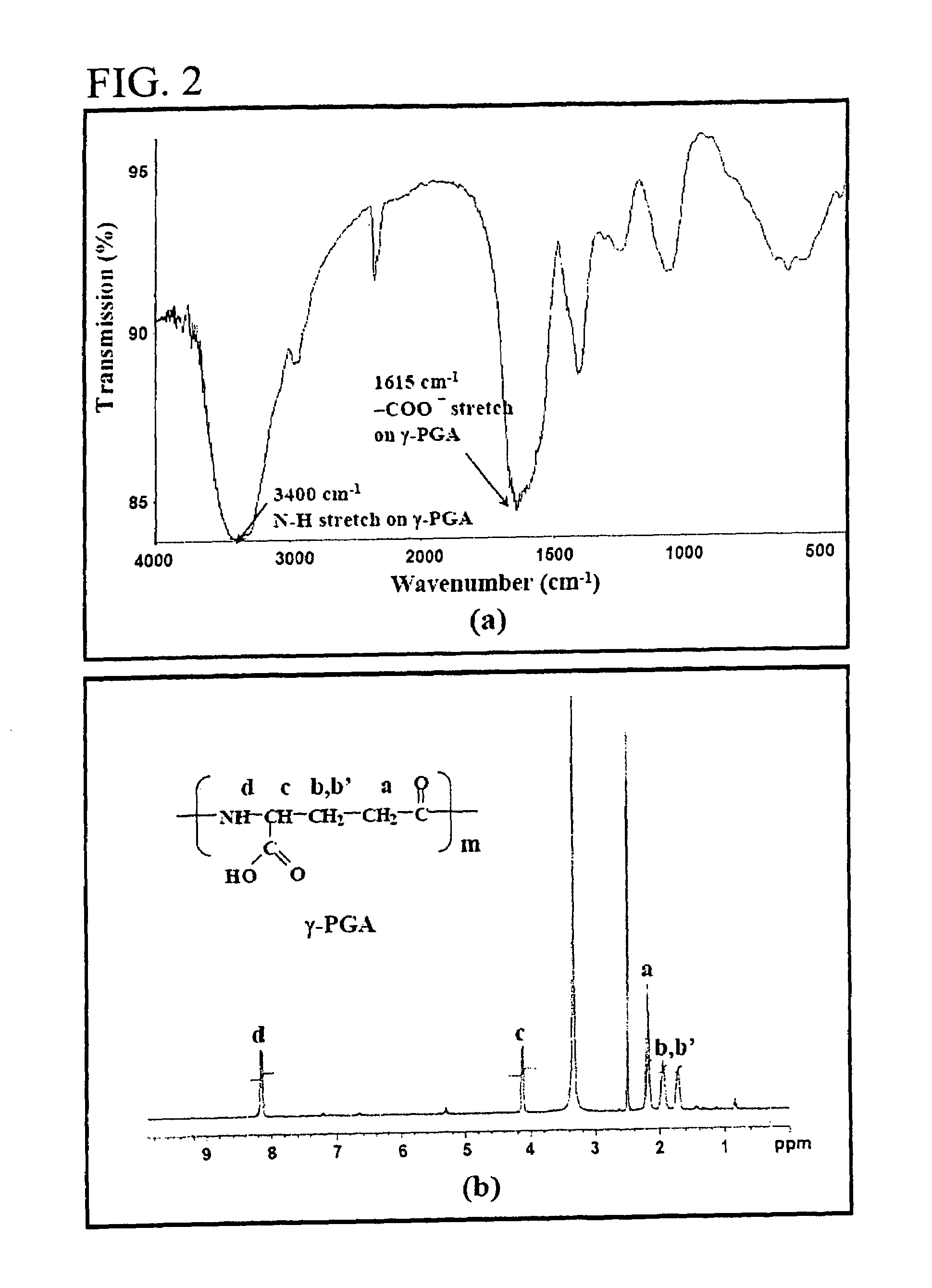
Production and Purification of γ-PGA

[0053]γ-PGA (FIG. 1) was produced by Bacillus licheniformis (ATCC 9945, Bioresources Collection and Research Center, Hsinchu, Taiwan) as per the method reported by Yoon et al. with slight modifications (Biotechnol. Lett. 2000; 22:585-588). Highly mucoid colonies (ATCC 9945a) were selected from Bacillus licheniformis (ATCC 9945) cultured on the E medium (L-glutamic acid, 20.0 g / l; citric acid, 12.0 g / l; glycerol, 80.0 g / l; NH4Cl, 7.0 g / l; K2HPO4, 0.5 μl; MgSO4.7H2O, 0.5 g / l, FeCl3-6H2O, 0.04 g / l; CaCl2.2H2O, 0.15 g / l; MnSO4.H2O, 0.104 g / l, pH 6.5) agar plates at 37° C. for several times. Subsequently, young mucoid colonies were transferred into 10 ml E medium and grown at 37° C. in a shaking incubator at 250 rpm for 24 hours. Afterward, 500 μl of culture broth was mixed with 50 ml E medium and was transferred into a 2.5-1 jar-fermentor (KMJ-2B, Mituwa Co., Osaka, Japan) containing 950 ml of E medium. Cells were cultured at 37° C. The p...

example no.3

Example No. 3

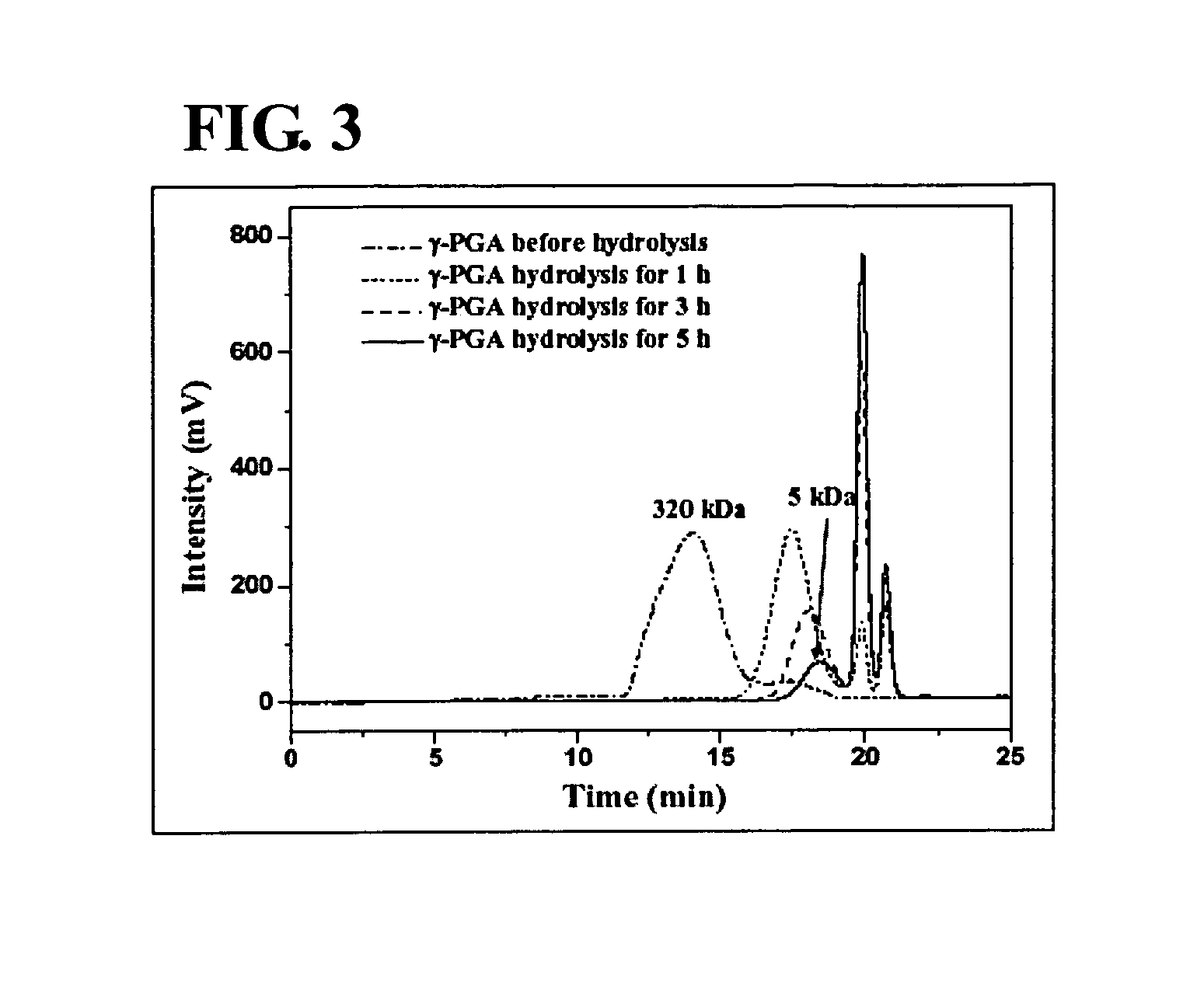
Hydrolysis and Analysis of γ-PGA

[0057]The average molecular weight (Mn) of the purified γ-PGA obtained via the previous procedure in Example No. 2 was about 320 kDa. The purified γ-PGA was then hydrolyzed in a tightly sealed steel container at 150° C. for distinct durations (J. Biol. Chem. 1973; 248:316-324). The average molecular weight along with the polydispersity of the hydrolyzed γ-PGA were determined by a gel permeation chromatography (GPC) system equipped with a series of PL aquagel-OH columns (one Guard 8 μm, 50×7.5 mm and two MIXED 8 μm, 300×7.5 mm, PL Laboratories, UK) and a refractive index (R1) detector (R12000—F, SFD, Torrance, Calif.). Polyethylene glycol (molecular weights of 106-22,000) and polyethylene oxide (molecular weights of 20,000-1,000,000) standards of narrow polydispersity (PL Laboratories, UK) were used to construct a calibration curve. The mobile phase contained 0.01M NaH2PO4 and 0.2M NaNO3 and was brought to a pH of 7.0. The flow rate of mob...


PUM

PropertyMeasurementUnit
particle sizeaaaaaaaaaa
particulate concentrationaaaaaaaaaa

Abstract

The nanoparticles composed of γ-PGA-PLA block copolymers that are conjugated with galactosamine as a potential drug delivery system and loaded with anticancer drugs for treating liver cancers.

Description

CROSS-REFERENCE TO RELATED APPLICATIONS[0001]This application is a continuation-in-part application of U.S. patent application Ser. No. 10 / 958,864 filed Oct. 5, 2004, entitled “Nanoparticles for Targeting Hepatoma Cells,” pending the entire contents of the co-pending application are incorporated herein by reference. The application also claims the priority benefits of U.S. Provisional Application No. 60 / 704,561 filed Aug. 2, 2005, entitled “Nanoparticles for Targeting Hepatoma Cells,” The entireties of the priority document are hereby incorporated by reference.FIELD OF THE INVENTION[0002]The present invention is related to nanoparticles composed of poly(γ-glutamic acid)-poly(lactide) block copolymers as a target drug delivery system, more particularly, the invention is related to nanoparticles loaded with bioactive agents and their uptake by HepG2 cells.BACKGROUND OF THE INVENTION[0003]Chemotherapy for cancers is usually limited by the toxicity of drugs to normal tissues. Additional...

Claims


Application Information

Patent Timeline
Patent Type & Authority Patents(United States)
IPC IPC(8): A61K9/16A01N43/04A61K9/14
CPCA61K9/5153A61K31/7008A61K31/704A61K47/48092A61K47/482A61K47/48315A61K47/48892A61K47/48915B82Y5/00C08G81/00A61K47/549A61K47/593A61K47/645A61K47/6931A61K47/6937
Inventor SUNG, HSING-WENLIANG, HSIANG-FATU, HOSHENG
Owner GP MEDICAL
Learn More