

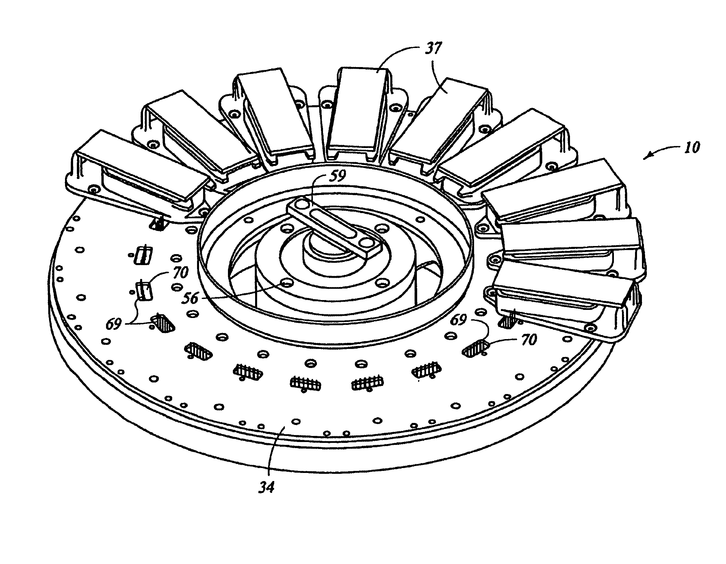
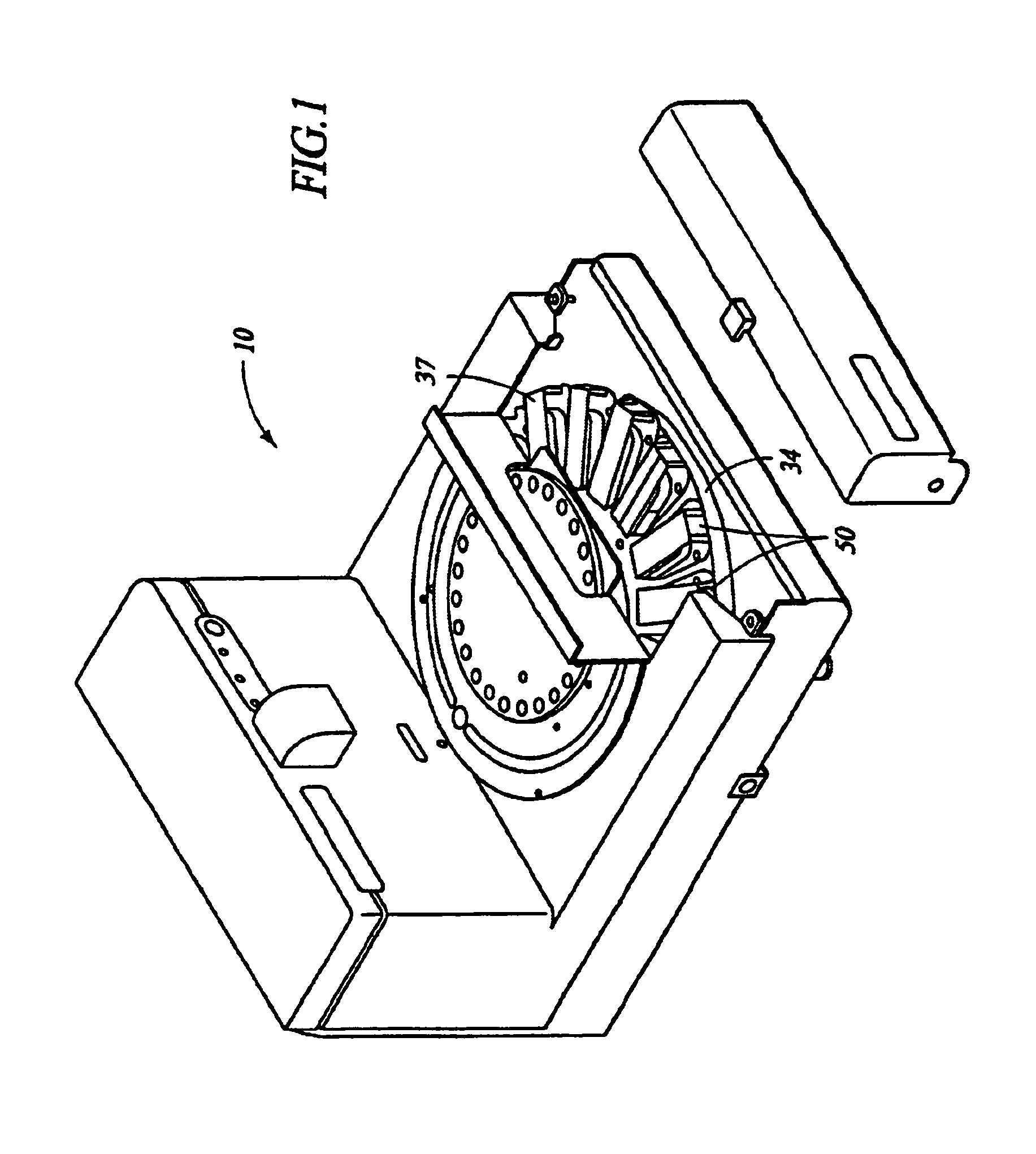
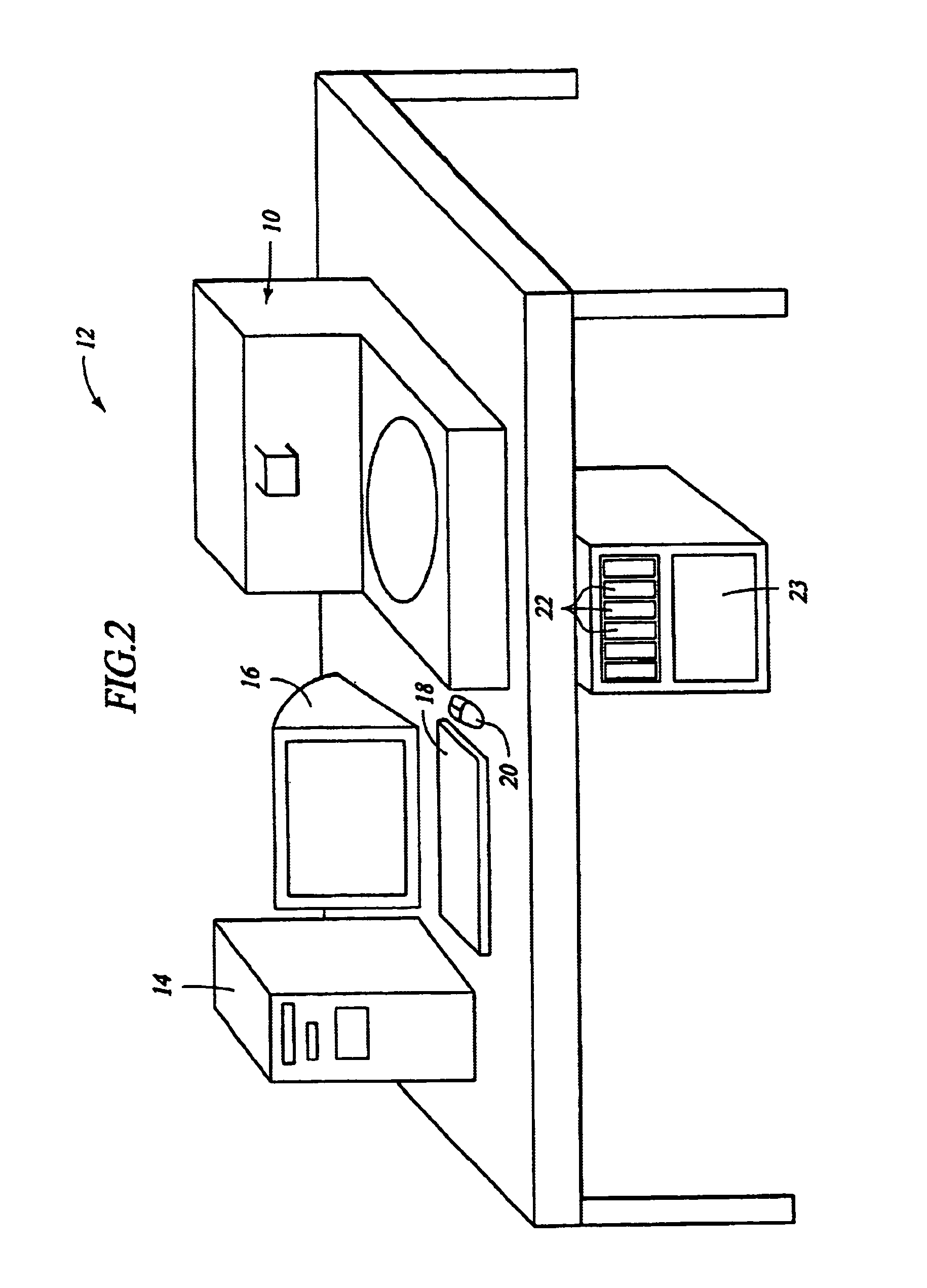
Automated molecular pathology apparatus having independent slide heaters
- Summary
- Abstract
- Description
- Claims
- Application Information
AI Technical Summary
Benefits of technology
Problems solved by technology
Method used
Image

Examples
examples
[0125]The following non-limiting examples further illustrate and detail uses and applications of the invention disclosed herein.
example i
Detection of High Risk Strains of Human Papaloma Virus (HPV) using Automated In-Situ Hybridization
[0126]A cervical smear, collected by standard collection devices such as a cytobrush or by a ThinPrep™ slide method (Cytec, Inc.) which would normally be stained by the Papanicolau stain (Pap smear), is subjected to in situ hybridization with a probe set specific to High Risk HPV types. The specimen slide is loaded into the slide holder of the apparatus according to the present invention (hereinafter referred to as “staining apparatus”). The system is set to run the HPV in situ program. This program performs all the steps of the in situ reaction with no interaction required from the user once the program has been started.
[0127]The staining apparatus first performs Slide Pretreatment: at room temperature 1.×APK. (10×APK. Ventana P / N 250-042) rinse and Coverslip™ application, then a Protease digestion for 4 minutes at 37° C. with Protease 1 followed by a rinse step in 2×SSC. Following the...
example ii
Detection of mRNA of Epstien Barr Virus (EBER) in Paraffin Embedded Tissue Using Automated In-Situ Hybridization
[0129]A 5 micron section was cut of spleen #EBV 37A and placed on a supper frost glass staining slide. The specimen slide was loaded into the slide holder of the apparatus according to the present invention (hereinafter referred to as “staining apparatus”). The staining apparatus was programmed to run the EBER in situ program. This program will perform all the steps of the in situ reaction with no interaction required from the user once the program has been started.
[0130]The staining apparatus first performs deparaffinization: slides were dry heated to 65° C. for 6 minutes, room temperature DI water rinses the slide, leaving 300 μL residual volume and 600 μL liquid coverslip, which prevents evaporation and protects the slide specimen from drying. The temperature remains at 65° C. to melt away the paraffin. Cell conditioning was done by rinsing the slide with DI water, then...
PUM
 Abstract
Description
Claims
Application Information