
Use of a biological material containing three-dimensional scaffolds of hyaluronic acid derivatives for the preparation of implants in arthroscopy and kit for instruments for implanting said biological material by arthroscopy

Inactive Publication Date: 2010-05-25
ANIKA THERAPEUTICS SRL
View PDF25 Cites 2 Cited by
  • Summary
  • Abstract
  • Description
  • Claims
  • Application Information

AI Technical Summary

Benefits of technology

[0054]Another advantage of the present invention lies in the fact that said biological material can be cryopreserved in order to preserve its characteristics of cell viability ready for implantation to be performed at a future time.
[0055]The kit of instruments for implanting biological material to be used for arthroscopy according to the present invention was constructed using materials with the preferred characteristic listed below:
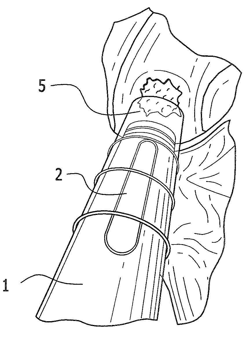
[0056]The cannula previously listed at item (b) used as a guide to give access in arthroscopy to the set of instruments listed hereafter and represented in FIGS. 6(I), 6(II), 8 (indicated with the number 5), is in Aise 316 steel and presents an inside diameter of 11.5 mm and is 111 mm long. In the aforementioned figures the sterile valves are not indicated.
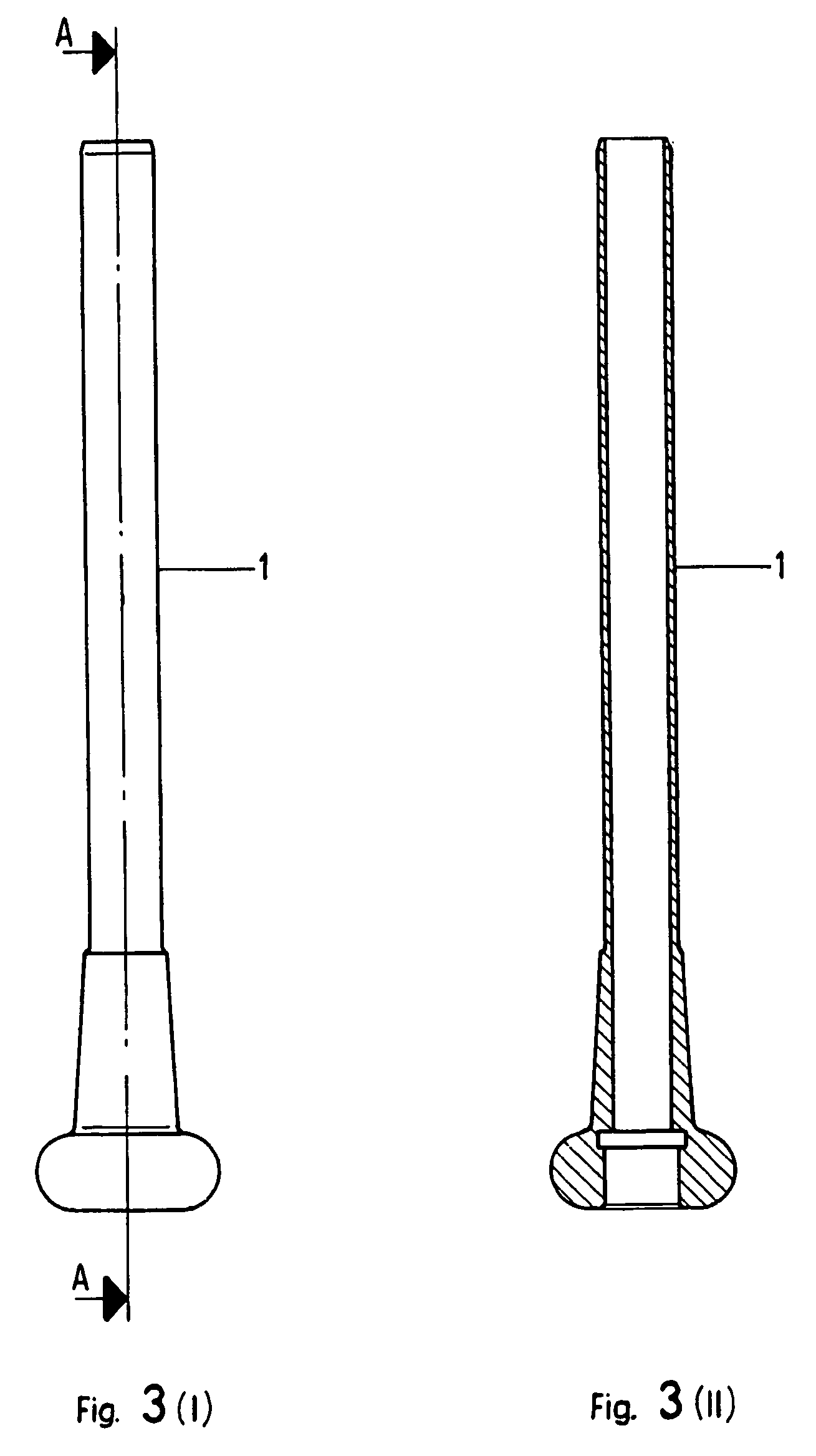
[0057]The mapper-sampler previously listed at item (c), being used to circumscribe the cartilage lesion by creating a circular imprint and to cut out and remove a piece of cartilage tissue of the same shape and size, and represented in the FIGS. 3, 6(I), 6(II) and 8 (indicated with the number 1) is a cannula in Aise 316 medical steel, 155 mm long, with an outer diameter of 10.5 mm and an inside diameter of 9 mm. The mappler sampler 1 in the kit according to the present invention is a hollow cylinder large enough to hold the plunger and is further characterised by having a concave tip like the mapper sampler and a control system by which the pressure exercised by the advancing plunger can be interrupted.
[0058]The guide wire previously listed at item (d), fixed with the aid of a drill to the centre of the lesion to stabilise the cutter during use, and represented in FIG. 7(I) (indicated with the number 3) has the diameter of 1 mm.
[0059]The concave hollow cutter-abrasor previously listed at item (e), used to create within the margins of the imprint left by the mapper 1 the site in which the biological material to be used in arthroscopy according to the present invention for subsequent grafting and represented in FIGS. 5 and 7(I) and 7(II) (indicated with 4) is in Aise 316 medical steel, is 162.5 mm long with an inside diameter of 1.2 mm and an outer diameter of 9.5 mm.

Problems solved by technology

The final phase of hyaline cartilage degeneration is accompanied by pain and limited mobility that may cause locking of the joint.
In the long term, the degenerative process may even cause the onset of complications such as osteoarthritis.
This is a costly procedure and is not even permanent because many prostheses have to be changed after about 10-15 years.
The resulting tissue does not, however, have the mechanical properties or physiological or structural characteristics of healthy, lasting joint cartilage.
Initially, this treatment triggers the development of hyaline cartilage, but the repair tissue does not take easily to the surrounding healthy tissues, and subsequently becomes ossified.
The surgical procedure is actually rather complex.
First of all, the operation requires open surgery.
Indeed, if the chamber is not perfectly sealed, the cells will leak out again and the operation will have failed.
To summarise, the main disadvantages of this procedure are that the operation is difficult to perform, the technique is invasive and the implanted cells are not perfectly differentiated.
Autologous and homologous osteochondral grafts involve techniques that are surgically invasive and complex and there is a risk of viral transmission with the latter.
The chief disadvantage of using said scaffolds is represented by an immune response directed towards the implanted material.
However, said cultures in gel do not provide suitable mechanical stability to remain adhered to the site and allow the reconstruction of the cartilage structure.
Lastly, although the gels constituted by substances such as agarose induce chondrocyte redifferentiation, the use of this compound has not been approved for internal application in humans.

Method used


Image

  • Use of a biological material containing three-dimensional scaffolds of hyaluronic acid derivatives for the preparation of implants in arthroscopy and kit for instruments for implanting said biological material by arthroscopy
  • Use of a biological material containing three-dimensional scaffolds of hyaluronic acid derivatives for the preparation of implants in arthroscopy and kit for instruments for implanting said biological material by arthroscopy
  • Use of a biological material containing three-dimensional scaffolds of hyaluronic acid derivatives for the preparation of implants in arthroscopy and kit for instruments for implanting said biological material by arthroscopy

Examples


example 1

Preparation of the Biological Material

[0077]The cell culture process is described in Brun et al., “Chondrocyte aggregation and reorganisation into three dimensional scaffolds”, J. Biomed. Mater. Res. 1998, 46, 337-346. Cartilage tissue taken from a non-weight-bearing area marginal to the lesion is detached by treatment with type-II collagenase, and the cells thus obtained are seeded in dishes containing HAM's F12 medium supplemented with foetal calf serum, 1% streptomycin-penicillin, 1% glutamine and with the following trophic factors, each of which in a quantity ranging between 1 and 10 ng / ml: TGF β1, recombinant human EGF, recombinant human insulin and recombinant human bFGF.

[0078]The cells are grown in vitro from one to four serial passages, then seeded on HYALOGRAFT®C (a non-woven fabric based on HYAFF®11t—total benzyl ester of hyaluronic acid), at a cell density of between 0.5×10⁶ and 4×10⁶ cells / cm2, and the culture medium described above. At each change of medium (2-10 ml eve...

example 2

Valuation of Cell Viability by the MTT Test

[0079]Cell viability of the biological material is determined by incorporation of the vital MTT dye (F. Dezinot, R. Lang “Rapid calorimetric assay for cell growth and is survival. Modification to the tetrazolium dye procedure giving improved sensitivity and reliability” J. Immunol. Methods, 1986, 22 (89), 271-277). A solution prepared by dissolving 0.5 mg / ml of MTT in phosphate buffer, pH 7.2 (PBS) is added to the test material and placed in an incubator set at 37° C. for 4 hours. Once incubation is complete, the MTT solution is aspirated, the material washed several times with PBS, after which 5 ml of extracting solution constituted by 10% dimethylsulphoxide in isopropyl alcohol is added. After centrifugation, the absorbance of the supernatant is determined by a spectrophotometric reading at 470 nm.

example 3

Verification of Cell Viability Recovery After Passage Through a Cannula

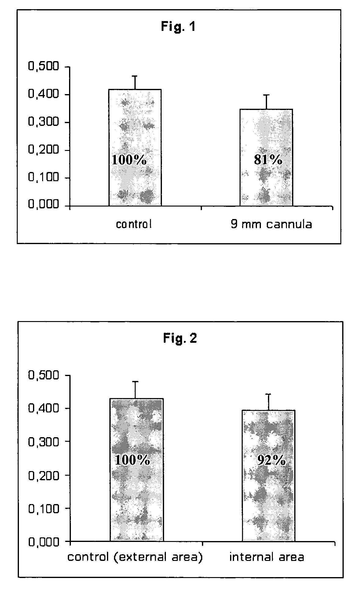
[0080]The MTT test as described in Example 2 is used to verify cell viability recovery after passage through a cannula, an instrument commonly used in arthroscopy and used here to place the biological material.

[0081]The results are reported in FIG. 1, which shows cell viability recovery relative to a bio-engineered cartilage construction (Hyalograft®C, dimensions 2×2 cm), prepared as described in Example 1. After 72 hours in its packaging, that is, in conditions of maximum metabolic stress, the biomaterial was gently extruded through a cannula with a diameter of 9 mm. It was found that the passage through the hollow of the cannula does not modify the cell viability of the present material.


PUM

PropertyMeasurementUnit
Timeaaaaaaaaaa
Cell viabilityaaaaaaaaaa

Abstract

The invention relates to use of a biological material containing cells supported on three-dimensional scaffolds containing at least one hyaluronic acid derivative for the preparation of grafts suitable for application by arthroscopy, and a kit of surgical instruments for implanting said biological material by arthroscopy.

Description

FIELD OF THE INVENTION[0001]The present invention concerns the use of biological material containing cells supported on three-dimensional scaffolds which comprise at least one hyaluronic acid derivative for the preparation of grafts suitable for application by arthroscopy, and a kit for surgical instruments for implanting said biological material by arthroscopy.BACKGROUND OF THE INVENTION[0002]The aim of joint cartilage repair is to restore the integrity of the joint surface, reduce pain and prevent any further deterioration of the tissues.[0003]Joint cartilage is a tissue which allows virtually frictionless movement of the joint. Its particular biological characteristics enable the joint to absorb forces at least five times greater than the body's weight. The joint cartilage, or hyaline, has a very limited capacity for self-repair, so the type of cartilage that is spontaneously regenerated after damage does not possess the same characteristics as the original tissue. It is known as...

Claims


Application Information

Patent Timeline
IPC IPC(8): A61F5/00A61F2/02C12N5/07A61L27/00A61L27/20A61L27/38C12N5/074
CPCA61L27/20A61L27/3804A61L27/3852C08L5/08
Inventor CALLEGARO, LANFRANCOMARCACCI, MAURILIODI FEDE, SERGIO
Owner ANIKA THERAPEUTICS SRL
Learn More