
Method for rapid typification of microorganisms and set of reagents used

a technology of microorganisms and reagents, applied in the direction of fluid pressure measurement, liquid/fluent solid measurement, peptides, etc., can solve the problems of large amount of buffer solution, and indirect measurement of the changes in the genetic background

Inactive Publication Date: 2010-06-08
CENT NACIONAL DE INVESTIGACIONES CIENTIFICAS (CINC)
View PDF7 Cites 1 Cited by
  • Summary
  • Abstract
  • Description
  • Claims
  • Application Information

AI Technical Summary

Benefits of technology

"The patent describes a process for quickly typing microorganisms using a method called PFGE (Pulsed Field Gel Electrophoresis). This process involves preparing samples of the microorganisms, designing optimal separation conditions, and running the PFGE to separate the DNA molecules. The resulting band patterns are then analyzed to identify and differentiate different strains of microorganisms. The technical effect of this patent is to provide a reliable and efficient method for typing microorganisms, which is important for identifying outbreaks of infection and monitoring vaccination programs."

Problems solved by technology

Thus, phenotypic methods have the disadvantage of giving an indirect measure of the changes in the genetic background.
Due to the large dimensions of the CHEF chamber, it requires large amount of buffer solution to cover its electrode platform.
Nevertheless, TAFE also requires large amount of buffer solution (3.5 liters) and biological sample.
As a conclusion, the commercially available equipments most frequently used to characterize the genome of microorganisms and parasites require long running time, and large amount of reagents and biological samples to resolve large DNA molecules in their band pattern.
Mobility inversion of DNA prevents correct size estimations and makes difficult the interpretation and comparison of the patterns given by numerous samples.
The main disadvantage of mobility inversion is the impossibility of identifying unambiguously the molecules present in a given band.
Hybridization notably increases the prices of the assay and is time demanding, thus the typing process will be more expensive and time consuming.
Bio-Rad, pp 11, 1995) by increasing the voltage applied to the electrodes is nearly impossible.
It is due to the limit of power supply output and the cooling system.
Current TAFE equipments have similar problems.
In addition, simple procedures to select miniequipment running parameters are not available.
However, spheroplasts need to be formed if said microorganisms possess cell wall, and the enzymes needed to form spheroplasts, as well as the proteases, are expensive.
In parallel, Higginson et al (Higginson D. et al., Anal. Lett., 1994, 27:7, 1255-1264) showed that S. cerevisiae DNA can be deproteinized using 8 M urea instead of proteinase K. However, the method described by Gardner is still expensive, because he used proteases to deproteinize the DNA, whereas, the method described by Higginson is cheap, but consumes 72 hours of incubation, which is a long time.
However, it consumes 6 hours and the authors attributed the protocol success to the activation of an autolysin of the Streptococcus.
Therefore, current methods are expensive and consume long times.
However, a general non-enzymatic method to treat microorganisms with cell walls prior to their embedding in agarose gels has not been presented yet.
However, intact DNA molecules are not obtained, because DNA is isolated in solution; and the authors did not propose the method to obtain the molecular karyotypes or the pulsetypes of the mentioned microorganisms.
Authors reported that the obtained DNA is suitable for hybridization, but they did not address the possibility of DNA restriction with endonucleases.
In conclusion, said method does not guarantee the obtaining of intact DNA molecules, whereas the possibility of digesting them with restriction enzymes remains unknown.
Therefore, a general procedure has not been proposed yet for the preparation of immobilized intact DNA from yeast, gram-positive and gram-negative bacteria and parasites by non-enzymatic methods in short times.
The use of disposable molds requires continuous supply, which can be a limiting factor in laboratories with low budget.
However, cutting the ribbons or noodles provokes plugs of non-homogeneous sizes and dimensions, which influence the quality of the electrophoresis patterns.
Consequently, the comparison of the patterns obtained in different lanes of the gel is difficult.
Difficulties in the comparisons of band patterns represent a disadvantage in microorganism typing.
However, it is a disadvantage: if the plugs are kept inside the mold during the incubations, the time needed to obtain the samples suitable for PFGE analysis is notably lengthened.
The selection of the experimental conditions is complex.
In addition, if extremely wide size ranges are entered to the auto-algorithm, as well as to the interactive program, erroneous results, such as band inversion in the mid range of the gel, can be generated.
However, said expression is valid on specific experimental conditions alone, and it does not predict the resolution between two any molecules.
However, said function only permits to obtain rough estimates of the two mentioned variables, and does not provide the molecule migrations at any experimental condition.
They did not generate methods allowing the PFGE user to select and set the experimental conditions needed to separate the molecules under analysis.
However, the comparison of the restriction patterns remains a subjective process, and it cannot be totally reduced to rigid algorithms (Tenover F et al., J. Clin. Microbiol, 1995, 33(9):2233-2239).
However, PFGE is relatively new, and size markers for wide DNA size range are not always available.
Total process for microorganism typing needs long time and many resources.
A factor limiting the use of PFGE for microorganism typing is the time needed to complete the analysis of isolates, It is from 2 to 3 days, thus reducing the capacity of the laboratories to analyze many samples (Olive D M and Bean P, J. Clin. Microbiol, 1999, 37:6, 1661-1669).

Method used


Image

  • Method for rapid typification of microorganisms and set of reagents used
  • Method for rapid typification of microorganisms and set of reagents used
  • Method for rapid typification of microorganisms and set of reagents used

Examples


example 1

Mold Able to be Sterilized for the Preparation of Sample Miniplugs

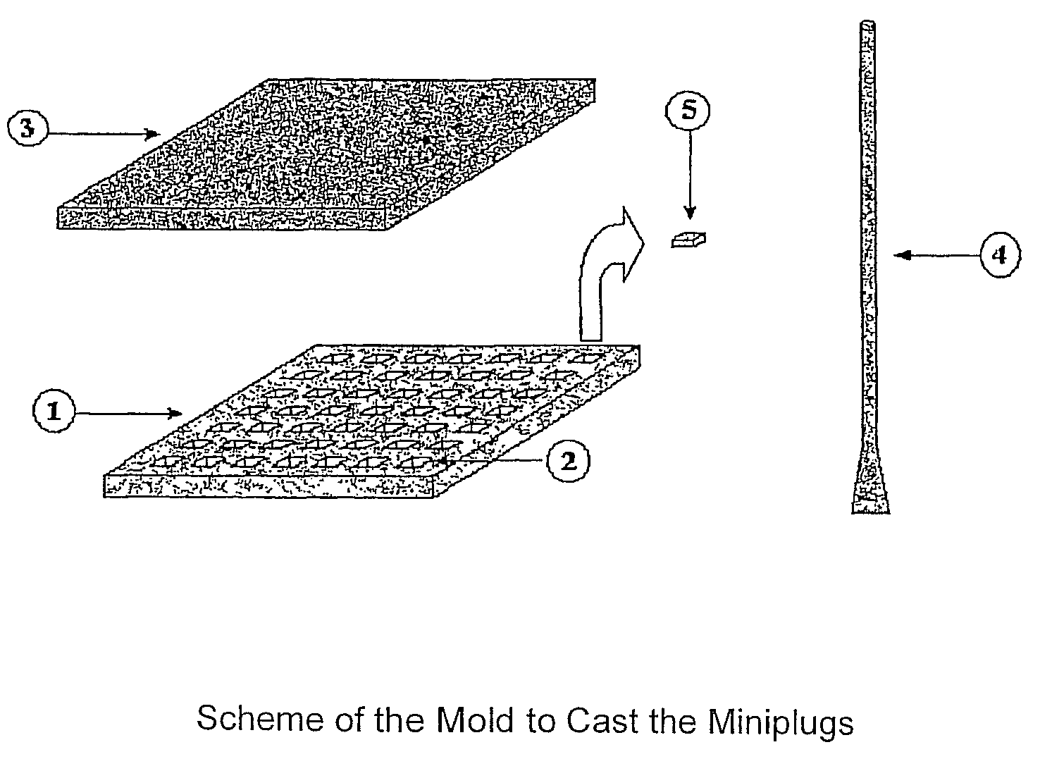
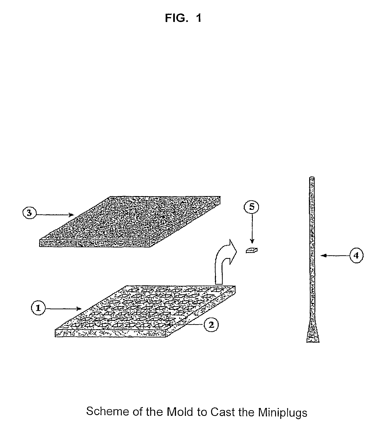
[0161]An example of the mold able to be sterilized and made of a flexible material (silicone, rubber or any other material) is shown in the scheme of FIG. 1. The mold is used to prepare agarose-embedded DNA samples. The sheet (1) has 49 depressions (2). The agarose-cell suspension is poured onto the mold or sheet and distributed in the mold with the especial spatula (4). Later, the sheet (1) is cover up with the lid (3), generating the miniplugs (5) containing embedded cells. The sheet of the real mold was made with melted silicone. It was poured into another mold until the sheet (1) was formed. In the example, miniplug dimensions are 3×3×0.7 mm (length, wide×thickness). To recover the miniplugs (5) from the sheet (1), said sheet is held by its ends and bent inside a container with a solution. Consequently, the miniplugs are released from the sheet and dropped into the solution.

[0162]In other mold variants, the depres...

example 2

Non-enzymatic Preparation of Agarose-embedded Intact Yeast DNA Starting from Cells Cultured in Broth Media

[0163]Yeasts (S. cerevisiae, H. polymorpha or P. pastoris) were grown in liquid YPG medium (YPG: 10 g yeast extract, 20 g glucose and 10 g peptone dissolved in one liter of distilled water) with shaking at 30° C. until late log phase. Cells were harvested by centrifugation and washed with 0.05 M EDTA, pH 7.5 (washing solution, Table I). Agarose-embedded intact DNA is obtained performing any of the two following variants:[0164]Variant 1: Cells are incubated prior to their embedding in agarose miniplugs.[0165]Variant 2: Cells are embedded in agarose miniplugs and further the miniplugs are incubated.

[0166]In both variants the cells are embedded in agarose by preparing a cell suspension of 1.3×10¹⁰ cell / ml in 1.5% low melting agarose which was first dissolved in 0.125 M EDTA. The cell suspension is poured onto the sheets (1) of the mold shown in FIG. 1, then, the mold is covered wit...

example 3

Non-enzymatic Preparation of Agarose-embedded Intact DNA from Pseudomonas Aeruginosa Grew in Broth and on Solid Media

[0173]Two colonies of P. aeruginosa were isolated from a blood-agar plate. One of them was inoculated in 5 ml of LB medium (10 g yeast extract, 5 g sodium chloride and 10 g bacto-triptone per liter of distilled water) and the other was streaked on a LB plate (LB plus 1.2% bacteriologic agar).

[0174]Both cultures were incubated overnight at 37° C. Broth culture was incubated with shaking and the plates were kept static. Cells, grown in broth, were collected by centrifugation, whereas the plates were washed with the proper washing solution (shown in Table I) and further collected by centrifugation.

[0175]Cells grew in broth or solid media were washed with the solution shown in Table I, collected by centrifugation and re-suspended at a concentration of 2×10⁹ cells per milliliter of 1.5% low melting agarose dissolved in 0.15 M NaCl. Agarose-cells mix was poured onto the she...


PUM

PropertyMeasurementUnit
pHaaaaaaaaaa
sizeaaaaaaaaaa
sizeaaaaaaaaaa

Abstract

A process for rapid typing of yeast, parasites and bacteria is provided. It comprises the following steps: a) Preparing immobilized intact DNA in 5 to 60 minutes aided by a method involving a reagent kit that only contains buffer solution, a detergent, a metal chelating agent and an agent to disrupt the hydrogen bonds. b) Separating intact DNA molecules or their restriction fragments in miniequipments for Pulsed Field Gel Electrophoresis of the CHEF (Contour Clamped Homogeneous Electric Field) and TAFE (Transversal Alternating Field Electrophoresis) systems in times comprised between 2.5 and 7 hours. c) Selecting the optimal conditions that should be set in miniCHEF with the aid of a method to simulate the electrophoresis patterns d) Providing reorientation times, migration velocities and sizes of the molecules calculated with the aid of a method to analyze the migrated distances without the use of size markers.

Description

[0001]This application is the national phase under 35 U.S.C. §371 of PCT International Application No. PCT / CU01 / 00008 which has an International filing date of Nov. 2, 2001, which designated the United States of America.DESCRIPTIVE MEMORYINTERNATIONAL PATENT CLASSIFICATION INDEX: C12N 1 / 00REFERENCE TO RELATED APPLICATION[0002]This application is a 371 National Stage Entry of PCT / CU01 / 00008 filed on Nov. 2, 2001.[0003]The present invention is related to molecular biology. In particular, a process is provided. Said process includes the use of methods and procedures, as well as a reagent kit for rapid microorganism typing. The microorganisms to be typed can be yeast, parasites and bacteria Gram-positive or Gram-negative. The microorganism typing is done by Pulsed Field miniGel Electrophoresis performed in miniequipments of CHEF or TAFE system.BACKGROUND OF THE INVENTION[0004]During the past years, infectious diseases have increased and multi-drug resistance microorganisms have arisen (...

Claims


Application Information

Patent Timeline
Patent Type & Authority Patents(United States)
IPC IPC(8): G01N27/447C12N1/06C12Q1/68
CPCG01N27/44773C12Q1/6806
Inventor LOPEZ CANOVAS, LILIARIVERON ROJAS, ANA MARIAHIGGISON CLARKE, DAVIDSANCHEZ ALONSO, AXELOROZCO OROZCO, ESTHERARENCIBIA DIAZ, OSCARARIOSA ACUNA, MARIA CONCEPCIONCLARK DONDERIZ, HILDA TERESAGIGATO PEREZ, REGNAR
Owner CENT NACIONAL DE INVESTIGACIONES CIENTIFICAS (CINC)
Learn More