High resoultion digital imaging apparatus

a digital imaging and high-resoultion technology, applied in the field of high-resoultion digital imaging apparatus, can solve the problems of increasing yield, reducing cost, and sensors still needing to be well calibrated, and achieves low noise detectors, small capacitance, and large thickness

Inactive Publication Date: 2012-02-21
NOVA R&D
View PDF21 Cites 52 Cited by
  • Summary
  • Abstract
  • Description
  • Claims
  • Application Information

AI Technical Summary

Benefits of technology

[0013]The hybrid silicon pixel detector (SiPD) is fabricated from two sections which are a two-dimensional silicon PIN diode array and a front end readout electronics chip with time delayed integration (TDI) based upon a charge coupled device (CCD) type read out. Both sections are designed to have matching pixel geometry and the two sections are electrically connected together using an indium bump bonding technique. This allows each diode to be directly connected to its readout electronics and allows fabrication of small capacitance and low noise detectors. The active detector section of the PIN diode array, can be made with no dead volume and with larger thickness to increase quantum efficiency. The diode array detects x rays directly without a scintillator or fluorescent screen. The diode may be fabricated with 44×44 micron2 pixels and 1.5 mm to 2 mm thickness. When higher resistivity silicon substrates are available, the thickness of the PIN diode array can be increased to 2 mm to further improve the quantum efficiency. Also smaller pixel sizes (≧30 micron) can be used to improve resolution if needed. The silicon PIN diode array and the readout electronics chip can be fabricated using Orbit's 2.0 micron CMOS CCD process. This is a well developed low noise process with good resistance to radiation.
[0014]The scanning digital mammography system uses a hybrid pixel detector system. A scanning linear detector array is mounted on an accurate swing arm and the array can be scanned sideways under the patient's breast. Sideways scanning is preferred as it allows the linear array to achieve uniform speed before starting to image and slow down only after the imaging is finished. It also allows coming close to the chest wall not to miss any breast tissue.
[0015]The scanning linear array detector system is read out using the time delayed integration (TDI) based on a CCD type readout. Standard TDI circuitry is designed and built using a CMOS CCD process. This reduces the complexity of the scanned imaging system. The TDI technique brings an important advantage to the proposed system since it greatly reduces the required data bandwidth, and it is also intrinsically immune to a small fraction of imperfect pixels. This increases the yield and decreases the cost. However, the sensors still must be well calibrated to allow for the non functioning pixels. The width of the hybrid chip is made from about 128 pixels (approximately 0.6 cm) to reduce x-ray tube loading and the length 512 pixels (approximately 2.2 cm) to minimize the depth (curvature) effect. The TDI is applied over the 128 pixels.

Problems solved by technology

This increases the yield and decreases the cost.
However, the sensors still must be well calibrated to allow for the non functioning pixels.

Method used


Image

  • High resoultion digital imaging apparatus
  • High resoultion digital imaging apparatus
  • High resoultion digital imaging apparatus

Examples


Embodiment Construction

Advantages of the Invention

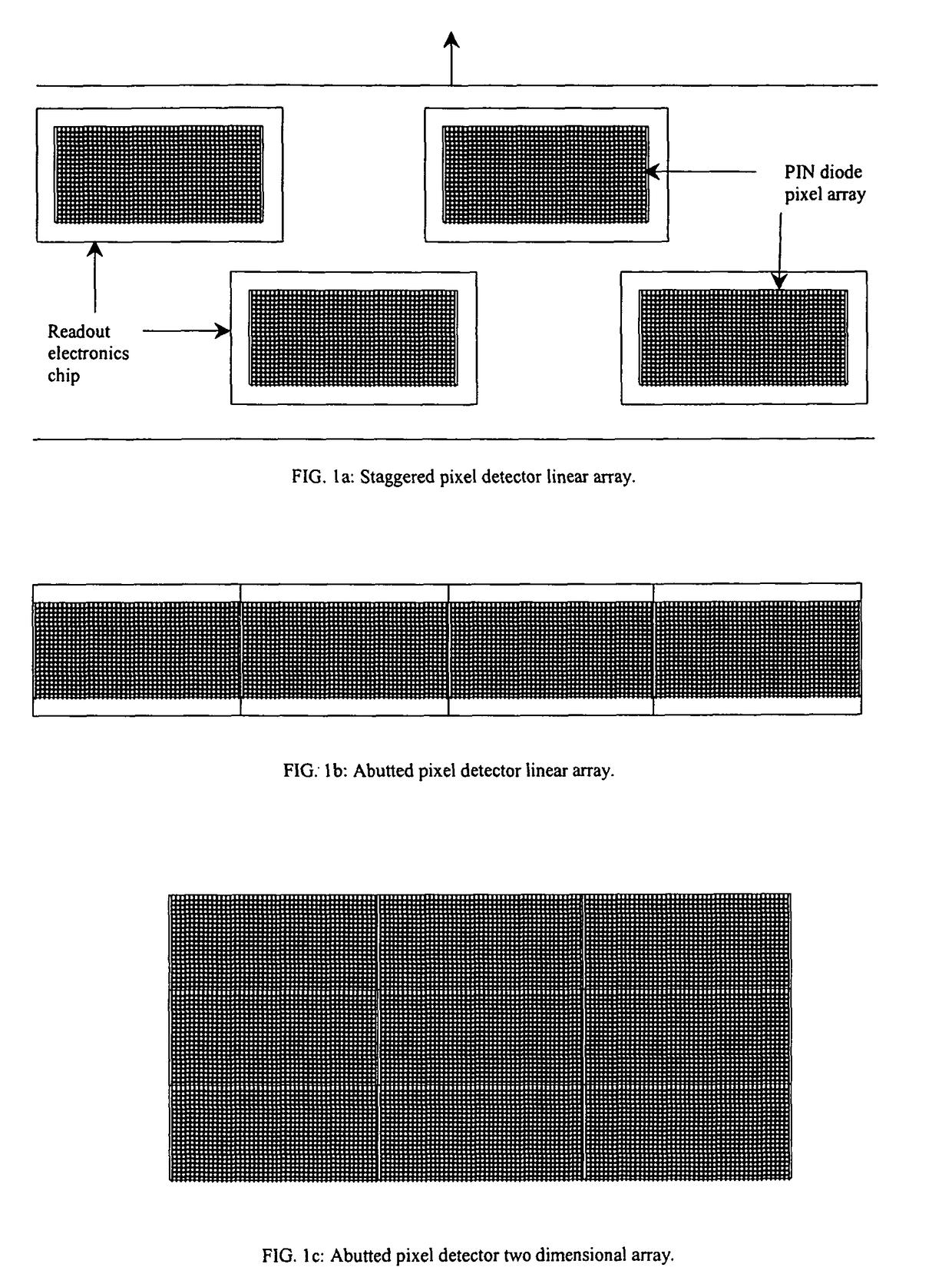
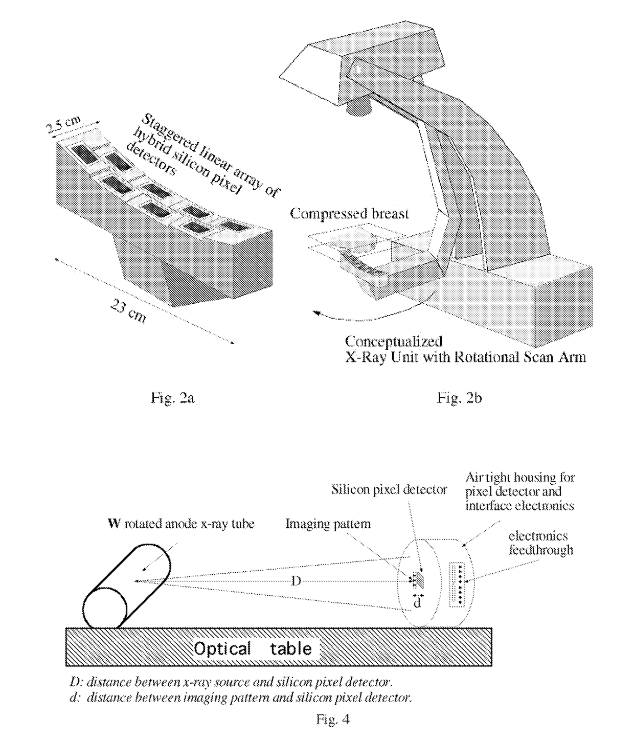
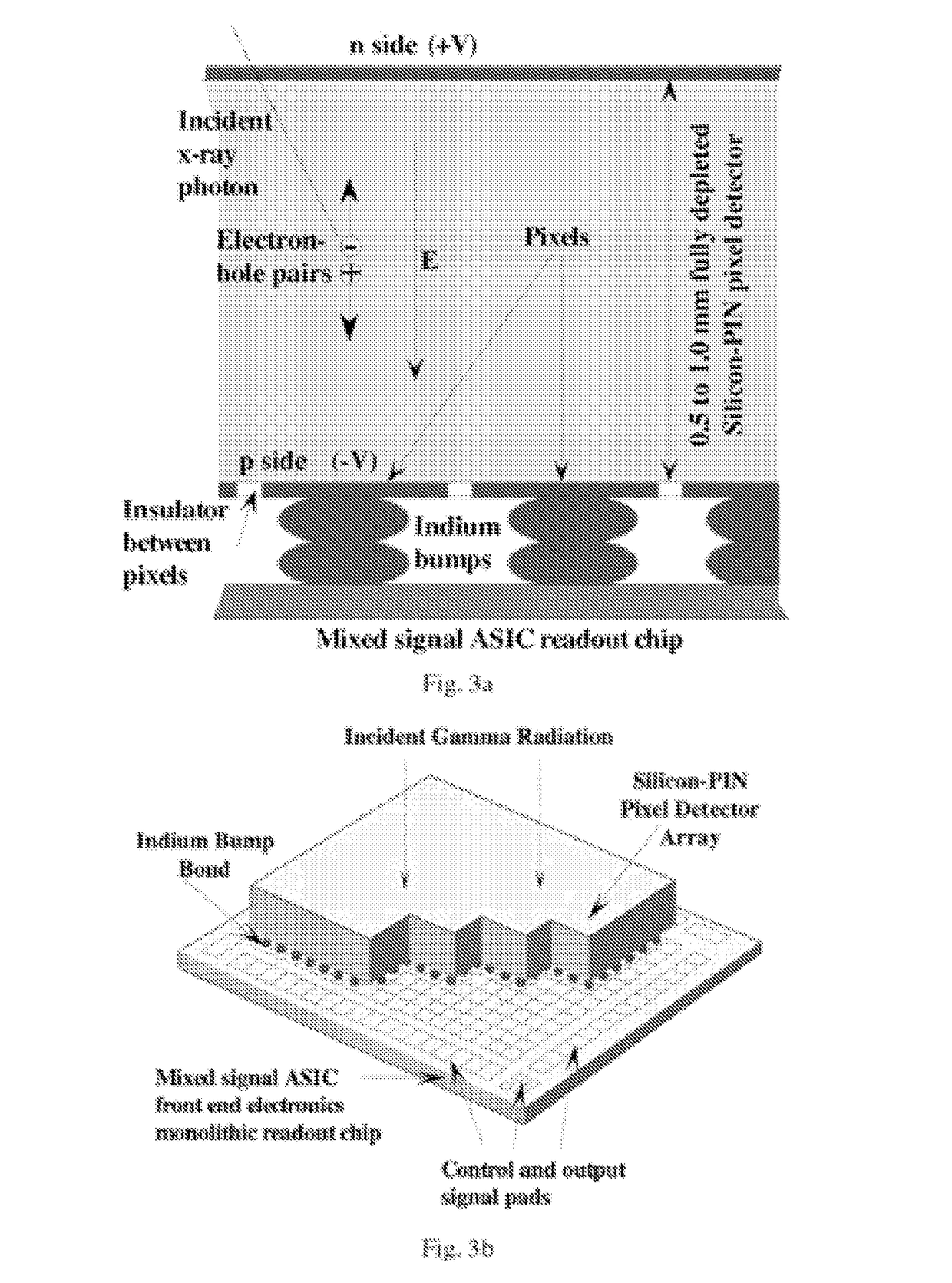
[0051]The SiPDs have excellent properties which can lower the radiation dose, improve resolution and overcome the limitations of the present mammography systems. The digital mammography system of this invention using SiPDs provides the following advantages:[0052]1. High spatial resolution: A silicon pixel detector with pixel size of 44×44 micron2 or 50×50 micron2 is conservatively planned for the proposed prototype. Smaller pixel sizes to 25×25 micron2 with TDI capability may be feasible if necessary. This will allow high resolution fine detail imaging.[0053]2. Low-dose rate: The interaction probability of SiPDs can be reasonably high because the depletion region can be made thick. The proposed SiPDs have excellent photo-absorption efficiency for 15 to 25 kev x rays. The present technology can produce 1.5 mm thick pixel detectors, which give about 95%, 78% and 53% quantum efficiencies for x-ray energies of 15, 20 and 25 keV. Larger thickness pixel detector...


PUM


Abstract

An integrated application specific integrated circuit having a detection layer, a time delayed integration capability, data acquisition electronics, and a readout function is provided for detecting breast cancer in women. The detection layer receives x-ray radiation and converts the received energy to electron pairs, one of which is received by pixels. The time delay integration is on the chip and a part of the readout architecture. The detector may be a hybrid silicon detector (SiPD), a CdZnTe detector, or a GaAs detector.

Description

CROSS REFERENCE TO PROVISIONAL PATENT APPLICATION[0001]This application claims the benefit of the filing date of U.S. Provisional Patent Application 60 / 128,407 filed Apr. 8, 1999, the disclosure of which is incorporated herein by reference.GOVERNMENT RIGHTS NOTICE[0002]This invention was made with U.S. Government support under Contract Numbers DAMD17-97-1-7255, 1R43CA61420-03, and CA66015 awarded by the Department of Defense, National Institute of Health and National Cancer Institute, and the National Institute of Health, respectively. The U.S. Government has certain rights in this invention.FIELD OF THE INVENTION[0003]Mammography has attained widespread acceptance as an important tool for the screening and early diagnosis of breast cancer. Current mammography systems are screen-film based and have limited x ray detection efficiency and limited ability to display fine details. The x-ray film also needs to be photographically developed before it can be viewed, which introduces extra ...

Claims


Application Information

Patent Timeline
Patent Type & Authority Patents(United States)
IPC IPC(8): H04N3/14H01L27/146
CPCH01L27/14634H01L27/14659H01L27/14661H01L27/14676H01L27/1469H04N5/32H04N5/372H04N25/71
Inventor TUMER, TUMAY OYIN, SHIMAEDING, DALE G.ASBROCK, JAMESYAFFE, MARTINMAWDSLEY, GORDONMAINPRIZE, JAMES
Owner NOVA R&D
Why Eureka
Try Eureka