
Ionic complex nanoparticles for detecting heparanase activities and method for preparing the same

a technology heparanase activity, which is applied in the field of ionic complex nanoparticles for detecting heparanase activity, can solve the problems of difficult to detect the heparanase activity expressed in cells and in vivo, and to detect diseases early by using the sensor, and achieves strong fluorescence, prevent over-expression of heparanase, and high quenching

Inactive Publication Date: 2012-12-04
KOREA INST OF SCI & TECH
View PDF0 Cites 7 Cited by
  • Summary
  • Abstract
  • Description
  • Claims
  • Application Information

AI Technical Summary

Benefits of technology

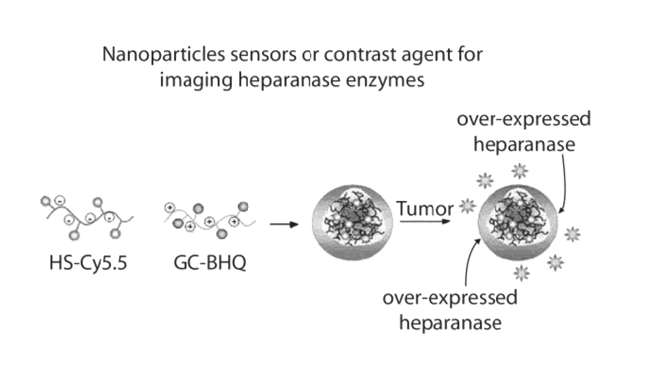
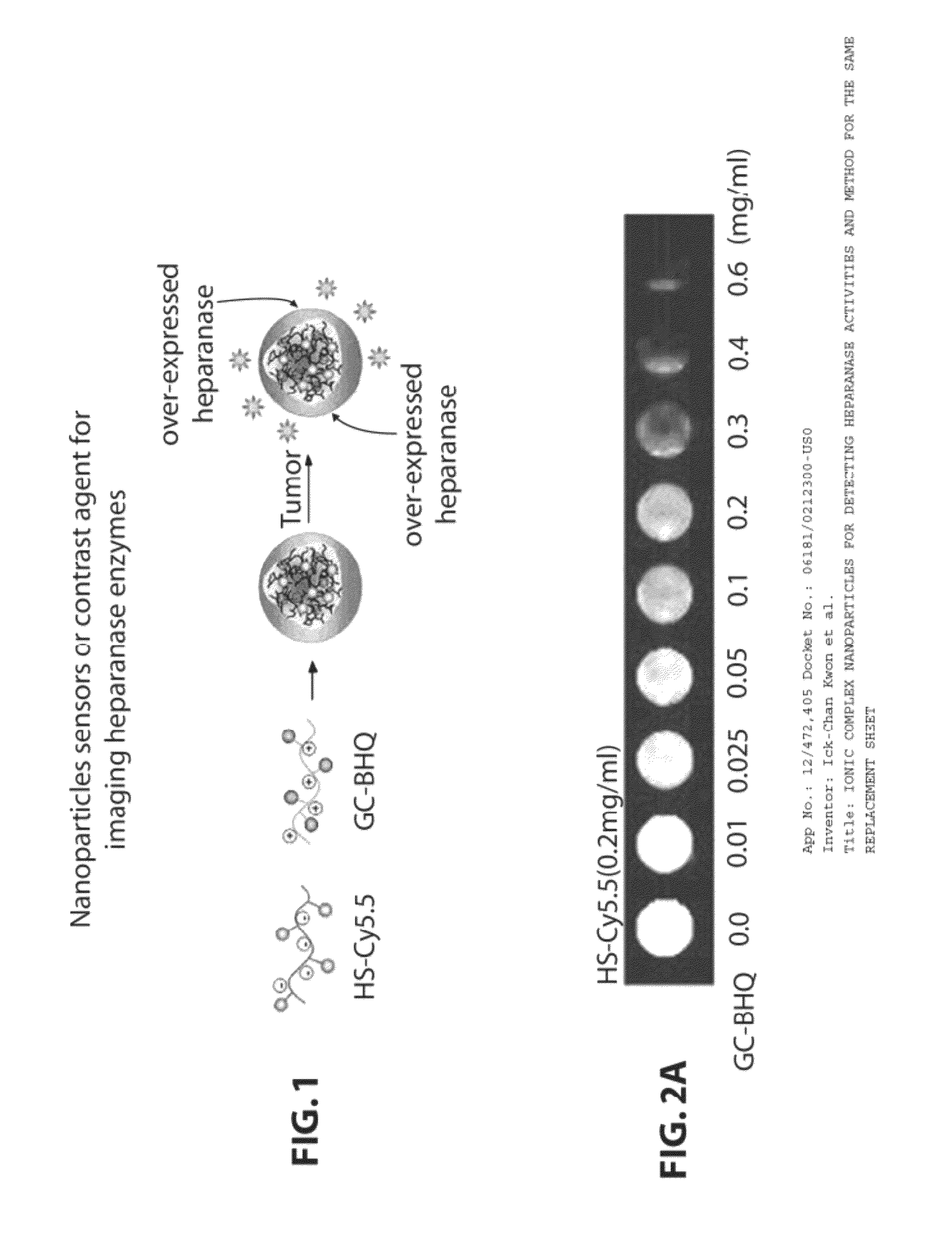
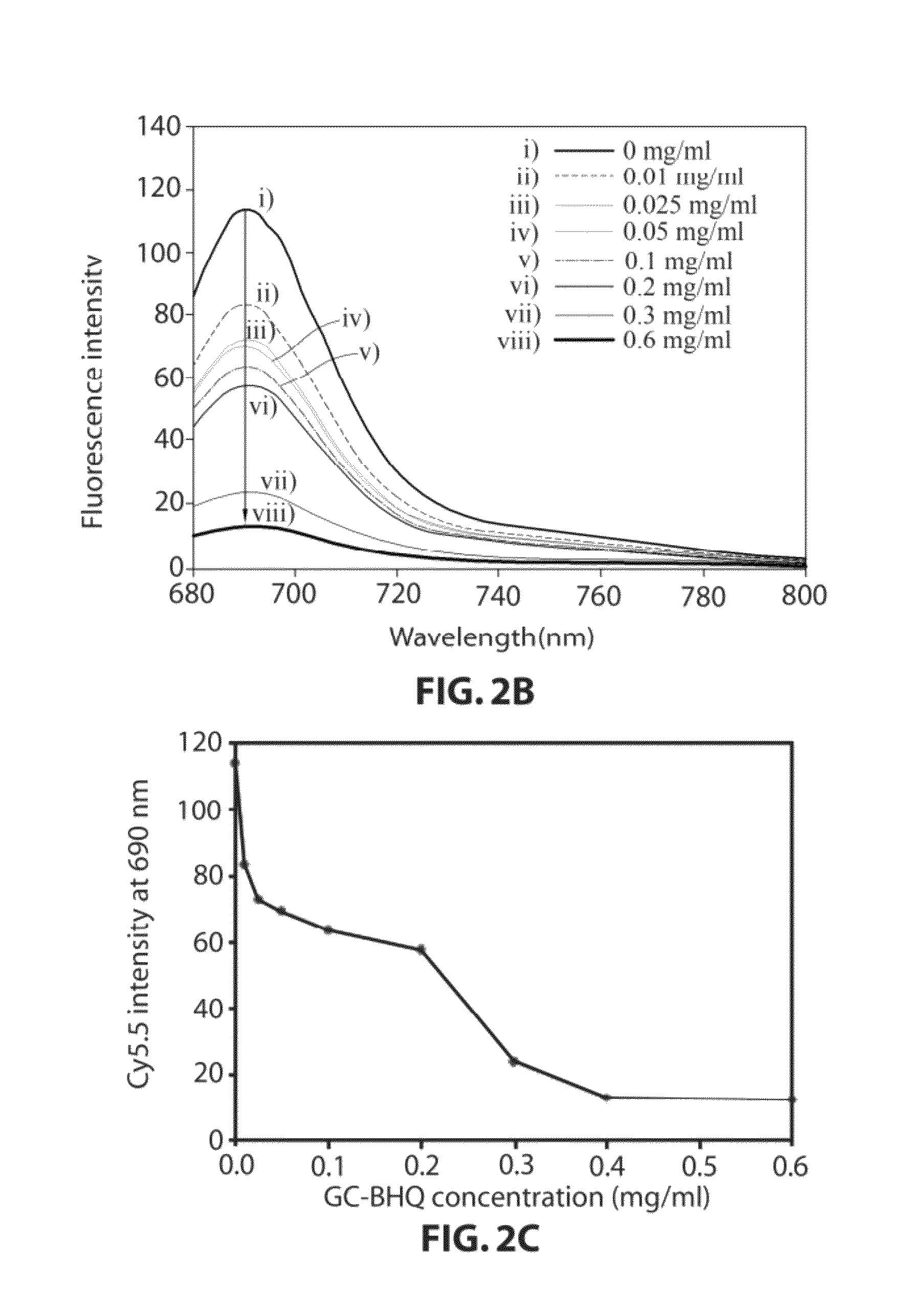
The present invention provides an ionic complex nanoparticle for detecting heparanase activities and a method for preparing the same. The nanoparticle consists of a negative-ion substrate polymer specifically degraded by heparanase, a positive-ion biocompatible polymer that can bind to the substrate polymer, and a fluorophore or quencher that can bind to each other. The nanoparticle can be used for imaging heparanase in cells and tissues, and for quantitative analysis of heparanase. The invention also provides a contrast agent composition for imaging heparanase expressed in cells and tissues. The negative-ion substrate polymer can be heparan sulfate, heparin, chondroitin sulfate, dermatan sulfate, keratan sulfate, or hyaluronic acid. The positive-ion biocompatible polymer can be glycol chitosan, poly-L-lysine, poly beta-amino ester polymer, poly beta-amino ester polymer, polyethylenimine, poly (amidoamine) dendrimer, or derivatives thereof. The nanoparticle has sizes of 50-800 nm and the distance between the fluorophore and quencher is 20 nm or less. The invention provides a useful tool for detecting and diagnosing diseases such as cancers, strokes, and autoimmune diseases.

Problems solved by technology

However, due to the lack of research on a method for imaging and analyzing activities and expressions of the heparanase, or a method for non-invasively imaging an expressed degree of the heparanase in vivo, the related technologies are required.
However, these methods require multi-step for measuring protocols, resulting in inefficiencies in the economic and time aspects when screening plenty of drugs in the process of developing novel drugs.
However, it is difficult to detect the heparanase activities expressed in cells and in vivo, or to early diagnose diseases by using the sensor.

Method used


Image

  • Ionic complex nanoparticles for detecting heparanase activities and method for preparing the same
  • Ionic complex nanoparticles for detecting heparanase activities and method for preparing the same
  • Ionic complex nanoparticles for detecting heparanase activities and method for preparing the same

Examples


preparation example 1

Method for Preparing Ionic-Complex Nanoparticles Sensor

1-1. Synthesis of Heparan Sulfate-Cy5.5 (HS-Cy5.5) and Glycolchitosan-BHQ3 (GC-BHQ3)

[0086]As one example of fluorophores-negative ion substrate polymers, HS-Cy5.5 was synthesized as shown in the following reaction formula 1.50 mg of heparan sulfate and 9 mg of Cy5.5-NHS were dissolved in 0.1M of NaHPO4 (5 ml, pH 9.0), and were reacted with each other at a room temperature for 6 hours in dark place. Next, the solution was dialyzed in distilled water for two days, and then was lyophilized. It was observed through UV that about 2 mol of Cy5.5 had bound to 1 mol of heparan sulfate.

[0087]As one example of positive ion biocompatible polymers-quenchers, glycol chitosan-BHQ3 was synthesized as shown in the following reaction formula 1. 100 mg of glycol chitosan and 11.5 mg of BHQ3-NHS were dissolved in 10 ml of DMSO, and then were reacted for 4 hours. Next, the reaction mixture was dialyzed in a co-solvent of water and methanol (1v:1v) ...

experimental example 1

Tests for the Ability of Heparanase Inhibitor to Inhibit Heparanase, by Using Ionic-Complex Nanoparticles of HS-Cy5.5 / GC-BHQ3

[0093]By using the ionic-complex nanoparticles of HS-Cy5.5 / GC-BHQ3 prepared in the preparation example 1, heparanase activities with or without heparanase inhibitor treatment were imaged.

[0094]The ionic-complex nanoparticles of HS-Cy5.5 / GC-BHQ3 prepared in the preparation example 1 were treacted with ‘heparanase’ and ‘heparanase+inhibitor’ at a temperature of 37□ for one hour, and then fluorescence emission therefrom by an enzyme degradation reaction was observed. After lapse of one hour, the fluorescence of the nanoparticles was observed by using a Kodak Image Station 4000MM Digital Imaging System mounted with a charge coupled device (CCD) camera. As a result, as shown in FIG. 3, the fluorescence intensity was increased when the heparanase was treated, whereas the fluorescence intensity was decreased when the ‘heparanase+inhibitor’ were treated. From the resu...

experimental example 2

Imaging of Heparanase Expression in Cancer Models Using Ionic-Complex Nanoparticles of HS-Cy5.5 / GC-BHQ3

[0095]To establish a cancer model, 1×10⁶ SCC7 cancer cells were implanted in nude mice. Here, the SCC7 are epithelial cancer cells, and commercially available from American Type Culture Collection (ATCC). Next, when the cancer has a size of about 5-8 mm, the ionic-complex nanoparticles of HS-Cy5.5 / GC-BHQ3 were injected into veins of the mice tails. Then, heparanase expression was imaged by using an eXplore Optix image device.

[0096]It is known that the SCC7 cancer tissues express a large amount of heparanase. Accordingly, in order to test the ability of a heparanase inhibitor to inhibit heparanase, 20 mg / kg of heparin was injected into cancer tissues. After lapse of 20 minutes, the ionic-complex nanoparticles of HS-Cy5.5 / GC-BHQ3 were injected into veins of the mice tails. Then, decrease of heparanase activities was imaged by using the explore Optix image device.

[0097]As shown in FIG...


PUM


Abstract

Disclosed are Ionic complex nanoparticles for detecting heparanase activities and a method for preparing the same. More specifically, disclosed are Ionic complex nanoparticles for detecting heparanase activities, wherein negative-ion substrate polymers specifically degraded by heparanase and positive-ion biocompatible polymers ionically bind to each other, and fluorophores or quenchers bind to each of the polymers. The ionic complex nanoparticles for detecting heparanase activities may be applied to a method for screening novel drugs such as inhibitors that prevent over-expression of heparanase. Various cells and tissues where over-expression of heparanase occurs may be non-invasively imaged in cancer cells, cancer tissues, and tissues of various inflammatory diseases. Accordingly, the ionic complex nanoparticles for detecting heparanase activities may be effectively used to early diagnose various diseases and incurable diseases including autoimmune diseases such as cancers, osteoarthritis, rheumatoid arthritis, and dementia.

Description

RELATED APPLICATION[0001]The present disclosure relates to subject matter contained in priority Korean Application No. 10-2009-0020303, filed on Mar. 10, 2009, which is herein expressly incorporated by reference in its entirety.BACKGROUND OF THE INVENTION[0002]1. Field of the Invention[0003]The present invention relates to ionic complex nanoparticles, and particularly, to ionic complex nanoparticles for detecting heparanase activities and a method for preparing the same.[0004]2. Background of the Invention[0005]An extracellular matrix (ECM) serves to fill extracellular spaces of each organ and tissue, and constitutes a complex network of macromolecules for cells inside each organ. The main components of a basement membrane include collagen (type IV collagen), laminin, and heparan sulfate proteoglycan (HSPG). The HSPG is present on cell surfaces as well as in the ECM. And, the HSPG is composed of proteoglycan (coreprotein+glycosaminoglycan), and heparan sulfate (HS). The HSPG has a r...

Claims


Application Information

Patent Timeline
Patent Type & Authority Patents(United States)
IPC IPC(8): A61B5/00
CPCA61K49/0054C12Q1/34G01N2333/924B82Y5/00C12Q1/25G01N33/533G01N33/573
Inventor KWON, ICK-CHANKIM, KWANG-MEYUNGCHOI, KUI-WONPARK, KYEONG-SOONLEE, SEUL-KI
Owner KOREA INST OF SCI & TECH
Learn More