Electrospun nerve guides for nerve regeneration designed to modulate nerve architecture

a nerve architecture and nerve guide technology, applied in the field of nerve guide, can solve the problems of reducing the efficiency and fidelity of axon targeting, affecting the function of the nerve guide, and affecting the regeneration process, so as to achieve the effect of functional recovery and functional recovery

Inactive Publication Date: 2017-02-21
VIRGINIA COMMONWEALTH UNIV
View PDF8 Cites 10 Cited by
  • Summary
  • Abstract
  • Description
  • Claims
  • Application Information

AI Technical Summary

Benefits of technology

[0019]The invention provides nerve guides formed from electrospun fibers for use in the directed regeneration of damaged or severed peripheral nerves. The technique of static air-gap electrospinning is used to generate cylindrical, seamless three dimensional (3D) arrays of highly aligned electrospun fibers oriented in parallel with the long axis of the construct. The air-gap technique allows the fabrication of cylinders of aligned fibers without “rolling” a sheet of fibers, and thus no potentially weakening or irregular seams or inordinately large “pores” are present in the guide, and the guide does not contain a “hollow” central channel. Instead, the guide comprises multiple open channels formed by elongated gaps between the fibers, and the channels are lined with aligned fibers that are oriented along the long axis of the guide and bounded by elongated, oriented fibers. The fibers of these structures are integrated on a true nano-scale. And, unlike other nerve guides that may contain hundreds to thousands of channels that are designed to support axon regeneration. A similar sized electrospun 3D nerve guide (depending on its size and fiber characteristics) produced by air gap electrospinning may contain hundreds of thousands to tens of millions of individual channels, each of which is “lined” with nano-to-micron scale diameter fibers that are designed to provide guidance cues. When the proximal end of a truncated nerve is introduced into one end of the nerve guide, the nerve tissue grows into the guide and along the channels in a directed manner. Thus, an axon which grows through the guide emerges from the other end of the guide at a desired, predictable location, e.g. in close proximity to the other, distal end of the severed nerve. This insures that the severed nerve ends will grow with a minimum of excess or random nerve tissue growth, permitting reconnection of the severed nerve to its distal target tissues, and restoration of functionality. The nerve guides of the invention thus closely mimic and have the structural advantages of natural nerve grafts but without the undesirable side effects of nerve disintegration. Experimental results presented herein show that animals treated with the nerve grafts of the invention showed levels of functional recovery after 7 weeks that are normally observed in animals treated with autologous grafts after 10-14 weeks. In some embodiments, the nerve guides also comprise gradients of therapeutic substances.
[0020]It is an object of this invention to provide an architectural arrangement that mimics the structure of the native autologous graft by depositing nano-to-micron scale electrospun fibers into a seamless, cylindrical structure. The axons are guided to grow in between the aligned fiber arrays in the channels provided between the individual fibers. This design provides physical guidance cues to direct the axons to grow along a specific direction and along a specific plane in order to reconstitute the topography of the nerve that existed before an injury. Maintaining normal topographical relationships is intended to increase the fidelity of axon targeting and increase the likelihood that an axon will emerge from the distal end of the graft in a position that approximates its position prior to injury. In turn, this type of regenerative patterns is designed to increase the probability that the regenerating axon returns to the distal target tissue (sensory and motor) that it innervated prior to injury. In addition, the seamless nature of the guide provides a far more uniform profile of pore spaces (i.e. the channels between the individual fibers) than a rolled sheet of material, reducing the tendency, of axons to cluster and grow as a jumbled disorganized mass along the seams, a pattern of growth in a regenerating nerve which is associated with reduced axon targeting. Because the air gap electrospinning process is very rapid it is possible to place substances of interest, e.g. cells, therapeutic agents, gradient threads (as described herein), etc. within the scaffold as the fibers are depositing into the fiber arrays. The supplementation of the guides with growth factors and / or cells, etc. can be used to further regulate the regenerative environment afforded by the scaffolds.

Problems solved by technology

In more extreme injuries where a long segment of the nerve is crushed or completely lost, treatment is greatly complicated.
This not-so subtle “spilling effect” jumbles normal nerve topography (this term refers to the relative position of the individual axons to each other within the nerve) and greatly reduces the efficiency, and fidelity, of axon targeting.
Once the injury gap exceeds this threshold, the regeneration process will be compromised to varying degrees.
Typically, in these injuries only a limited number of axons will actually traverse the wound bed and the efficiency of targeting to the distal tissues is poor, resulting in limited functional recovery.
As these degenerative changes become entrenched, the prospects of meaningful functional recovery are greatly diminished, even if a large number of axons are efficiently targeted to these sites.
However, use of this autologous tissue comes at the expense of donor site morbidity and results in the transplantation of a “nerve guide” that is packed with axonal fragments.
This degeneration process slows regeneration by initially impairing the penetration of the nascent axons into the autograft, a complication that clearly exacerbates the effects of long term muscle de-innervation and tissue atrophy.
Due to the lack of a solid substrate, this late-stage pathological endpoint represents a physical gap that impedes axonal regeneration and functional recovery.
At the neuronal level, axotomy of motor and sensory neurons results in the loss of trophic support by target tissue, which exasperates neuronal cell death.
Mono-therapies that address each of these obstacles to regeneration individually have resulted in only limited axonal regrowth and functional recovery.
To date, no single intervention has been devised to collectively address all of the known obstacles to axon regeneration in the spinal cord.
Despite these preliminary and encouraging results, it is difficult to fabricate a clinically relevant nerve guide using the conventional electrospinning process which uses a rapidly rotating target mandrel to produce aligned fibers.
These constructs are easily amenable to experimentation in vitro, however, a 2D sheet is less acceptable, or adaptable, for use in vivo.
A flat 2D sheet can be spun into a very thick structure and then cut into strips that resemble “square cylinders.” These structures are technically seamless, however, fiber alignment tends to degrade the thicker a sheet becomes when fibers are processed under conventional 2D electrospinning conditions.
Cutting such a sheet also distorts the existing alignment along the cutting plane.
In part this is because air currents and complex electric fields induced by the rotation of the mandrel disturb the trajectory of the charged jet, fiber flight and fiber deposition onto the target, thereby limiting the extent of alignment that can be achieved.
While the fibers of this type of construct can be induced to exhibit a considerable degree of alignment when produced under these conditions, the fibers, unfortunately, are deposited onto the target mandrel in a circumferential orientation (i.e. the axis of alignment is ˜90° off with respect to the resulting long axis of the hollow tube).
While the nano-to-micron diameter fibers that form the wall of this type of construct do represent a barrier that reduces the risk of inflammatory cells penetrating into the hollow lumen of the guide (critical to the regeneration process [17,18]), this architectural pattern (i.e. the arrangement of electrospun fibers in a circumferential pattern) does not lend itself well to providing directional guidance cues to regenerating axons.
Unfortunately, the resulting tubes are hollow and thus fail to mimic the architecture of natural nerve growth.
Further, a flat sheet that is rolled into a tube must, of necessity, have “seams” where the edges of the rolled sheets are surface exposed, e.g. on the wall of the hollow center of the tube and / or on the surface of the tube, resulting in discontinuities and possible weak connective points in the structure.
Rolling a sheet can not truly integrate the fibers on the nano-scale that is necessary to make a uniform set of “pores”.
However, the guides are made from sheets of several layers of conduits which are rolled into cylinders and are thus hollow.
The scaffold is capable of directing the morphological pattern of attached and growing cells to form a helical pattern around the tube walls, but would not be suitable for use as a nerve guide, where the axons must grow straight down the guide and not wrap or change topology during growth.

Method used


Image

  • Electrospun nerve guides for nerve regeneration designed to modulate nerve architecture
  • Electrospun nerve guides for nerve regeneration designed to modulate nerve architecture
  • Electrospun nerve guides for nerve regeneration designed to modulate nerve architecture

Examples


example 1

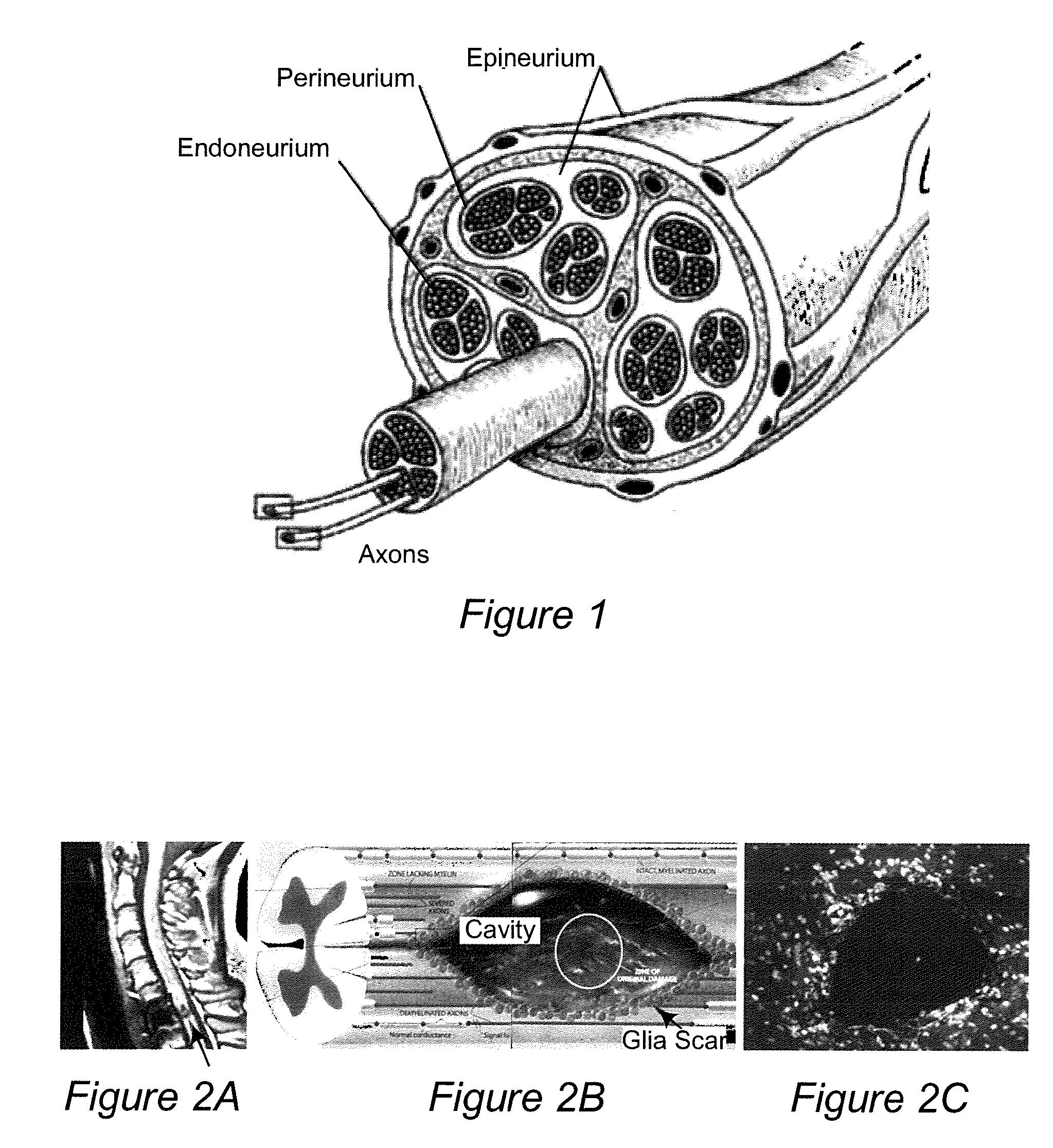
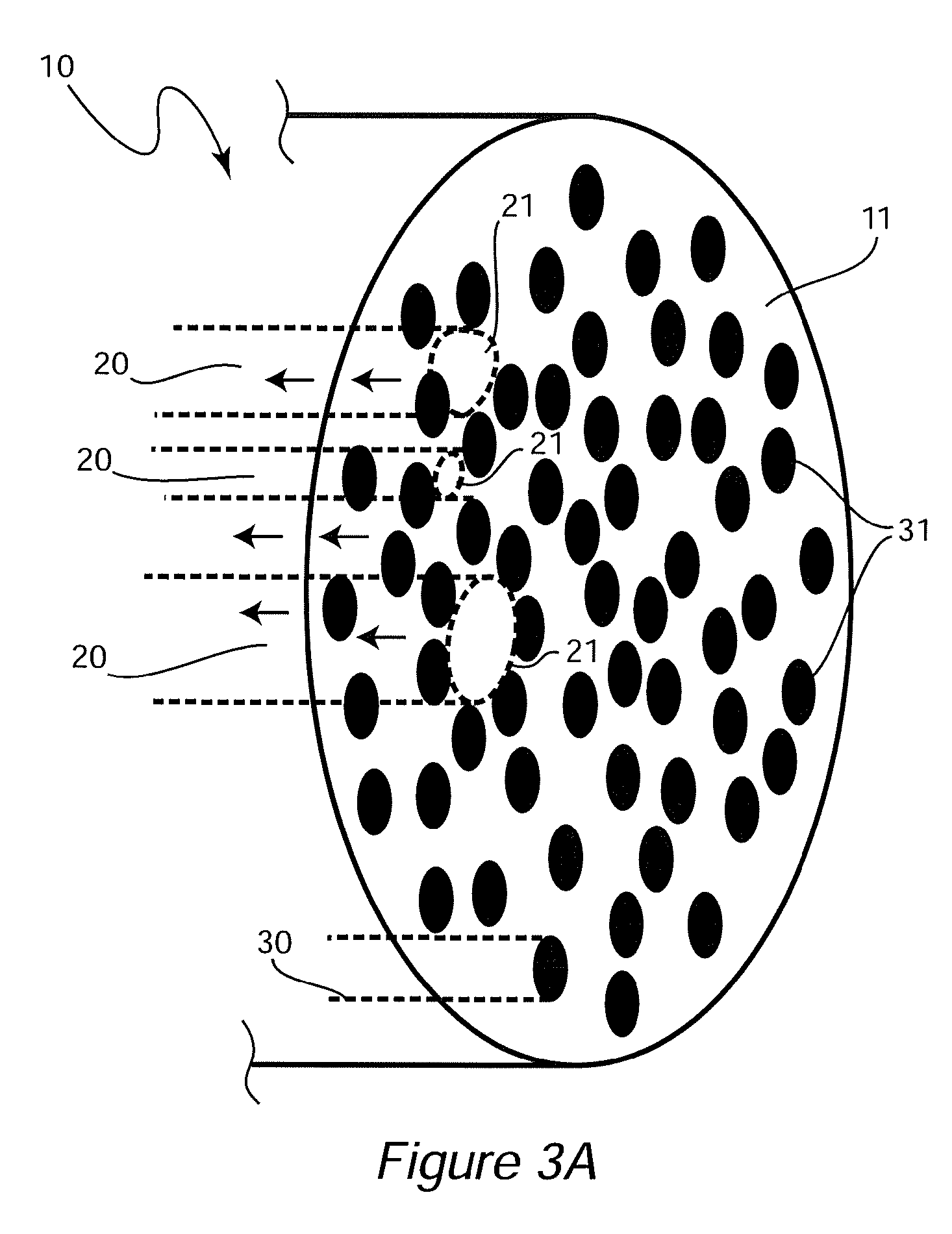
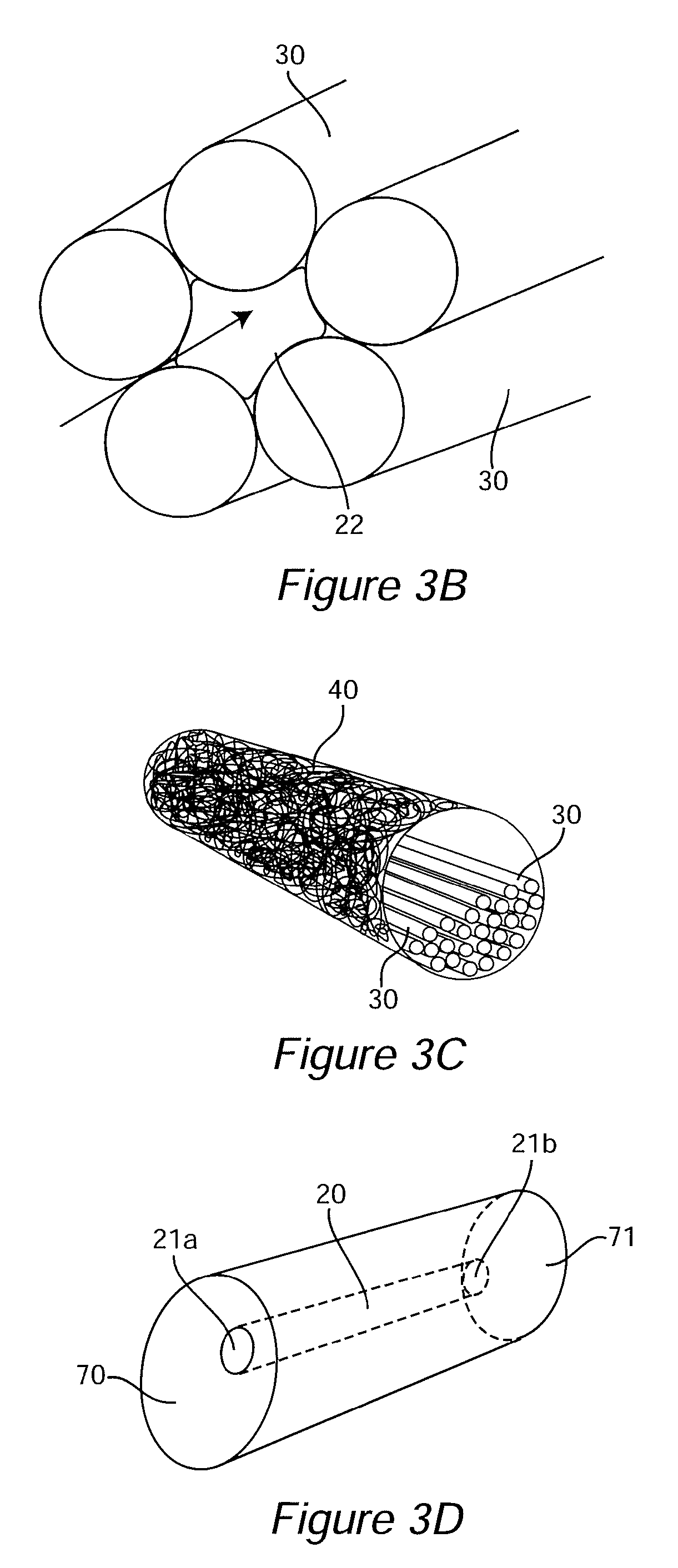
[0097]An ideal nerve guide configuration would closely mimic the structure of an autologous nerve graft. This idealized guide would display a cylindrical shape and be composed of dense three dimensional (3D) arrays of highly aligned electrospun fibers that were oriented in parallel with the long axis of the construct. Gaps and elongated spaces between the stacked fiber arrays would provide thousands of channels for directed axonal growth. This type of configuration can be approximated by rolling a 2D sheet of aligned fibers into a cylinder. Unfortunately, rolling a 2D sheet will result in a seam in the construct that could provide an avenue for the infiltration and penetration of inflammatory and interstitial cells into the construct. The seam could also represent a nexus for mechanical failure. Air gap electrospinning makes it possible to circumvent these limitations and produce cylindrical, seamless, and truly, 3D constructs composed of aligned arrays of electrospun fibers that ar...

example 2

Gradients of Therapeutic Substance

[0143]In human nerve injuries, where a segment of tissue has been injured to the point where a nerve guide must be used to re-establish continuity of the injured tissue (or otherwise direct the regeneration process) the actual physical dimensions of the nerve segment that has to be reconstructed may be relatively nominal in length. However, even the loss of a relatively short segment of tissue can represent a nearly insurmountable barrier to regeneration. There is considerable evidence that growth factor gradients established by various cells that migrate into the injury site play a critical role in directing the nerve regeneration process. Clearly, in circumstances where a nerve guide is used to reconstruct damaged tissue the regeneration process could be enhanced, and accelerated, if a specific growth factor gradient could be directly incorporated into a nerve guide prior to reconstructive surgery. However, recapitulating a growth factor gradient ...

example 3

Dissolution Dependent Gradients

[0146]As described above, the second type of gradient, the, Dissolution Dependent Gradient (DDG) is fabricated by varying the concentration of carrier (e.g. alginate) along the length of the thread like construct. This type of gradient can be fabricated using a constant concentration of therapeutic reagent or a variable concentration of therapeutic reagent. The basis for this type of gradient lies in the observation that alginate threads composed of low concentrations of this carbohydrate dissolve faster than alginate threads composed of higher concentrations of this carbohydrate. Thus, the rate of release of an active agent incorporated into the thread can be controlled by placing the agent within a segment of the gradient thread that will dissolve at or within a desired time frame, e.g. a first segment of the thread may dissolve over a period of days (e.g. 1-7 days), a second segment may dissolve over a period of e.g. 1-2 weeks, a third segment may d...


PUM

PropertyMeasurementUnit
total lengthaaaaaaaaaa
lengthaaaaaaaaaa
lengthaaaaaaaaaa

Abstract

Nerve guides which are formed from three dimensional (3D) arrays of highly aligned electrospun fibers are provided. The electrospun fibers are oriented parallel to the long axis of the guide, and gaps and elongated spaces between the stacked fiber arrays provide channels for directed axonal growth. In some embodiments, the nerve guides also comprise high precision gradients of beneficial substances such as growth factors, which aid in nerve regeneration and growth along the guide.

Description

CROSS-REFERENCE TO RELATED APPLICATIONS[0001]This application is a 371 of international application No. PCT / US2010 / 048744, filed Sep. 14, 2010, which claims benefit of U.S. provisional application No. 61 / 242,150, filed Sep. 14, 2009, and U.S. provisional application No. 61 / 323,387, filed Apr. 13, 2010, each of which is incorporated herein by reference.BACKGROUND OF THE INVENTION[0002]Field of the Invention[0003]The invention generally relates to nerve guides for use in nerve regeneration. In particular, the invention provides nerve guides which are formed from three dimensional (3D) arrays of highly aligned electrospun fibers that are oriented in parallel with the long axis of the seamless and cylindrically shaped constructs. Gaps and elongated spaces between the stacked fiber arrays provide channels for directed axonal growth.[0004]Background of the Invention[0005]A peripheral nerve is an enclosed, cable-like bundle of peripheral axons (long, slender projections of neurons, see FIG...

Claims


Application Information

Patent Timeline
Patent Type & Authority Patents(United States)
IPC IPC(8): A61L27/18A61F2/02A61B17/11
CPCA61L27/18A61B17/1128A61F2/02C08L67/04A61L2430/32
Inventor SIMPSON, DAVIDBOWLIN, GARYCOLELLO, RAYMONDCHOW, WOONJHA, BALENDU SHEKHAR
Owner VIRGINIA COMMONWEALTH UNIV
Why Eureka
Try Eureka