
Systems and method for engineering muscle tissue

a technology of muscle tissue and engineering methods, applied in the field of cell growth and tissue engineering, can solve the problems of inability to reproduce the multiscale effects of complex ecm structures, inability to fully understand the effect of cell and tissue function engineering control, and inability to fully understand the effect of cell and tissue function, etc., to achieve accurate recapitulation and high throughput format

Active Publication Date: 2018-06-12
UNIV OF WASHINGTON CENT FOR COMMERICIALIZATION
View PDF26 Cites 17 Cited by
  • Summary
  • Abstract
  • Description
  • Claims
  • Application Information

AI Technical Summary

Benefits of technology

[0008]In particular, the inventors have developed a nanotextured polymer platform that is configured to have the rigidity, flexibility and topography for the enhanced maturation of myogenic cells in an anisotropically and anatomically correct manner to mature these into an adult phenotype in vitro. In particular, the inventors demonstrate the mechanical and electrophysiological function of engineered cardiac tissue constructs, demonstrating that they faithfully mimic the highly organized function seen in normal heart tissue in vivo. Moreover, strikingly, the inventors demonstrate that the engineered tissue structure and function were highly sensitive to variation of the nanoscale topographic features of the platform, revealing an unexpected level of mechanical regulation.
[0065]Accordingly, the present disclosure relates to an ANP substrate which is able to generate aligned, anisotropic cultures of mature cardiomyocytes through the use of nanotextured cell culture substrates / scaffolds which contain nanotopographical surface cues and tunable rigidities. Its purpose is to generate more faithful models of human cardiac tissue for disease modeling and in vitro drug screens, using hiPSC-derived cardiomyocytes and other immature cardiomyocyte types. These anisotropic cultures of cardiomyocytes will more accurately recapitulate adult human cardiac tissue due to their incorporation of human cardiomyocytes and inherent organization mimicking that of physiological heart tissue.

Problems solved by technology

However, given the small scale of the underlying interactions, their effect on cell and tissue function is far from completely understood.
However, these initial attempts at engineering control of cell function have frequently not been bioinspired or biomimetic and have failed to reproduce the multiscale effects of complex ECM structures (e.g., networks of fibers and components of the basement membrane) and associated chemical ligands, which control integrated multicellular ensembles on scales ranging from a few nanometers to hundreds of micrometers (Geiger B, et al., (2001) Nat Rev Mol Cell Biol 2:793-805, Abrams et al., (2003) Urol Res 31:341-3469, 10).
Although the structure of cardiac tissue is highly organized in vivo, cardiomyocyte ensembles lose their native organization and adopt random distribution when cultured in vitro by using common culturing techniques, potentially compromising many of their physiological properties.
However, it is likely that the structure and function of the in vivo cardiac tissue are regulated by much smaller, nanoscale cues provided by the ECM, which might exercise complex, multiscale control of cell and tissue function.
In this regard, the inability to direct the differentiation of multipotent progenitors specifically to mature muscle cells remains a major obstacle for optimal in vivo cardiac myogenesis during cardiac repair following injury.
Furthermore, while methods of cell based therapy using cells on scaffolds exist, their use is limited benefit to the extent to which they support growth, differentiation and function of cells for a functional engineered cardiac tissue.
Thus, although these attempts have been made to organize the cardiomyocytes into anisotropic arrays, the controllability, scalability and bio-mimetic properties of such efforts remain uncertain and uncontrolled.
In addition, the structural and mechanical properties and the degree of biocompatibility of the materials commonly used as the cell substrata have limited both the therapeutic potential of the designed tissues and their ability to provide a fundamental understanding of the principles of cardiac tissue self-organization.

Method used


Image

  • Systems and method for engineering muscle tissue
  • Systems and method for engineering muscle tissue
  • Systems and method for engineering muscle tissue

Examples


example 1

[0656]The development of a novel “off-the-shelf” engineered heart tissue platform for high throughput drug screening would have wide applicability both for safety testing and the identification of new therapeutic compounds. Many promising compounds are found to have unanticipated cardiotoxic effects (e.g. pro-arrhythmic interference cardiac ion channels), and this remains the most common reason that otherwise efficacious drugs are removed from the market. Regulatory agencies require preclinical safety screens in heterologous systems (e.g. cultured cells genetically modified to overexpress a particularly susceptible ion channel, such as the HERG channel) and animal models, but both have important limitations. Heterologous systems lack the full constellation of ion channels and signaling pathways present in intact cardiomyocytes, while animal studies are very expensive, low-throughput and often limited to relatively short time-points. There are also important differences between human...

example 2

[0667]Development of a High Throughput and Patient-Specific Tissue Culture Platform with Different Nanotopographical Patterns and Stiffnesses.

[0668]Development of ANP Cell Culture Substrates with Tunable Topography and Stiffness.

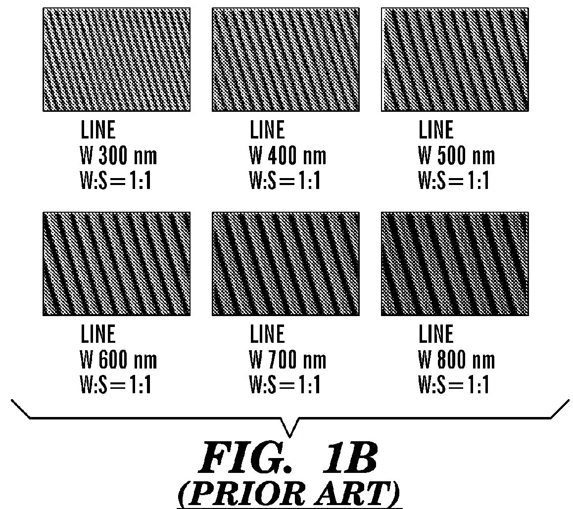
[0669]Using a PEG-based materials, the inventors have generated an ANP using well-established biocompatible hydrogel polymer whose mechanical and topographic properties can be modulated by UV-assisted capillary force lithography. In particular, the inventors synthesized polyethylene glycol-gelatin methacrylate (PEG-GelMA) composite prepolymers and then fabricated nanogroove structures on large areas (>10 cm2). Modulation of the concentration of PEG and GelMA allows for control of the Young's modulus of the material to match the rigidity of myocardium. The gelatin component of the composite biomaterial helps promote cell attachment. To account for the scale of the ECM cues and possible variability in the diameter of the ECM fibrils in the heart tissue, the wi...

example 3

[0672]Maintenance of Human iPSC Derived Cardiomyocytes in Long Term Culture on Substrates with Different Nanotopographical Patterns and Stiffnesses Sustains Cellular and Tissue-Level Electrophysiological and Contractile Function

[0673]Optimization of Cellular Function.

[0674]The inventors have identified an optimal combination of nanotopography and substrate stiffness that maximizes cell survival and maintains cellular function at both the single cell and monolayer level. Multi-well plates can be used for studies of confluent monolayers. As an additional biomimetic cue, electrical pacing can be applied to the cell culture for up to 3 months. Optical mapping is performed using voltage- and calcium sensitive dyes, thereby measuring action potential duration (APD), calcium transient amplitude (CaT), spontaneous beat rate, average conduction velocity (av CV), conduction anisotropy ratio (AR), maximum capture rate (MCR), and heterogeneity index of action potential duration (HI) for compari...


PUM

PropertyMeasurementUnit
widthaaaaaaaaaa
depthaaaaaaaaaa
widthaaaaaaaaaa

Abstract

The present invention generally relates to the field of cell growth and tissue engineering, in particular, tissue engineered compositions comprising a nanotextured substrate which is structurally configured for growth of cells in an anatomically correct adult phenotype in vitro. In particular, described herein are nanotextured substrates which are structurally configured for the anisotropic organization, maturation, and growth of in vitro-differentiated muscle cells, such as cardiomyocytes, and methods for the production and use thereof in varying sizes, nanotextures and substrate rigidities. In vitro-differentiated cardiomyocytes grown on the nanotextured substrates described herein are better-differentiated and more closely mimic adult cardiac tissue than the same cells grown on a non-textured substrate of the same composition. The nanotextured substrate / cell constructs provide a platform for screening to predict the effect of test agents or drugs on, for example, human cardiac tissue, including patient-derived tissue, or for the identification of agents that effect various cardiac functional parameters.

Description

CROSS REFERENCE TO RELATED APPLICATIONS[0001]This application is a 35 U.S.C. § 371 National Phase Entry Application of International Application No. PCT / US2013 / 032237 filed Mar. 15, 2013, which designates the U.S., and which claims benefit under 35 U.S.C. 119(e) of U.S. Provisional Patent Application Ser. No. 61 / 620,301, filed on Apr. 4, 2012, the contents of which is incorporated herein in its entireties by reference.FIELD OF THE INVENTION[0002]The present invention generally relates to the field of cell growth and tissue engineering, in particular, a tissue engineered composition comprising a nanotextured platform which is structurally configured for growth of cells in an anatomically correct adult phenotype in vitro.BACKGROUND OF THE INVENTION[0003]Living cells and tissues can display high sensitivity to local micro- and nanoscale molecular and topographic patterns, including those provided in vivo by complex and well-defined structures of extracellular matrix (ECM) (Stevens M et...

Claims


Application Information

Patent Timeline
Patent Type & Authority Patents(United States)
IPC IPC(8): A61K48/00C12M3/00C12M1/12A61L27/18A61L27/14C12N5/0735A61L27/38C12N5/074C12N5/077B01L3/00A61L27/50
CPCC12N5/0606A61L27/14A61L27/18A61L27/3826A61L27/3834A61L27/3873A61L27/50B01L3/5085C12M21/08C12M25/14C12N5/0657C12N5/0658C12N5/0696C08L67/04C12N2535/00A61K48/00A61L2400/12A61L2400/18B01L2300/0851B01L2300/0896B01L2400/086C12N2503/02C12N2533/30C12N2533/40
Inventor KIM, DEOK-HOLAFLAMME, MICHAELMURRY, CHARLESGUPTA, KSHITIZYOO, HYOKJIAO, ALEX
Owner UNIV OF WASHINGTON CENT FOR COMMERICIALIZATION
Learn More