
mRNA (messenger ribonucleic acid) quick-extraction kit
A kit and cellulose technology, applied in the biological field, can solve the problems of high price and achieve the effect of simple operation and low cost
- Summary
- Abstract
- Description
- Claims
- Application Information
AI Technical Summary
Problems solved by technology
Method used
Image

Examples
Embodiment 1
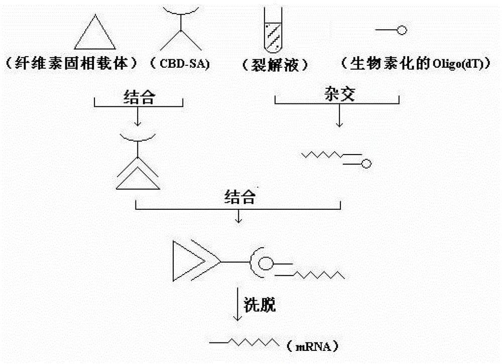
[0039] Example 1. Activation and assembly of cellulose filter paper (i.e. preparation of bioactivated cellulose solid phase carrier)
[0040] First prepare the fusion protein CBD-SA (see Chinese patent CN101640085B).
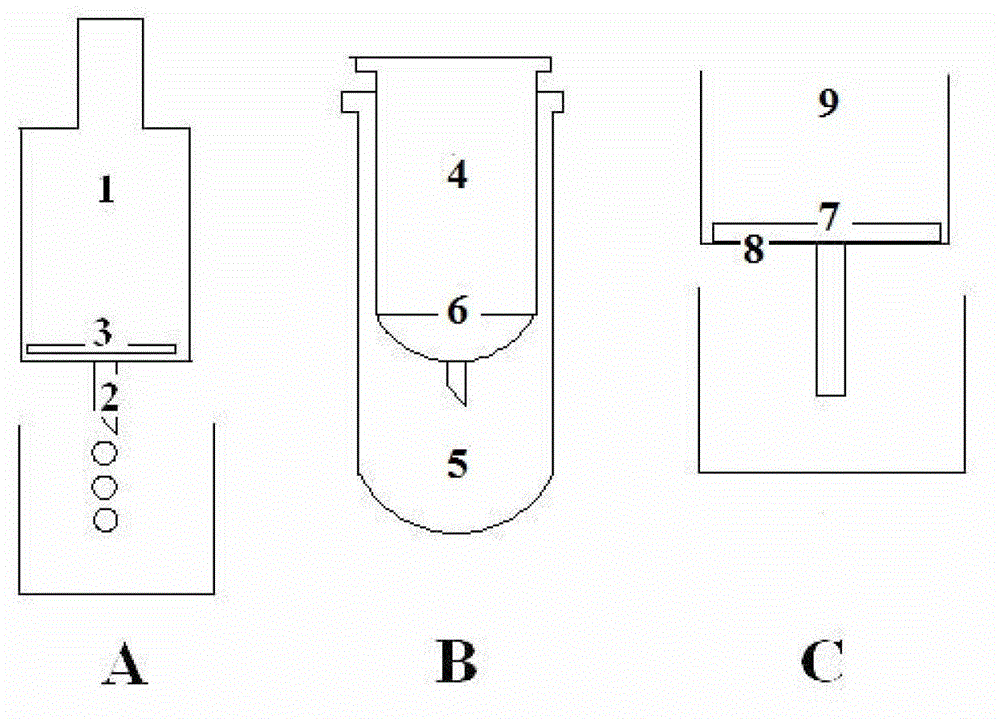
[0041] Then, at 2-8°C, take a certain amount of cellulose filter paper, add the fermented lysate of the recombinant fusion protein CBD-SA, oscillate for sufficient reaction, rinse with PBS buffer repeatedly for 8 times, and then dry at room temperature Dry it. by figure 2 The double-layer microcentrifuge tube of B is taken as an example. The bioactivated cellulose filter paper is cut into a circle with a suitable diameter for the inner tube, and placed flat on the bottom of the inner separation tube and on the upper end of the microporous membrane or screen-like structure. , with a thickness of 1 mm, about 5 layers of filter paper are placed.
Embodiment 2
[0042] Activation and assembly of embodiment 2 cellulose absorbent cotton
[0043] Referring to Example 1, at 2-8°C, take a certain amount of absorbent cotton, add the fermented cell lysate of the recombinant fusion protein CBD-SA, oscillate for sufficient reaction, rinse with PBS buffer repeatedly for 8 times, and then dry at room temperature , and pressed into flakes. Cut the above-mentioned bioactivated absorbent cotton into a circle suitable for the diameter of the inner tube, lay it flat and tightly fix the bottom of the inner separation tube, and the upper end of the microporous membrane or screen-like structure, with a thickness of about 2mm.
Embodiment 3
[0044] The extraction of mRNA in the tissue sample of embodiment 3
[0045] The rat was anesthetized and dissected, and 50 mg of heart tissue was taken and chopped, placed in a lysate (4M guanidine isothiocyanate, 25 mM sodium citrate and 1% β-mercaptoethanol, pH7.0), homogenized and pulverized, and then added 1mL binding buffer (75mM NaCl, 8.5mM sodium citrate, pH 7.2), mix by inversion, then add 60pM biotinylated Oligo(dT) probe, mix well, and incubate at 70°C for 5min. Then at room temperature, 12000rpm, centrifuge for 10min, carefully collect the supernatant, transfer to the inner layer separation tube of Example 1 activated with cellulose filter paper, and centrifuge at room temperature for 1min at 12000rpm. Discard the waste liquid in the outer tube; add 600 μL of SSC buffer solution to the inner tube, centrifuge at 12000 rpm at room temperature for 30-60 s, discard the waste liquid in the collection tube, and repeat this step twice. Finally, put the inner tube into a n...
PUM
 Abstract
Description
Claims
Application Information