Mycoplasma bovis diagnosis reagent and its application

A technology of Mycoplasma bovis and diagnostic reagents, applied in the field of animal diagnostic medicine, can solve problems such as inability to accurately reflect pathogen infection, lag in basic research on Mycoplasma bovis, long duration of antibodies, etc. The effect of small difference and low detection cost

Inactive Publication Date: 2013-06-26
重庆市动物疫病预防控制中心 +1
View PDF2 Cites 19 Cited by
  • Summary
  • Abstract
  • Description
  • Claims
  • Application Information

AI Technical Summary

Problems solved by technology

For Mycoplasma bovis, a more prominent problem is that due to the relative lag in the basic research of Mycoplasma bovis, the advantage and conserved antigens that induce the body to produce antibodies after infection with this pathogen have not yet been clarified—that is, the rapid induction of antibodies and the The duration of antibody production is long, and th...

Method used


Image

  • Mycoplasma bovis diagnosis reagent and its application
  • Mycoplasma bovis diagnosis reagent and its application
  • Mycoplasma bovis diagnosis reagent and its application

Examples


Embodiment 1

[0060] Embodiment 1 multi-epitope fusion antigen

[0061] Construction of a gene vector for pathogenicity-associated protein P33 and specific membrane protein P30 of Mycoplasma bovis

[0062] 1pMD18-T-P33 and pMD18-T-P30 constructs

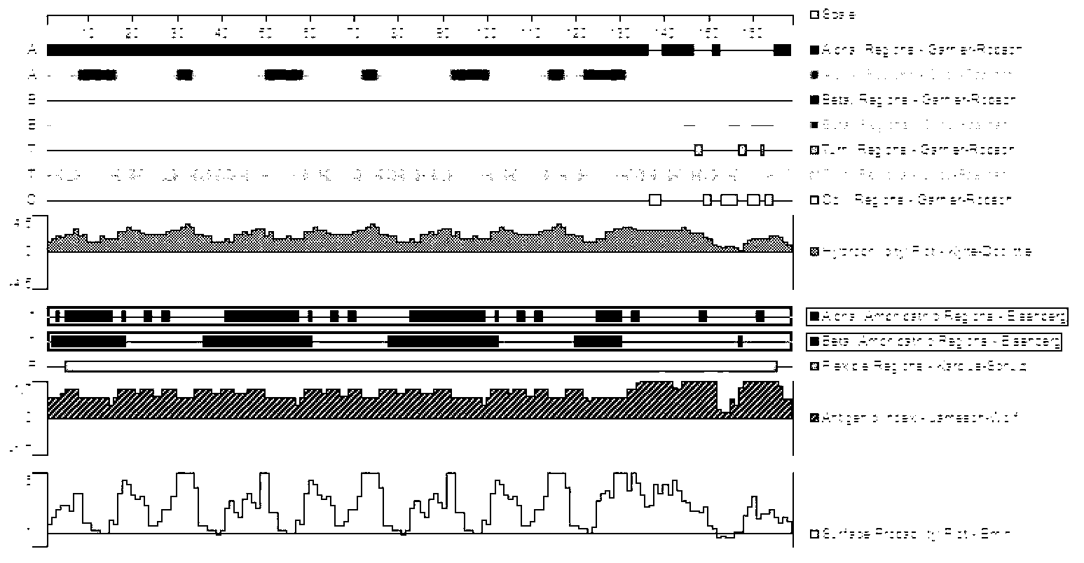
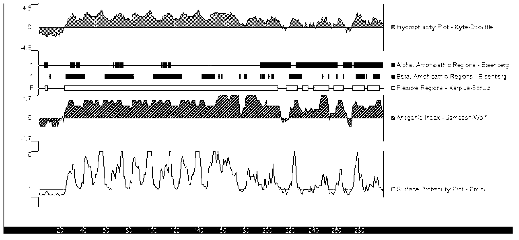
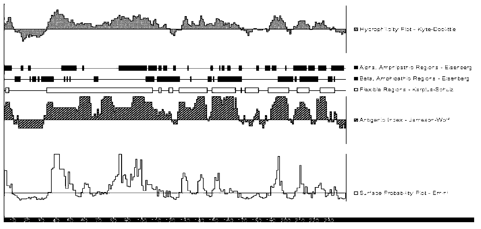
[0063] The full length of the P33 gene fragment is 903bp, and its nucleotide sequence is shown in SEQ ID NO: 3, which contains 5 TGA codons encoding tryptophan. For the B cell epitope analysis of P33, see figure 2 ;

[0064] SEQ ID NO.3:

[0065] “ggatccatattattgacaacactatcgccaattatctcattgccatttttatctgctagttg catcaccgaagcaaaatcagataacaaaatggaaaaagatattaagataaacgaaaatacagatgaaa aaaattcttctgaaacaatgaataacaaacaaaaacaagataaaagcagtatagattcaaagatggaa gaaaaagcagataacaaaacggaaaaagatattaagataaacgaaaatacagatgaaaaaaattcttc tgaaacaatgaataacaaacaaaaacaagataaaagcagtatagattcaaagatggaagaaaaagcag ataacaaaacggaaaaagatattaagataaacgaaaatacagatgaaaaaaattcttctgaaacaatg aataacaaacaaaaacaagataaaagcagtatagaatcgaaaatgaaagaaaaaacagaaaagcaaga ttcaaaaactaactcagaaaaacaagatt...

Embodiment 2

[0086] Example 2 Preparation of multi-epitope fusion antigen P39 kit for detecting Mycoplasma bovis antibodies

[0087] The composition of an indirect ELISA detection kit

[0088] 1 Antigen-coated plate:

[0089] 1) Coating: Multi-epitope fusion antigen was diluted to 5 μg / mL with 50 mM carbonate buffer (pH 9.3), 100 μL was added to each well, and incubated overnight at 4°C.

[0090] ●Carbonate buffer (5×, pH9.3) 1L

[0091] Component Amount and Final Concentration

[0092] Na2CO3 (MW105.99) points 7.949g (75mM)

[0093] Pure analysis

[0094] Analysis of NaHCO3 (MW84.01) 14.702g (175mM)

[0095] pure

[0096] Dissolve in ultrapure water, dilute to 1L, sterilize by filtration, and store at 2-8°C. Dilute 5 times with ultrapure water before use.

[0097] 2) Blocking: 1% casein, 300 μL / well, incubate at 37°C for 2 hours, seal and store at 2-8°C.

[0098] 2 Sample diluent: 0.5% casein, prepared with PBS (1×, pH 7.4). For the preparation method of PBS, refer to page 1570 ...


PUM


Abstract

The invention relates to the diagnostic medicine of animals, especially relates to a mycoplasma bovis diagnosis technology, and concretely relates to a multi-epitope fusion antigen having an amino acid sequence represented by SEQ ID NO:1 or SEQ ID NO:2, and its application in the preparation of a mycoplasma bovis diagnosis reagent. The diagnosis reagent can be used as a solid phase vector coating antigen of an indirect ELISA kit and is combined with its specific antibody, a horseradish peroxidase coupled anti-cattle IgG antibody is added and incubated, and a color development reaction is carried out, and the color development degree is proportional to the amount of the anti-mycoplasma bovis antibody in a sample to be measured. The technology has the advantages of simple operation, no need of complex equipment, low technical requirements on the laboratorial conditions and experiment personals, low detection cost, and suitableness for the large-scale development in the basic level and the culture farm; the multi-epitope fusion antigen has a low making cost and is suitable for large-scale application; and has the advantages of high sensitivity and specificity, small batch difference, and high detection result consistence because of the adoption of multi-epitope as a target.

Description

technical field [0001] The invention relates to animal diagnostic medicine, in particular to the diagnosis technique of Mycoplasma bovis pneumoniae. Background technique [0002] Mycoplasma bovis (MB) belongs to the kingdom of prokaryotes, Firmicutes, Mollicula, Mycoplasma, Mycoplasma family, Mycoplasma genus, and is the smallest and simplest prokaryote that can replicate autonomously. In the 1960s, it was first reported that Mycoplasma bovis caused pneumonia and mastitis in cattle. After that, people found that MB could also cause arthritis, keratitis, otitis, reproductive tract inflammation, abortion and other diseases in cattle. According to foreign reports, at least a quarter of the annual economic loss caused by bovine respiratory diseases is caused by Mycoplasma bovis, especially in the cattle transportation link, the incidence rate of the whole herd is as high as 90%, and the duration lasts for more than three months. affect the growth and development of cattle. At ...

Claims


Application Information

Patent Timeline
IPC IPC(8): C07K19/00G01N33/68G01N33/569G01N33/543
Inventor 熊仲良曾政贺德华严俊蔺露谢建华凌洪权欧武海丁平
Owner 重庆市动物疫病预防控制中心
Why Eureka
Try Eureka