Injectable medicated particle-inlaid porous composite microsphere preparation and preparation method thereof

A technology of porous composite microspheres and porous microspheres, applied in the field of medicine, can solve the problems of sudden release of growth factors or chemical drugs, difficulty in controlling the pore structure, and poor mechanical strength of the gel, so as to reduce systemic side effects and avoid sudden release , Improve the effect of repairing curative effect

Inactive Publication Date: 2014-01-08
SECOND MILITARY MEDICAL UNIV OF THE PEOPLES LIBERATION ARMY
View PDF8 Cites 20 Cited by
  • Summary
  • Abstract
  • Description
  • Claims
  • Application Information

AI Technical Summary

Problems solved by technology

However, the complex of drug-containing microspheres and gels still has many disadvantages: it cannot be used as a carrier for cell expansion in vitro; in vivo gelation time and scaffold shape are difficult to control; gel mechanical strength is poor and pore structure is difficult to control, which is easy to cause Leaky cells are bad for proliferation
However, the porous structure will lead to the burst release of growth factors or chemical drugs, which needs to be resolved
For example: Chinese patent application CN200810044593.6, the title of the invention is "a preparation method and application of degradable polymer porous microspheres", the publication number is CN101249077, which discloses a preparation method of degradable polymer porous microspheres, The prepared porous microspheres use the adsorption method to allow the drug to enter the porous structure of the microsphere, but the disadvantage is that the adsorbed drug is released quickly
Another example: Chinese patent CN200810161673.X, the title of the invention is "a double microsphere preparation of hydrophilic drug and its prep...

Method used


Image

  • Injectable medicated particle-inlaid porous composite microsphere preparation and preparation method thereof
  • Injectable medicated particle-inlaid porous composite microsphere preparation and preparation method thereof
  • Injectable medicated particle-inlaid porous composite microsphere preparation and preparation method thereof

Examples


Embodiment 1

[0067] Example 1: Preparation of PLGA porous composite microspheres containing model drug bovine serum albumin (Bovine serum albumin, BSA) chitosan microspheres

[0068] A, prepare chitosan microspheres containing BSA:

[0069] According to literature reports (Kim SE, Park JH, Cho YW, Chung H, Jeong SY, Lee EB, et al. Porous chitosan scaffold containing microspheres loaded with transforming growth factor-beta1: implications for cartilage tissue engineering. Journal of controlled release: official journal of the Controlled Release Society.2003;91:365-74), prepared by the following method: weigh 120mg chitosan (molecular weight 100000, Zhejiang Jinke Biochemical Co., Ltd.), add 3mL 2% (V / V) acetic acid aqueous solution After vortexing to dissolve, add 1 mL of 0.5% (W / V) model drug bovine serum albumin (Amresco, USA) aqueous solution, mix well and drop into 90 mL of n-octanol (containing 4% Span 80, V / V), after stirring at 1200 rpm for 30 minutes, 10 mL of 5% (W / V) sodium tripol...

Embodiment 2

[0072] Example 2: Morphological observation of PLGA porous composite microspheres embedded with BSA-containing chitosan microspheres

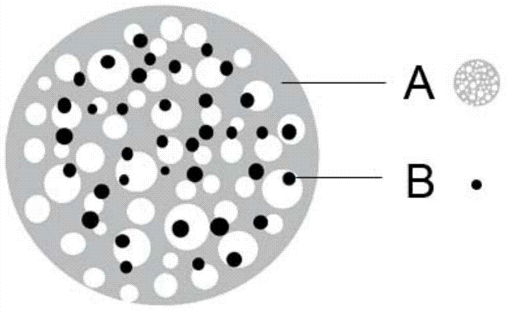
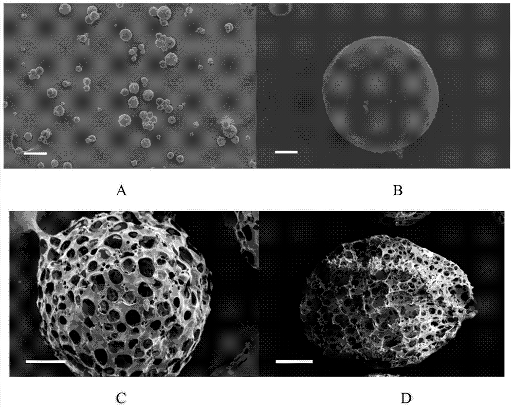
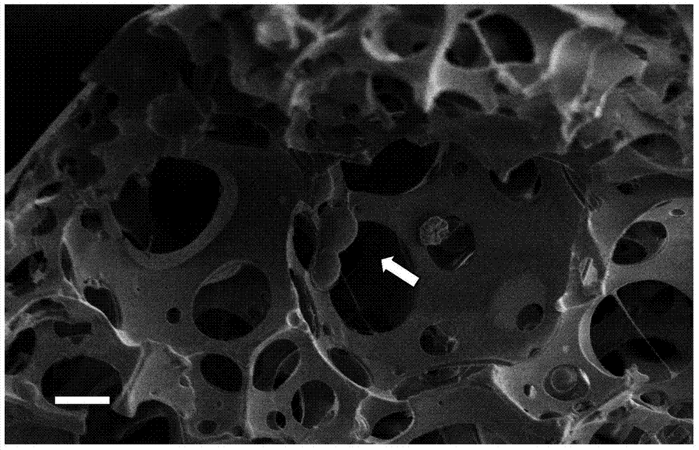
[0073] Get the BSA chitosan microspheres prepared in a certain amount of embodiment 1 and the PLGA porous microspheres containing BSA chitosan microspheres, place on the sample plate that is glued with double-sided adhesive tape, after gold plating, scan electron microscope ( ZESSMA10, Germany) was observed to obtain chitosan microspheres ( figure 2 A, B, the scales are 50 μm and 5 μm respectively) and PLGA porous composite microspheres containing BSA chitosan microspheres ( figure 2 C, D, scale 100 μm) Morphology and distribution of chitosan microspheres in porous microspheres ( figure 2 , scale bar 10 μm).

[0074] The shape of chitosan microspheres is round and the surface is smooth, as shown in Figure 2A and B. The shape of PLGA porous microspheres is round, and a large number of pores are randomly distributed on the surface, such as ...

Embodiment 3

[0075] Embodiment 3: the particle size determination of the PLGA porous composite microspheres embedded with BSA chitosan microspheres

[0076] The PLGA porous composite microspheres embedded with BSA chitosan microspheres prepared by Example 1 are dispersed in deionized water, and their particle diameters are measured with Malvern Masersizer2000 ( Figure 4 A). Randomly select 3 microspheres from the scanning electron microscope photos of porous microspheres, measure their surface pore size (negligible if the pore size is less than 5 μm), and take the average value. The pore size distribution diagram is as follows: Figure 4 Shown in B. Measure the particle diameter of the microspheres on the scanning electron microscope photo containing BSA chitosan microspheres, get the average value ( Figure 4 C).

[0077] Such as Figure 4 As shown in A, the average particle size of the PLGA porous composite microspheres embedded with BSA-containing chitosan microspheres is 353.32±35...


PUM

PropertyMeasurementUnit
The average particle sizeaaaaaaaaaa
Average pore sizeaaaaaaaaaa
The average particle sizeaaaaaaaaaa

Abstract

The invention belongs to the technical field of medicines. The invention provides an injectable medicated particle-inlaid porous composite microsphere preparation. The injectable medicated particle-inlaid porous composite microsphere preparation is prepared by the following steps: wrapping polypeptide and protein medicines or chemical medicines by using hydrophilic materials such as chitosan, collagen or albumin to form medicine-loading particles; constructing microspheres with three-dimensional (3D) porous structures by using medicated particles and porous microsphere materials; and inlaying the medicated particles in the porous microspheres. The invention also provides a preparation method of the porous composite microsphere preparation and an application of the porous composite microsphere preparation in preparation of tissue defect repair materials. The porous composite microsphere preparation provided by the invention can realize slow release of the medicines and is favorable for proliferation and differentiation of seed cells.

Description

technical field [0001] The invention belongs to the technical field of medicine, and in particular relates to an injectable porous composite microsphere preparation inlaid with drug-containing microparticles and a preparation method thereof. Background technique [0002] Tissue damage caused by factors such as trauma and pathology is a huge challenge for clinicians. Conventional surgical treatment often brings unbearable pain to patients, and the treatment effect is unpredictable, often failing to form good new tissue. Using tissue engineering, using degradable polymer materials to make tissue engineering scaffolds to repair tissue damage is a research hotspot in recent years. Tissue engineering is a discipline that applies the principles and technologies of life science and engineering to study biological substitutes for repairing, maintaining and promoting the function and shape of human tissues or organs after damage. With the need of non-surgical treatment methods such ...

Claims


Application Information

Patent Timeline
IPC IPC(8): A61L27/22A61L27/20A61L27/18A61L27/56A61L27/54
Inventor 陈琰钟延强陶春黄景斌鲁莹邹豪俞媛张翮孙治国刘俊杰高静
Owner SECOND MILITARY MEDICAL UNIV OF THE PEOPLES LIBERATION ARMY
Why Eureka
Try Eureka