Pep-1 peptide modified gliomas targeted nano drug delivery system and preparation method thereof

A technology of glioma and pep-1, applied in the direction of anti-tumor drugs, pharmaceutical formulations, medical preparations with non-active ingredients, etc., can solve the problem of single function

Inactive Publication Date: 2014-03-26
NANJING MEDICAL UNIV
View PDF4 Cites 20 Cited by
  • Summary
  • Abstract
  • Description
  • Claims
  • Application Information

AI Technical Summary

Problems solved by technology

[0007] The purpose of the present invention is also to target the Pep-1 peptide that can not only penetrate the blood-brain barrier but also specifically home to brain gliomas in view of the defect of single function in the glioma targeted

Method used


Image

  • Pep-1 peptide modified gliomas targeted nano drug delivery system and preparation method thereof
  • Pep-1 peptide modified gliomas targeted nano drug delivery system and preparation method thereof
  • Pep-1 peptide modified gliomas targeted nano drug delivery system and preparation method thereof

Examples


Embodiment 1

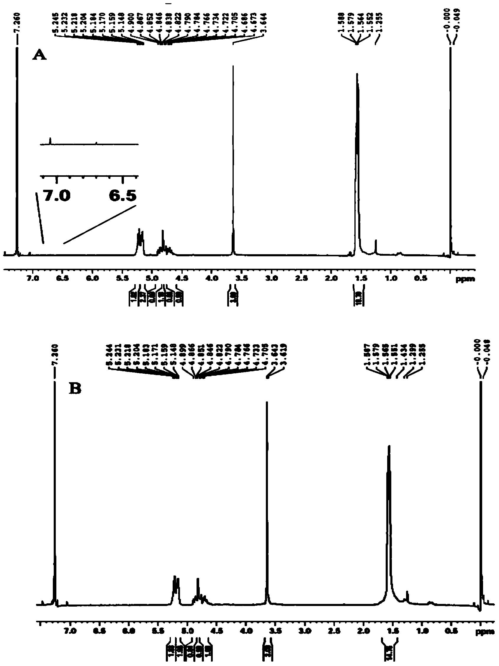
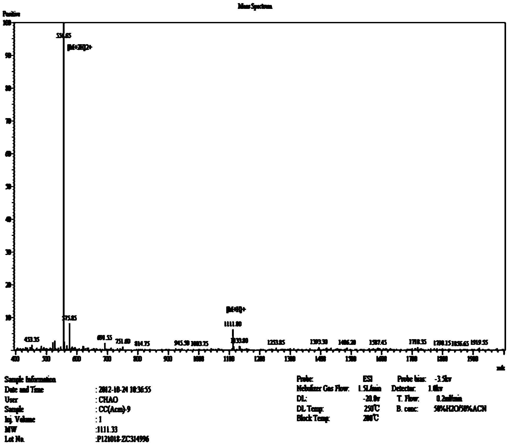
[0040] Example 1: Synthesis of Pep-PEG-PLGA

[0041] Weigh 20mg Mal-PEG(3500)-PLGA(38000), dissolve it in 2mL DMF, add dropwise to 8mL phosphate buffer (pH: 7.0), and stir to form a nanoparticle solution. Weigh the Pep-1 (Acm) polypeptide and dissolve it in 2 mL of phosphate buffer (pH: 7.0). Add the polypeptide solution dropwise to the nanoparticle solution (the molar ratio of Male to Pep-1 (Acm) is 1:3) , Nitrogen protection, stirring, reaction for 12h. The nanoparticle solution after the reaction is dialyzed to remove unreacted polypeptides. Add 1 mL of acetic acid to the dialyzed nanoparticle solution, add dropwise a proper amount of iodine in methanol solution, protected by nitrogen, and react for 0.5 hours. After the reaction, the nanoparticle solution is dialyzed and lyophilized to obtain Pep-PEG-PLGA.

Embodiment 2

[0042] Example 2: Emulsification / solvent evaporation method

[0043] Add 1mg PTX and MePEG(2000)-PLGA(38000): Pep-PEG-PLGA(38000)=9:1 to ethyl acetate to dissolve, add an appropriate amount of 1% poloxamer 188 aqueous solution, and intermittently ultrasonic (200W, 5min) form an oil-in-water (O / W) emulsion, disperse it into an appropriate amount of 0.5% poloxamer 188 aqueous solution, stir, rotate and evaporate at 40°C to remove ethyl acetate, filter with 0.45μm and 0.22μm microporous membranes respectively, Get nanoparticles.

[0044] The nanoparticle solution is clear in appearance and has obvious blue opalescence. The laser particle size analysis showed that the obtained nanoparticles showed a normal distribution with an effective diameter of 94.25nm and a polydispersity of 0.117. Observed under a scanning electron microscope, the nanoparticles have a regular spherical appearance, are well dispersed in the solution, and have good stability. The nanoparticle encapsulation rate ...

Embodiment 3

[0045] Example 3: Solvent diffusion method

[0046] Add 1mg PTX and MePEG(2000)-PLGA(38000): Pep-PEG-PLGA(38000)=9:1 to acetone to dissolve, add dropwise to an appropriate amount of 1% poloxamer 188 aqueous solution, stir, 50℃ The acetone was removed by rotary evaporation and filtered with 0.45μm and 0.22μm microporous membranes respectively to obtain nanoparticles.

[0047] The appearance of the nanoparticles is clear, with obvious blue opalescence. Laser particle size analysis shows that the obtained nanoparticles have a normal distribution with 106.45nm as the effective diameter, and the polydispersity is 0.165. Observed under a scanning electron microscope, the nanoparticles have a regular spherical appearance, are well dispersed in the solution, and have good stability. The nanoparticle encapsulation rate was 23.4%, and the drug loading was 1.14%.


PUM


Abstract

The invention discloses a Pep-1 peptide modified gliomas targeted nano drug delivery system and a preparation method thereof. The nano drug delivery system comprises polymer nano particles prepared by using an amphiphilic block copolymer (Pep-PEG-PLGA) as a carrier material, paclitaxel wrapped and carried by the polymer nano particles, and modified ligand Pep-1 polypeptide on the surfaces of the polymer nano particles. The amphiphilic block copolymer (Pep-PEG-PLGA) is composed of Male-PEG-PLGA and MePEG-PLGA, Pep-1 is adopted as a molecule with targeting function, the amphiphilic block copolymer PEG-PLGA is taken as a carrier material, the Pep-1 polypeptide is modified on the carrier material via covalent binding, and gliomas targeted polymer nano particles are prepared. According to the Pep-1 peptide modified gliomas targeted nano drug delivery system, gliomas targeting can be voluntarily performed, and the uptaking and accumulation of an anti-tumour drug in the gliomas part can be improved, as a result, the gliomas therapeutic effect is improved.

Description

Technical field [0001] The invention belongs to the technical field of tumor targeting and sustained-release drug delivery systems, and relates to a targeted nano-delivery system modified by glioma homing peptides, and in particular to a new type of peptide modified polymer nanoparticles loaded with paclitaxel in brain glial Tumor targeting nano-delivery system and preparation method thereof. Background technique [0002] Glioma is the most common malignant tumor of the central nervous system, accounting for about 80% of all primary nervous system tumors. Glioma has a high degree of malignancy, a very poor prognosis, and unsatisfactory treatment effects. The current treatment methods for malignant tumors mainly include surgery, chemotherapy, and radiotherapy. Among them, chemotherapy is a very common treatment method in clinical applications, but it is absolutely Most chemotherapeutic drugs not only kill tumor cells, but also destroy normal tissues and cells, causing serious sid...

Claims


Application Information

Patent Timeline
IPC IPC(8): A61K9/51A61K31/337A61K47/42A61K47/34A61P35/00C08G65/48C08G63/91
Inventor 辛洪亮徐群为王宝彦吕玲燕王中元吴琳赵越
Owner NANJING MEDICAL UNIV
Why Eureka
Try Eureka