Micro-nanofiber tissue engineering scaffold and preparation method thereof

A fibrous tissue and microfiber technology is applied in the field of micro-nanofiber tissue engineering scaffold and its preparation, and achieves the effects of simple preparation method, favorable adhesion, and simple and easy preparation method

Inactive Publication Date: 2014-04-02
EAST CHINA JIAOTONG UNIVERSITY
View PDF6 Cites 20 Cited by
  • Summary
  • Abstract
  • Description
  • Claims
  • Application Information

AI Technical Summary

Problems solved by technology

However, so far there is no relevant disclosed technology that combines bact

Method used


Image

  • Micro-nanofiber tissue engineering scaffold and preparation method thereof
  • Micro-nanofiber tissue engineering scaffold and preparation method thereof

Examples


Embodiment 1

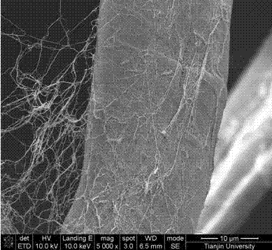
[0022] As an embodiment of the micro-nanofibrous tissue engineering scaffold of the present invention, it includes micron fibers, bacterial cellulose nanofibers, micropores and bacterial cellulose nanopores; the diameter of the micron fibers is 5 microns, and the bacterial cellulose The diameter of the nanofiber is 10 nanometers; the diameter of the micropore is 100 micrometers, and the diameter of the bacterial cellulose nanopore is 10 nanometers.

[0023] A preparation method for preparing micro-nanofibrous tissue engineering scaffolds using cellulose as microfiber scaffolds, cutting the cellulose microfiber scaffolds into square pieces with a size of 20×20×0.5 mm, and preparing Acetobacter xylinum culture medium, that is, using deionized Water is the solvent, and the mass fractions of each solute are: 2.5% glucose, 0.75% yeast powder, 1.0% peptone, 1.0% Na2HPO4, stir in a beaker at room temperature until the solute is completely dissolved, and adjust the pH of the medium to ...

Embodiment 2

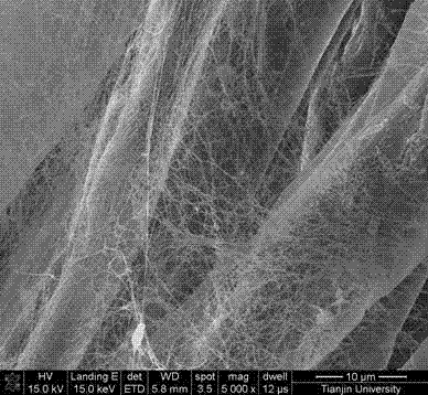
[0026] As an embodiment of the micro-nanofibrous tissue engineering scaffold of the present invention, the difference from Embodiment 1 is that in this embodiment, the diameter of the micron fibers is 500 microns, and the diameter of the bacterial cellulose nanofibers is 100 microns. nanometer; the diameter of the micrometer pore is 500 microns, and the diameter of the bacterial cellulose nanopore is 100 nanometers.

[0027] A method for preparing micro-nanofibrous tissue engineering scaffolds using gelatin as a microfiber scaffold, cutting the gelatin microfiber scaffold into square pieces with a size of 20×20×2mm, and preparing Acetobacter xylinum culture medium (that is, using deionized water as a solvent , the mass fractions of each solute are: 2.5% glucose, 0.75% yeast powder, 1.0% peptone, 1.0% Na2HPO4), stir in a beaker at room temperature until the solute is completely dissolved, adjust the pH of the medium to 5 by adding acetic acid dropwise; Put the plain microfiber ...

Embodiment 3

[0030] As an embodiment of the micro-nanofibrous tissue engineering scaffold of the present invention, the difference from Embodiment 1 is that in this embodiment, the diameter of the micron fibers is 250 microns, and the diameter of the bacterial cellulose nanofibers is 50 microns. nanometer; the diameter of the micrometer pore is 300 microns, and the diameter of the bacterial cellulose nanopore is 60 nanometers.

[0031] Step 1 in the preparation method of micro-nanofibrous tissue engineering scaffold: prepare bacterial fermentation medium: use deionized water as solvent, and the mass fractions of each solute are: glucose 2.5%, yeast powder 0.75%, peptone 1.0%, Na2HPO41 .0%, stir in a beaker at room temperature until the solute is completely dissolved, and adjust the pH of the medium to 4.5 by adding acetic acid dropwise.


PUM

PropertyMeasurementUnit
Diameteraaaaaaaaaa
Diameteraaaaaaaaaa
Diameteraaaaaaaaaa

Abstract

The invention relates to the technical field of biological materials, in particular to a micro-nanofiber tissue engineering scaffold and a preparation method thereof. The micro-nanofiber tissue engineering scaffold comprises microfibers, bacterial cellulose nanofibers, micropores and bacterial cellulose micropores, wherein the microfibers are 5-500 microns in diameter, the bacterial cellulose nanofibers are 10-100 nm in diameter, the micropores are 200-500 microns in diameter, and the bacterial cellulose micropores are 10-100 nm in diameter. Compared with the prior art, the micro-nanofiber tissue engineering scaffold is high in bionic degree, stable in structure and proper in porosity and aperture, and can provide a proper external environment for cells; the preparation method is simple and easy to implement, low in cost, wide in raw material source, environment-friendly and pollution-free.

Description

technical field [0001] The invention relates to the technical field of biomaterials, in particular to a micro-nano fiber tissue engineering scaffold and a preparation method thereof. Background technique [0002] Whether it is tissue engineering or regenerative medicine, the performance of scaffold materials determines the ultimate success or failure. From the perspective of geometric characteristics, the extracellular matrix in vivo (hereinafter referred to as ECM) is a three-dimensional fibrous structure, which not only contains micro-scale pores and fibers, but also has nano-scale pores and fibers, that is, ECM has multi-scale fibers (50-500 mm in diameter). Nano) and porous structures, these nanoscale structures work in concert with microstructures to control the behavior of cells and the function of tissues and organs. Therefore, from the perspective of bionics, it is necessary to design and manufacture fibrous biomimetic scaffolds with appropriate geometric properties...

Claims


Application Information

Patent Timeline
IPC IPC(8): A61L27/56A61L27/20A61L27/18A61L27/24A61L27/22
Inventor 熊光耀万怡灶罗红林李秋萍麻春英李德英
Owner EAST CHINA JIAOTONG UNIVERSITY
Why Eureka
Try Eureka