
Surface plasma resonance (SPR) sensor chip as well as preparation method and application thereof

A surface plasmon and sensor chip technology, applied in the field of medical detection, can solve the problems of low expression abundance, lack of common features, difficult detection, etc., and achieve the effects of good test effect, stable test effect and simple use method.

Inactive Publication Date: 2014-04-02
UNIV OF SCI & TECH BEIJING
View PDF3 Cites 9 Cited by
  • Summary
  • Abstract
  • Description
  • Claims
  • Application Information

AI Technical Summary

Problems solved by technology

However, traditional SPR detection methods are difficult to directly detect molecules with low molecular weight and ultra-low concentration.
miRNAs have short sequences, lack of common features, low content, and low abundance in cells, so it is difficult to detect directly using traditional SPR technology
Most SPR sensors are based on spherical gold nanoparticles, and there are less studies on gold nanorods (GNRs)

Method used


Image

  • Surface plasma resonance (SPR) sensor chip as well as preparation method and application thereof
  • Surface plasma resonance (SPR) sensor chip as well as preparation method and application thereof
  • Surface plasma resonance (SPR) sensor chip as well as preparation method and application thereof

Examples


Embodiment 1

[0046] 1. The method of sputtering a gold film with a thickness of 40-60 nm on the glass slide is:

[0047] (1) Place the BK7 slides at 80°C in piranha (H 2 o 2 with concentrated H 2 SO 4 Mixed solution with a volume ratio of 1:3) for 30 minutes, after cooling to room temperature, the slides were rinsed with deionized water, N 2 blow dry;

[0048] (2) Place the cleaned glass slide in a vacuum coating machine for coating:

[0049] a) First coat a layer of 2 nm thick chromium film;

[0050] b) Continue to plate a 50 nm thick gold film on the chromium film.

[0051] 2. Chip preparation:

[0052] (1) Rinse the clean gold film with 100% ethanol and then with deionized water, and finally wash it with N 2 Air dry for later use.

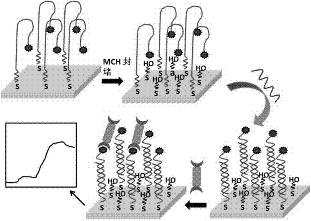
[0053] (2) Prepare 0.1 μM biotin-miRNA probe solution, which contains 2 μM TCEP in PBS solution (0.1 M, pH 7.4).

[0054] (3) Drop 25 μL of biotin-miRNA probe solution onto the prepared gold membrane and leave it overnight at 4°C.

[0055] (4) Aft...

Embodiment 2

[0080] 1. Preparation of gold film:

[0081] (1) Place the BK7 slides at 80°C in piranha (H 2 o 2 with concentrated H 2 SO 4 Mixed solution with a volume ratio of 1:3) for 30 minutes, after cooling to room temperature, the slides were rinsed with deionized water, N 2 blow dry;

[0082](2) Place the cleaned glass slide in a vacuum coating machine for coating:

[0083] a) First coat a layer of 2 nm thick chromium film;

[0084] b) Continue to plate a 50 nm thick gold film on the chromium film.

[0085] 2. Preparation of chip: Same as Example 1.

[0086] 3. Preparation of GNRs-streptavidin

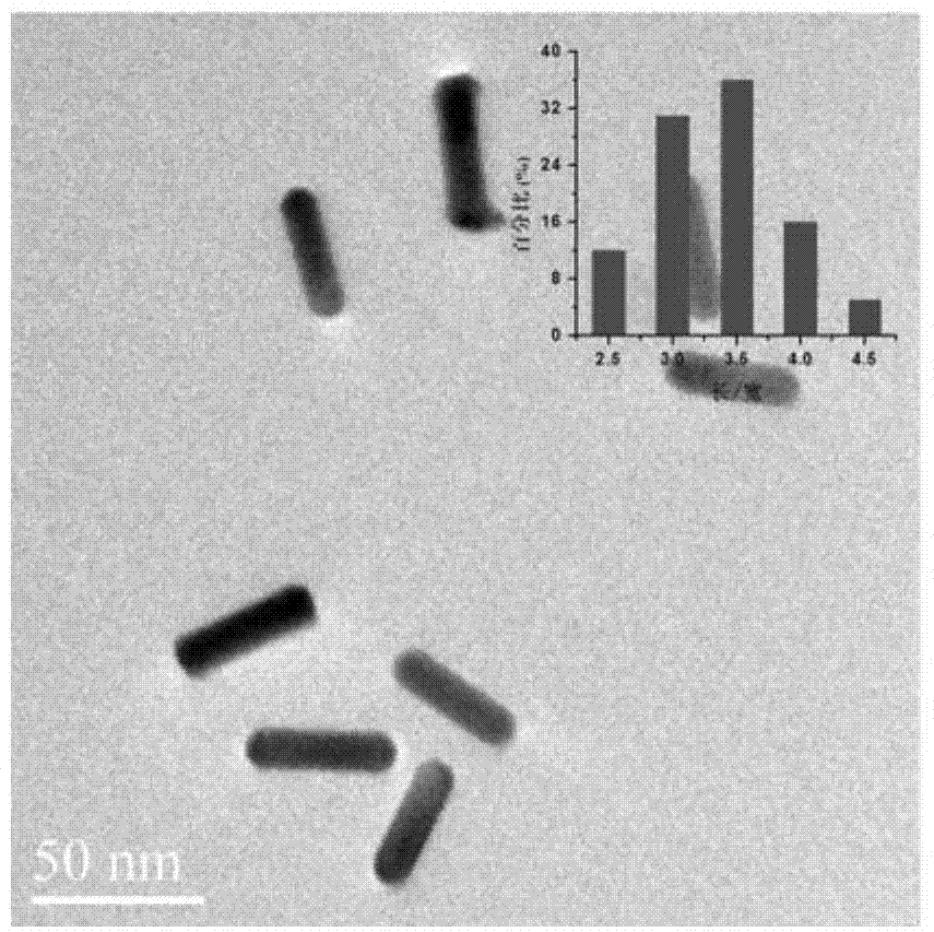
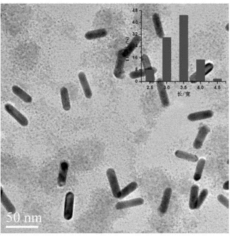
[0087] Use 1-ethyl-(3-dimethylaminopropyl) carbodiimide hydrochloride (EDC) to activate the carboxyl group of lipoic acid (TA), then combine with the amine group of streptavidin, the The procedure for end-binding of GNRs and streptavidin is as follows: first, 3 mL of redispersed GNRs solution was added dropwise to 6 mL of diluted streptavidin (2×10 -8 M) solution, shake lightly for ...

Embodiment 3

[0097] The steps for specific detection using the miRNA sensor obtained in Example 1 are:

[0098] (1) Inject 20 nM of target miRNA sequence, single-base mismatch sequence and three-base mismatch sequence on the surface modified by biotin-miRNA probe respectively, the steps are the same as 4 in Example 2, and record the corresponding SPR response signal.

[0099] (2) The regeneration steps of the sensor refer to Embodiment 2.


PUM

PropertyMeasurementUnit
Thicknessaaaaaaaaaa
Thicknessaaaaaaaaaa

Abstract

The invention relates to a surface plasma resonance (SPR) sensor chip as well as a preparation method and application thereof and belongs to the field of medical detection. A structure of the SPR sensor chip is as follows: the surface of a substrate glass sheet is coated with a chromium film and a gold film in sequence, a biotin-miRNA probe is modified on the surface of the coated gold film; the biotin-miRNA probe is used for reducing sulfydryl by applying tri(2-carboxyethyl) phosphine (TCEP (Trichloroethyl Phosphate)), and 6-sulfydryl 1-hexyl alcohol (MCH) is used for plugging. The surface plasma resonance (SPR) sensor chip has the beneficial effects of being used for directly detecting molecules with low molecular weight and ultra-low concentration; moreover, the invention discloses the preparation method of the SPR sensor chip, according to the preparation method, the preparation cost is low and the test effect is stable; the invention discloses application of the sensor chip, the application method of the chip is simple, special equipment is not needed; moreover, test materials are reproducible and can be recycled, and the test effect is good.

Description

technical field [0001] The invention belongs to the field of medical detection, in particular to a detection method for microRNA (miRNA), specifically an ultrasensitive and label-free rapid detection method for microRNA (miRNA) based on surface plasmon resonance (SPR) and gold nanorod signal amplification, belonging to field of medical testing. [0002] technical background [0003] MicroRNAs (miRNAs) are a class of non-coding small single-stranded RNAs that are highly conserved in evolution, play a vital role in gene post-transcriptional regulation, and are widely present in animals and plants. More and more evidence shows that miRNAs Participate in many important physiological and pathological processes, such as development, organ formation, apoptosis, cell proliferation, tumorigenesis, etc. Therefore, the analysis and detection of miRNAs has become a key technology for us to further study their biological functions, disease diagnosis and treatment, and the development o...

Claims


Application Information

Patent Timeline
IPC IPC(8): G01N21/552B82Y40/00
Inventor 郝开红董海峰金时代文浩
Owner UNIV OF SCI & TECH BEIJING
Learn More