
Monoclonal antibody against extracellular domain of goose CD3 epsilon chain and application of monoclonal antibody in detection of goose CD3<+> T lymphocytes

A technology of monoclonal antibody and extracellular region, applied in anti-receptor/cell surface antigen/cell surface determinant immunoglobulin, receptor/cell surface antigen/cell surface determinant, application, etc. The occurrence and development of goose T lymphocytes, the rules of the body's immune defense, and the constraints on the development of basic immune theories of waterfowl, etc.

Active Publication Date: 2014-12-24
NORTHEAST AGRICULTURAL UNIVERSITY
View PDF3 Cites 4 Cited by
  • Summary
  • Abstract
  • Description
  • Claims
  • Application Information

AI Technical Summary

Problems solved by technology

[0007] However, the research on the cloning and expression of the goose CD3ε chain gene and the preparation of corresponding polyclonal and monoclonal antibodies is still blank, which seriously restricts the development of the basic immune theory of waterfowl, resulting in the inability to understand the occurrence and development of goose T lymphocytes. The role of immune defense, for some diseases that cause huge economic losses to the goose industry, how to evaluate whether the pathogen activates T lymphocytes, how to activate T lymphocytes, what role the activated T lymphocytes play, and how to use pathogens and T lymphocytes The relationship between the immune function of lymphocytes guides the diagnosis of diseases, the development of vaccines, etc., thus providing a theoretical basis and material basis for scientific prevention of epidemic diseases

Method used


Image

  • Monoclonal antibody against extracellular domain of goose CD3 epsilon chain and application of monoclonal antibody in detection of goose CD3&lt;+&gt; T lymphocytes
  • Monoclonal antibody against extracellular domain of goose CD3 epsilon chain and application of monoclonal antibody in detection of goose CD3&lt;+&gt; T lymphocytes
  • Monoclonal antibody against extracellular domain of goose CD3 epsilon chain and application of monoclonal antibody in detection of goose CD3&lt;+&gt; T lymphocytes

Examples


Embodiment 1

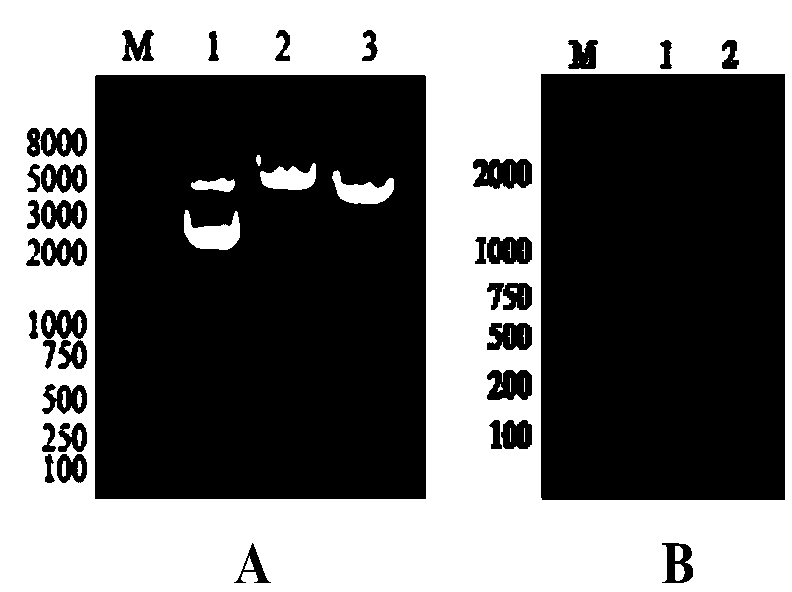
[0067] Example 1 Cloning of Goose CD3ε Extracellular Region Gene

[0068] Method:

[0069] 1. Cloning of goose CD3ε gene

[0070] 1.1 Primer design

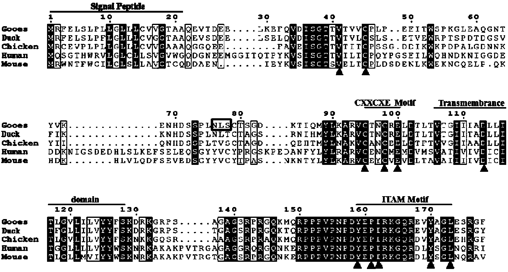
[0071] Refer to the mallard CD3ε gene sequence published on GenBank (accession number: AF378704), and use Primer premier5.0 molecular biology software to assist in the design of a pair of specific primers (CD3-S and CD3-A, shown in Table 1) for amplification Goose CD3ε gene, primers were synthesized by Shanghai Shenggong Biological Engineering Technology Service Co., Ltd.

[0072] Table 1 Primers for amplifying goose CD3ε gene

[0073]

[0074] 1.2 Cloning and identification of goose CD3ε gene

[0075] (1) Gene cloning, purification and recovery

[0076] Using the goose thymus cDNA amplified by Dr. Wei Shuangshi in the laboratory as a template, add each solution in the following order to establish a 50μL PCR reaction system: 10×Pfu Buffer 5μL; dNTP (2.5mM) 4μL; upstream and downstream primers (10μM) each 1μL, Template 1μL, Pfu (2.5U / μL) 1μL...

Embodiment 2

[0124] Example 2 Preparation and identification of rabbit anti-goose CDε extracellular domain polyclonal antibody

[0125] Method:

[0126] 1. Preparation of rabbit anti-goose CD3ε extracellular domain polyclonal antibody

[0127] Two 2.5kg New Zealand white rabbits were selected, and blood was collected from the ear vein before immunization and the serum was separated as a negative control. Take 200 ug of rGoCD3ε protein purified in Example 1 and an equal volume of Freund’s complete adjuvant, and after fully emulsifying, inject multiple subcutaneous injections into the back of New Zealand white rabbits for immunization. The immunization interval is two weeks. A total of four immunizations are performed in the same way. For the first immunization, but the adjuvant was Freund's incomplete adjuvant, 10-14 days after the last immunization, blood was collected from the rabbit's heart, serum was collected, and stored at -20°C.

[0128] 2. The titer determination of rabbit anti-goose CD3ε ...

Embodiment 3

[0149] Example 3 Preparation of monoclonal antibodies to the extracellular region of goose CD3ε

[0150] Method:

[0151] 1. Preparation of immunogen

[0152] Using the eukaryotic expression plasmid pcDNA3.1-CD3εex prepared in 4 in Example 2 as the immunogen, the recombinant plasmid was extracted and purified first, and the method for mass preparation of plasmids was carried out according to the "Molecular Cloning Experiment Guide". The purified plasmid was subjected to single and double enzyme digestion and PCR identification with BamHI and XhoI. Store at -20℃ for later use.

[0153] 2. Animal immunity

[0154] First 1-3 days before immunization, 4-6 weeks old BALB / c mice were pretreated with bupivacaine hydrochloride, and then the recombinant eukaryotic expression plasmid pcDNA3.1-CD3εex was diluted with sterile PBS , Immunized separately, 100μg / mouse, injected into the quadriceps femoris muscle of the hind leg. The immunization interval is 2 to 3 weeks. After three immunizations,...


PUM


Abstract

The invention discloses a monoclonal antibody against an extracellular domain of a goose CD3 epsilon chain and an application of the monoclonal antibody in detection of goose CD3<+> T lymphocytes. Amplification is performed by adopting goose thymus cDNA as a template to obtain a goose CD3 epsilon gene, according to the characteristics of a GoCD3 epsilon extracellular domain gene, a pair of specific primers are designed, amplification is performed to obtain the goose CD3 epsilon extracellular domain gene (CD3 epsilon ex) and the CD3 epsilon ex is expressed by virtue of escherichia coli; the recombinant plasmid containing the CD3 epsilon ex is adopted as an immunogen to immunize BALB / c mice, and the recombinant protein rGoCD3 epsilon is used for booster immunization to obtain a hybridoma cell strain (3C11) capable of stably secreting the monoclonal antibody against the extracellular domain of the goose CD3 epsilon chain. The monoclonal antibody can react with the purified recombinant protein rGoCD3 epsilon and can have a specific reaction with separated goose peripheral blood lymphocytes. Therefore, a novel and effective technical means is provided for detecting the goose CD3<+> T lymphocytes.

Description

Technical field [0001] The invention relates to a monoclonal antibody and its application, in particular to a monoclonal antibody to the extracellular region of goose CD3 epsilon chain and its application in the detection of goose CD3 + The application of T lymphocytes belongs to the field of biotechnology. Background technique [0002] T lymphocytes (referred to as T cells) are important immunocompetent cells, which have a key role in directly mediating cellular immune responses and regulating the immune response of the body. At different developmental stages of T cells, different types of receptors and surface antigens are expressed on the cell surface. The study of the biological characteristics of T cell surface antigens is the basis for understanding the development and function of T cells and identifying T cell subsets. The preparation of monoclonal antibodies to corresponding T cell surface antigens, and on this basis, the establishment of different subgroups of T cell de...

Claims


Application Information

Patent Timeline
IPC IPC(8): C07K14/705C12N15/12C12N5/20C07K16/28G01N33/577G01N33/569
CPCC07K14/7051C07K16/2809G01N33/56966
Inventor 马波张雪莲李洪涛高明春张文龙魏双施王君伟
Owner NORTHEAST AGRICULTURAL UNIVERSITY
Learn More