

Fluorescent-CT bimodal imaging probe and preparation method thereof
A-CT, dual-mode technology, applied in fluorescence-CT dual-mode iodine-doped carbon quantum dots and its application in biomedical imaging, fluorescence-CT dual-mode imaging probes and their preparation fields, can solve the problem of Short residence time, lack of tissue-specific viscosity and osmotic pressure, etc., to avoid side effects, reduce the risk of adverse reactions, and strong X-ray attenuation
- Summary
- Abstract
- Description
- Claims
- Application Information
AI Technical Summary
Problems solved by technology
Method used
Image

Examples
Embodiment 1
[0029] Dilute 0.1 g of glycine and 0.9 g of iodixanol to 20 mL of double-distilled water, and stir magnetically at room temperature (15-25°C) to fully dissolve to obtain a uniform and transparent solution. Add the above solution into a hydrothermal reaction kettle with polytetrafluoroethylene, heat at 180°C for 4 hours, and after the solution is naturally cooled, filter with medium-speed filter paper to remove insoluble black precipitates, centrifuge at 15,000 g for 30 minutes to remove large particles, and collect The supernatant was injected into a dialysis bag with a molecular cut-off of 2000 Da for dialysis. The dialysis time was 72 h, and the water was changed every 12 h. The dialyzed product was rotovaped to obtain a concentrated solution. The concentrated solution is freeze-dried at -50° C. to powder to obtain gadolinium-doped carbon quantum dots. Yield is 1.2%.
Embodiment 2
[0031] Dilute 0.3 g of glycine and 0.7 g of iodixanol to 20 mL of double-distilled water, and stir magnetically at room temperature (15-25°C) to fully dissolve them to obtain a uniform and transparent solution. Add the above solution into a hydrothermal reaction kettle with polytetrafluoroethylene, heat at 180°C for 4 hours, and after the solution is naturally cooled, filter with medium-speed filter paper to remove insoluble black precipitates, centrifuge at 15,000 g for 30 minutes to remove large particles, and collect The supernatant was injected into a dialysis bag with a molecular cut-off of 2000 Da for dialysis. The dialysis time was 72 h, and the water was changed every 12 h. The dialyzed product was rotovaped to obtain a concentrated solution. The concentrated solution is freeze-dried at -50° C. to powder to obtain gadolinium-doped carbon quantum dots. The yield was 2.6%.
Embodiment 3
[0033]Dilute 0.5 g of glycine and 0.5 g of iodixanol to 20 mL of double-distilled water, and stir magnetically at room temperature (15-25°C) to fully dissolve to obtain a uniform and transparent solution. Add the above solution into a hydrothermal reaction kettle with polytetrafluoroethylene, heat at 180°C for 3 hours, and after the solution is naturally cooled, filter with medium-speed filter paper to remove insoluble black precipitates, centrifuge at 15,000 g for 30 minutes to remove large particles, and collect The supernatant was injected into a dialysis bag with a molecular cut-off of 2000 Da for dialysis. The dialysis time was 72 h, and the water was changed every 12 h. The dialyzed product was rotovaped to obtain a concentrated solution. The concentrated solution is freeze-dried at -50° C. to powder to obtain gadolinium-doped carbon quantum dots. The yield was 4.3%.
PUM
| Property | Measurement | Unit |
|---|---|---|
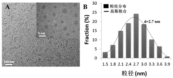
| Particle size | aaaaa | aaaaa |
| Width at half peak | aaaaa | aaaaa |
Abstract
Description
Claims
Application Information